












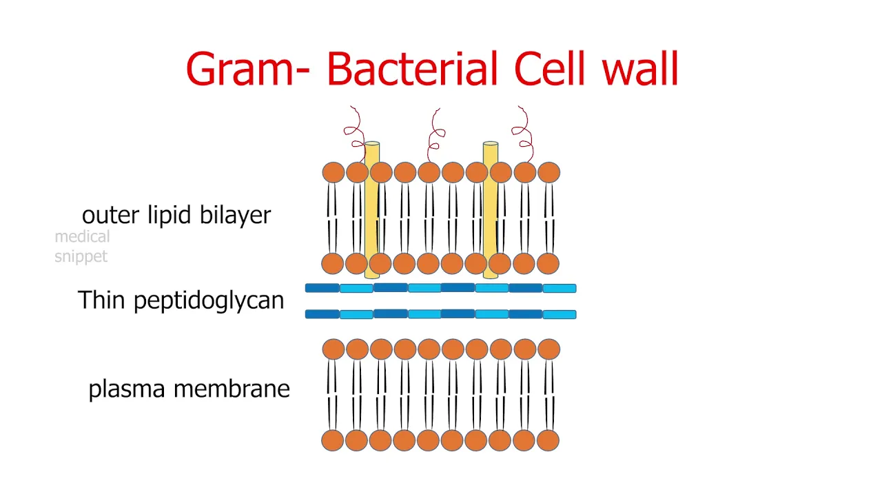












Bacteria are classified as gram-positive or gram-negative based on their cell wall structure revealed through the Gram stain technique; gram-positive bacteria have a thick peptidoglycan layer (up to 30 layers) that retains the crystal violet dye, appearing purple, while gram-negative bacteria have a thin peptidoglycan layer sandwiched between two lipid bilayers, causing them to lose the primary stain and appear pink after counterstaining with safranin.
Gram Positive vs Gram Negative Bacteria: Cell Wall Structure & Gram Staining
Added:Basic prokaryotic cell anatomy, specifically distinguishing the functions of the cell membrane from the outer cell wall.
The chemical structure of biological macromolecules, particularly how amino acids and sugars link to form complex polymers like peptidoglycan.
The fundamental principles of light microscopy, including why contrast-enhancing techniques like biological staining are necessary to visualize microbes.
The concept of osmotic pressure and how a rigid cell wall protects bacterial cells from osmotic lysis in hypotonic environments.
The mechanism of action of beta-lactam antibiotics (such as penicillin) and how cell wall structural differences dictate bacterial susceptibility.
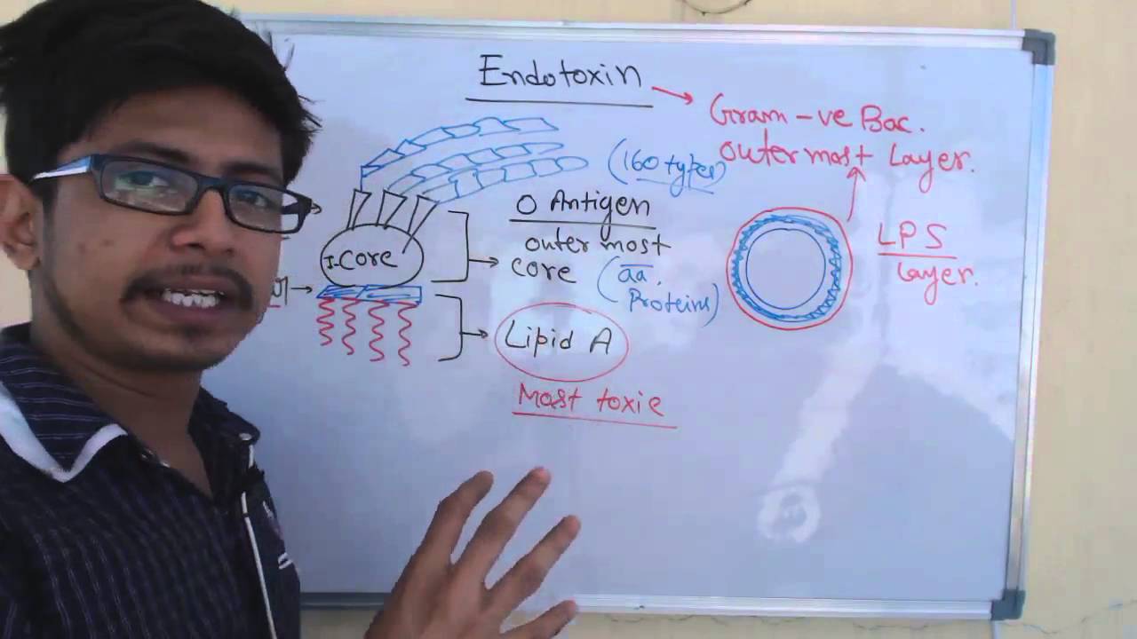
The role of Lipopolysaccharide (LPS) as an endotoxin in Gram-negative bacteria and its clinical significance in triggering septic shock.
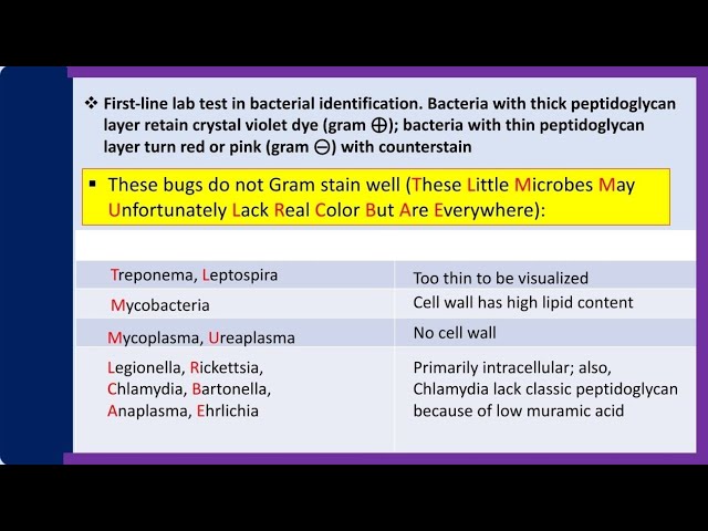
Atypical bacterial groups that do not conform to standard Gram staining, such as acid-fast Mycobacteria and cell-wall-deficient Mycoplasma.
The workflow of clinical microbiology diagnostics, utilizing Gram stain results to guide rapid, empirical antibiotic selection for patient infections.
3.2M views44.6Klikes3:09Original Release: 2019-02-25
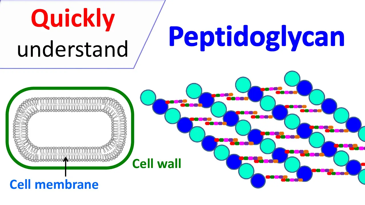
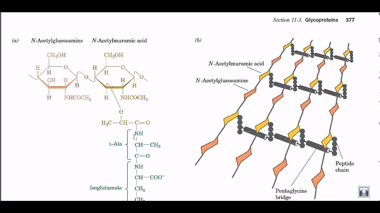
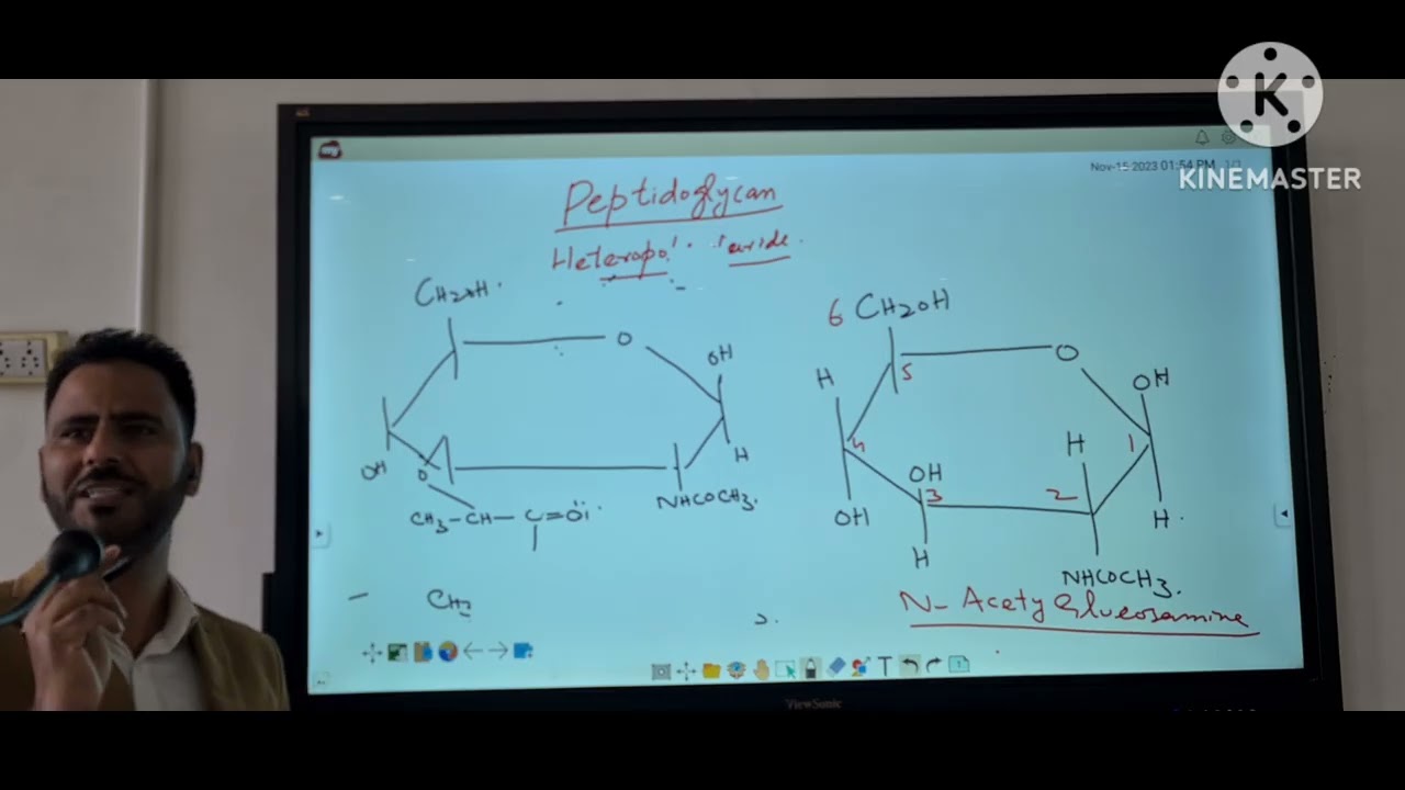
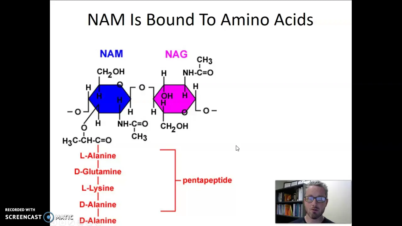
bacteria have cell walls made up of polysaccharides that give them strength and rigidity this is important since bacteria often experience variations in osmotic pressure due to the different solutions they encounter and it is their cell walls which prevent them from shrinking or swelling as a reminder as Moses is the process by which solvent molecules pass through a semipermeable membrane from a less concentrated solution to a more concentrated one equalizing the concentration on either side of the membrane nearly all bacterial cell walls have a peptide polysaccharide layer called peptidoglycan also known as moraine peptidoglycan is a polymer made up of sugars and amino acids which forms a kind of mesh bacteria can be classified based on their reaction to the Gram stain which identifies them as gram positive or gram negative based on the chemical and physical properties of their cell walls gram positive bacteria have a thick cell wall which consists of up to around 30 layers of peptidoglycan this cell wall surrounds a mono derm which is a single plasma membrane gram-negative bacteria have a much thinner cell wall consisting of a single layer of peptidoglycan this layer of peptidoglycan is sandwiched between two lipid bilayer membranes called dye terms we can differentiate between gram-positive and gram-negative bacteria by dyeing them with crystal violet and then washing them with a decolorizing solution then a counter stain is added for example safranin or fruit scene gram positive bacteria will retain the crystal violet dye and remain purple while the gram negative bacteria will be stained pink note that gram positive bacteria also pick up the pink color of the counter stain however this is not visible when they are dyed with the darker purple color of the crystal violet stain the reason for these staining differences is due to differences in cell wall structure which is the chief difference between gram-positive and gram-negative bacteria the Gram stain detects peptidoglycan and since gram positive bacteria have a thick multi-layered peptidoglycan layer they retain the crystal violet dye gram-negative bacteria do not retain the dye for two reasons they have an outer membrane getting in the way of the crystal violet and they lack peptidoglycan to retain the stain although both gram positive and gram negative bacteria can be pathogenic gram-negative bacteria are more resistant to antibodies because of their impenetrable cell wall unfortunately these bacteria also develop resistance more quickly not all bacteria can be reliably classified through gram staining acid-fast bacteria and gram variable bacteria for example do not respond to gram staining acid-fast bacteria are bacteria whose cell walls retain stains particularly well although they aren't closely related to gram positive bacteria they can't appear purple after the Gram stain test Gram variable bacteria show a mix of pink and purple cells when stained if you liked this video like and subscribe you can also support me by following the link to my patreon if you have any topics you'd like me to cover please leave a comment
Up Next
22:38
Antibiotic Resistance Mechanisms & Gene Spread | Microbiology Lecture
@asmicrobiology
38.9K views•2017-04-26

50:12
Bacterial Communication: Quorum Sensing and Biofilm Formation Explained
@JHUAAP
12.6K views•2011-06-28

5:35
Enteric Nervous System Explained: The Gut's Brain | Neurobiology Lecture
@alumniu6029
438 views•2018-09-12

7:08
Bacteriophages: Earth's Deadliest Killers and Future Antibiotics
@kurzgesagt
34.6M views•2018-05-13
Related Study Plans & Knowledge Roadmaps
Structured learning paths in Biology
biology
RNAi: Biogenesis, RISC & Gene Silencing
01Molecular Biology Foundations: DNA to Protein
02Introduction to RNA Interference (RNAi)
03Molecular Biogenesis of miRNA and siRNA
+ 2 more learning modules
10h5 modules
Explore Roadmap
biology
Mycorrhizae: Signaling, Biophysics & Ecology
01Foundations of Mycorrhizal Symbiosis
02Molecular Signaling and Root Colonization
03Biophysics and Bioenergetics of Nutrient Exchange
+ 1 more learning modules
11h4 modules
Explore Roadmap
biology
Circadian Clocks: TTFL, SCN & Periphery
01Foundations of Molecular Biology & Gene Expression
02Introduction to Chronobiology and Actograms
03The Core TTFL: Molecular Architecture of the Clock
+ 3 more learning modules
14h6 modules
Explore Roadmap
biology
Population Genetics: Math, Drift & Selection
01Genetic & Probability Foundations
02The Hardy-Weinberg Principle and Baseline Models
03Mathematical Modeling of Natural Selection
+ 3 more learning modules
18h6 modules
Explore Roadmap