







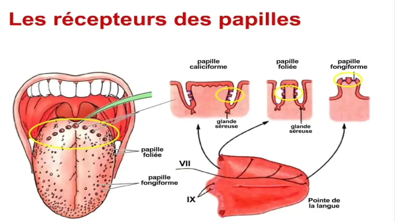



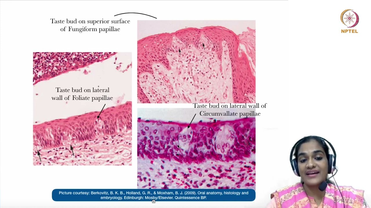
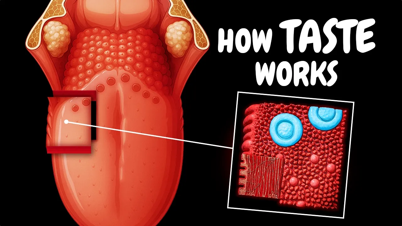

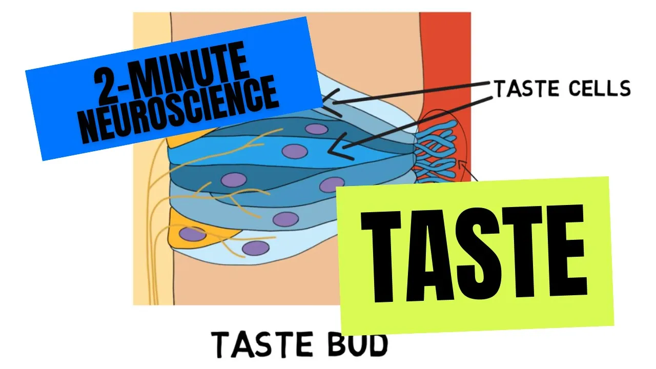

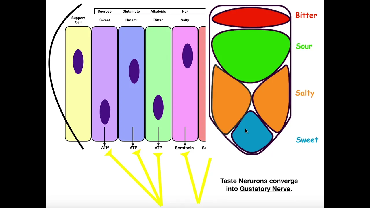







The tongue contains four types of lingual papillae (filiform, fungiform, foliate, and circumvallate), which are localized projections of connective tissue (lamina propria) that elevate the epithelium, while taste buds are microscopic sensory structures embedded within the epithelium of these papillae; filiform papillae are the most numerous and contain keratin but no taste buds, whereas fungiform, foliate, and circumvallate papillae contain taste buds, with circumvallate papillae featuring specialized clefts that trap food particles for taste detection before flushing them away via serous gland secretions.
Histology of Taste Buds and Lingual Papillae
Added:Gross anatomy of the tongue, including its major regions (anterior two-thirds and posterior one-third) and surface features.
Basic tissue histology, specifically the characteristics of stratified squamous epithelium (both keratinized and non-keratinized).
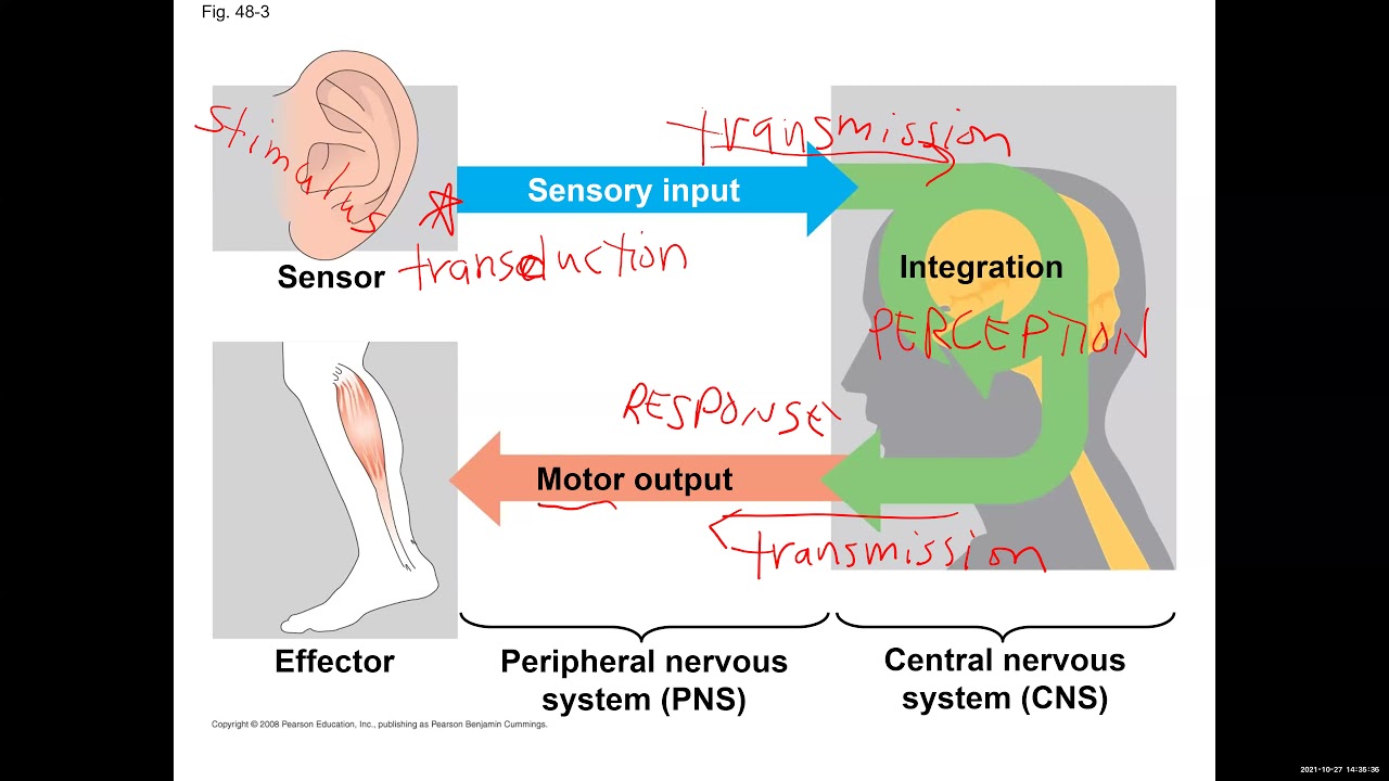
An introduction to the nervous system and how specialized sensory receptor cells transduce external stimuli into neural signals.
The fundamental concept of gustation (the sense of taste) and the distinction between taste (chemical sensation) and flavor (multisensory perception).
The physiological mechanisms of taste transduction, including G-protein coupled receptors (GPCRs) and ion channels involved in detecting the five basic tastes.
The neural pathways of taste, tracing signals from cranial nerves (VII, IX, and X) to the solitary nucleus and the primary gustatory cortex.
Clinical correlation of tongue pathology, such as geographic tongue, hairy tongue, glossitis, and taste disorders like ageusia and dysgeusia.
The role of olfaction (smell) and trigeminal somatosensory inputs (texture, temperature, spiciness) in modulating the overall perception of flavor.
79.9K views1.4Klikes12:15Original Release: 2017-05-13
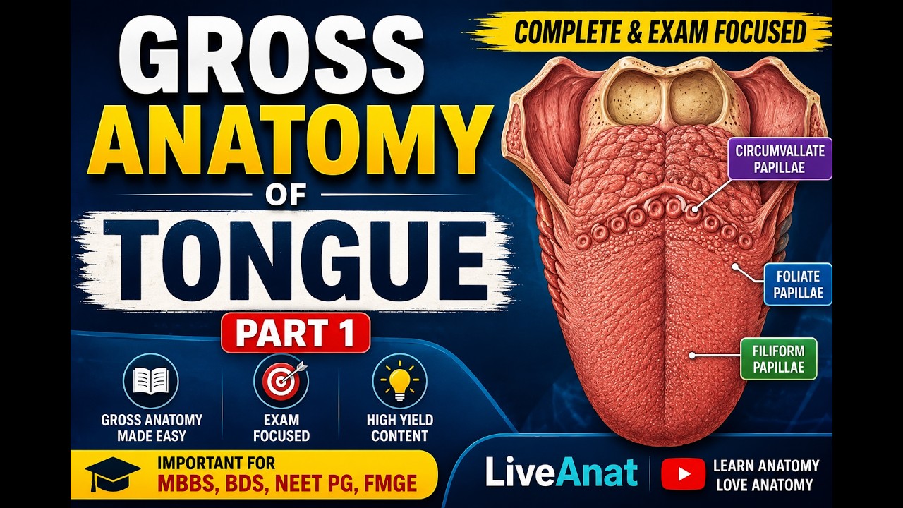
hey guys it's Ryan uh in this video we're going to talk about hystology of the tongue and specifically different differentiate between taste buds and these things called lingual Pilla so let's talk about the tongue in general terms tongue has three main regions the root or the base of the tongue attaches posteriorly to the high bone and the mandible via muscles like the hyoglossus and genioglossus um has the Apex or the tip of the tongue uh touches the inzer teeth and then every point in between there is the dorsum or the dorsal surface of the tongue which is further divided into an anterior 2/3 and a posterior third um and you can notice that they're divided by this uh v-shaped terminal uh terminal sulcus also called sulcus terminalis and so everything anterior to that uh V is the anterior 2/3 everything posterior to it is the posterior third so uh this Fingal part or uh the post sulcus posterior third houses the lingual tonsil um and has no pill on it which we'll talk about in in just a little bit and the uh in contrast the oral part or the preus anterior 2/3 is where all of the Pilla are confined to and we're going to talk about all four of these types so I want to make this really important distinction between Pilla and taste buds some people say that all these little bumps here are the taste buds but that's not true uh the bumps are actually called Pilla and they are localized projections of underlying connective tissue so this underlying layer of connective tissue called blamin appropria elevates the outer layer called epithelium above the base level in that spot and it forms a little uh tongue bump and again uh these Pilla are limited to the pre sucus oral part of the dorsum now taste buds are embedded within the epithelium of these papill and they can even be found embedded in other parts other epithelium of the oral cavity like on a soft pallet and the epiglottis so they can be found uh in more places than just the tongue all right so the four types of papilla are filor fungiform foliate which are pretty minor and the circum valet so filor and Pilla are the most numerous they're the smallest in in size mostly conical in shape and arranged in rows and now they have a thick layer of keratin that that provides a rough surface of the tongue increasing friction um and it can also cause a whitish tint like if you've ever seen the tongue of a cat they have very heavy characterzation of filor Pilla so their tongue appears kind of like having white hairs on it and the filor pill uh do not have taste buds on them or in them I should say so here is a hystological image of filor pill that I found on Google and it's pretty nice it shows you the conle shape in cross-section you can see it's like arranged in a row when when the section was taken and we can um easily see this thick layer of kerot in here it's pretty much uh acellular or might have some nuclei but mostly not and um we can see right underneath it is a stratified squamous epithelium which is kind of like our default oral cavity EP thelium and then um importantly this lamin propria layer this loose connective tissue layer has these um projections upwards that are um perfectly correlated to the filor Pilla above so it's really like this volcanic eruption occurring down here is causing the epithelium to um bump up in the same positions um and then the the purpose of this uh these upward projections is that it increases the contact area between these two layers and so the lamin appropria which is so thick uh and filled with um vasculature and nerves can supply support and Supply nutrition to the uh overlying epithelium so it really just increases surface area so I mentioned a little bit about the lamon appropriate and I just wanted to talk about um these kind of helpful tool so it's universally uh thought that the lamin propria plus the epithelium together um it causes or creates the mucosa layer so um lamin propria plus epithelium equals mucosa also known as the muc mucus membrane which is the the moist lining of the respiratory tract urogenital tract and the GI tract including the oral cavity those places all have a mucosa layer so lamin appropria I remember it by this um little uh I guess kind of like pneumonic thing and I say lamina capria and I remember lamina it has lymphatics here lamina for nerves and then cap for capillaries and I for immune cells and immune cells include lymphocytes plasma cells and especially these B cells that secrete um imunoglobulin a so it has like the I a here so if you remember and you say it like lamina capria you can kind of fill in the rest of the um components here I think it's just a a silly little way that might help you remember uh quickly what's what you're going to find in this layer all right so the next pill is the fungiform Pilla they fewer of these in uh number they're somewhat larger and they're rounded kind of mushroom is shape um they have a deep red color CU they lack um they don't have that thick keratin layer and they do contain uh taste buds this time so uh fungiform pill here they are they're these kind of uh rounded shapes you can see again the lamin appropria here um project upwards and also here we're going to talk more about it later but here are the taste buds just a sneak peek hiding embedded in this epithelium layer all right and then we have the the folate or the the minor type of pill there aren't that many of them they're exclusively located on the posterior lateral border of the tongue which is interesting because uh the posterior lateral border border of the tongue is a highrisk site for oral cancer caner and so an inflamed or swollen foliate Pilla can often look like something worse um and then they also contain taste buds the circum Valley Pilla are probably the most um prominent because they form I'm kind of jumping ahead of myself but they form this terminal sulcus that we talked about before they're arranged in this neat little row about 8 to 12 in number they are defin mushroom shaped they're the largest ones we have and um they're arranged in a V technically just in front of the terminal sulcus so I said that this is like um the terminal sulcus itself well it's kind of like the terminal sulcus is just behind the circum Valley Pilla so that these Pilla can be considered as part of the anterior 2/3 of the tongue basically um and they also contain taste buds so here's a picture of a circum Valley Pilla histologically and then we have a couple layers here uh we have that cariz stratified squamous layer here we have a taste bud embedded in the epithelium have the underlying lamin propria projection then we have a thing called a cleft which is this as you have um the circum Valley Pilla located next to each other and you have this divot in the epithelium this Valley created uh that's important when thought in conjunction with these Von ebner's gland um there's a bunch of these glands located at the base of the cfts and they are sear secreting which is a watery um kind of proteinaceous liquid material and so what happens is let's say we eat something and we have is nice tasty uh piece of burger and the this piece of food will get trapped in the CFT and as it travels down it's getting um all the taste buds are right here so they're right next door to the food particle so they're able to uh receive and uh experience the taste of whatever food you're eating and then so it doesn't get stuck there we have the sear secretion from the Von edner glands that flushes the food back out of the CFT so I just I love that animation but hopefully that makes sense as to how this works and how um how cool this design is that when food comes down taste buds are there to taste it and then water the sear secretion is there to flush it out so it doesn't get uh stuck there and I just have a couple quick questions how many of these pill are there on a tongue oh roughly about 8 to 12 and how are they arranged in that v-shape uh which is just technically anterior to the the v-shaped terminal sulcus all right so now let's talk about taste buds so these are not the bumps on the tongue they're actually thousands of them per tongue and they're located within the epithelium of Pilla and as we said soft uh also soft pallet and elsewhere in the mouth so we have our tongue diagram we have our row of Valley pill we zoom in on that and we get this histological image and then we zoom in on this component and we get something like this so here's our epithelium and then all of these circular structures are the actual taste buds and so we have really cool here we can see the taste pore which is so as food slides down this CFT particles can be um received through the taste por um and then we have our taste receptor cells which are these columnar shaped cells um and then taste buds will have sort of Basil cuboidal cells at the bottom and then we can see we have a connective tissue layer uh deep to the epithelium layer housing the taste buds all right guys that's it for this video I hope you found it helpful and I'll see you all next time thanks for watching
Up Next

12:46
Olfaction Anatomy and Function | MCAT Biology | Khan Academy
@khanacademymedicine
419.2K views•2013-10-25

50:12
Bacterial Communication: Quorum Sensing and Biofilm Formation Explained
@JHUAAP
12.6K views•2011-06-28

5:35
Enteric Nervous System Explained: The Gut's Brain | Neurobiology Lecture
@alumniu6029
438 views•2018-09-12

7:08
Bacteriophages: Earth's Deadliest Killers and Future Antibiotics
@kurzgesagt
34.6M views•2018-05-13
Related Study Plans & Knowledge Roadmaps
Structured learning paths in Biology
biology
RNAi: Biogenesis, RISC & Gene Silencing
01Molecular Biology Foundations: DNA to Protein
02Introduction to RNA Interference (RNAi)
03Molecular Biogenesis of miRNA and siRNA
+ 2 more learning modules
10h5 modules
Explore Roadmap
biology
Mycorrhizae: Signaling, Biophysics & Ecology
01Foundations of Mycorrhizal Symbiosis
02Molecular Signaling and Root Colonization
03Biophysics and Bioenergetics of Nutrient Exchange
+ 1 more learning modules
11h4 modules
Explore Roadmap
biology
Circadian Clocks: TTFL, SCN & Periphery
01Foundations of Molecular Biology & Gene Expression
02Introduction to Chronobiology and Actograms
03The Core TTFL: Molecular Architecture of the Clock
+ 3 more learning modules
14h6 modules
Explore Roadmap
biology
Population Genetics: Math, Drift & Selection
01Genetic & Probability Foundations
02The Hardy-Weinberg Principle and Baseline Models
03Mathematical Modeling of Natural Selection
+ 3 more learning modules
18h6 modules
Explore Roadmap