Proteostasis: Folding, Ubiquitin & Autophagy
Learning Goal: Deconstruct the mechanisms of cellular protein homeostasis (proteostasis), focusing on chaperone-assisted folding, the ubiquitin-proteasome pathway, and autophagy.
- Prerequisites: Introductory biochemistry (understanding of amino acid properties, basic cell structure, and the central dogma).
- Estimated Total Study Time: 12 Hours
Module 1: Foundations of Protein Structure and Synthesis
This module establishes the foundational principles of protein biology. Before analyzing how cells degrade or repair misfolded proteins, you must understand how polypeptides are synthesized by ribosomes, how they fold, and the thermodynamic laws governing their transition from a disordered linear chain to a highly ordered, functional 3D conformation.
Recommended Videos

- Why this video: This video provides a highly visual and concise review of the four levels of protein structure (primary, secondary, tertiary, and quaternary). It introduces how hydrophobic interactions force nonpolar residues into the protein interior while polar residues remain exposed to the aqueous cytosol, a fundamental driving force in proteostasis.

- Why this video: A deeper, thermodynamic exploration of folding. It explains why protein folding is a spontaneous process driven by a negative change in Gibbs free energy (). You will learn about the thermodynamics of the hydrophobic effect, and how local entropy decreases as the protein folds, but overall system entropy increases due to the release of ordered water molecules.
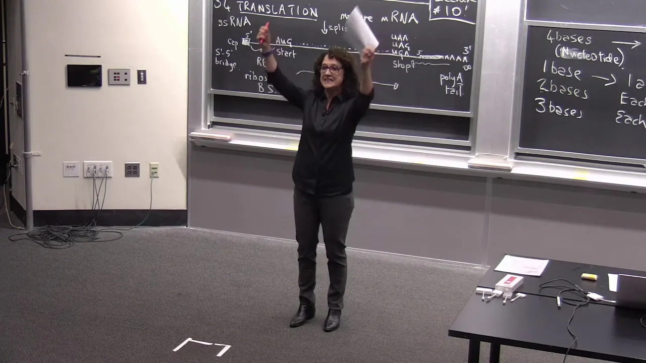
- Why this video: An academic lecture from MIT detailing the molecular mechanics of translation. It covers ribosomal structure, tRNA charging, codon recognition, and the catalysis of peptide bonds. This establishes the structural context of the nascent polypeptide chain emerging from the ribosomal exit tunnel—the exact point where proteostasis networks first engage.
Module 1 Knowledge Checkpoint
- Differentiate between the chemical forces driving secondary structure (hydrogen bonds along the peptide backbone) and tertiary structure (hydrophobic effects, ionic bonds, disulfide bridges, and Van der Waals forces among R-groups).
- Explain how a protein folding reaction can be spontaneous () when the conformational entropy of the folding polypeptide decreases dramatically.
- Describe the molecular events that occur at the A, P, and E sites of the ribosome during mRNA translation elongation.
Module 2: Chaperone-Assisted Protein Folding
While some small proteins fold spontaneously, the crowded macromolecular environment of the cytoplasm (up to 400 mg/mL of proteins) makes newly synthesized chains highly susceptible to premature aggregation. This module explores the molecular chaperones (Hsp70, GroEL/ES chaperonins) that bind to exposed hydrophobic residues to facilitate proper folding and prevent toxic cellular aggregation. It also covers the endoplasmic reticulum's stress response.
Recommended Videos

- Why this video: Led by legendary researcher Arthur Horwich, this video bridges the historical gap between Christian Anfinsen’s "in vitro self-assembly" experiments and the discovery of in vivo chaperone-assisted folding. It explains why cells need active machinery to fold complex proteins within crowded cellular environments.

- Why this video: Provides a highly detailed biochemical and structural breakdown of the GroEL-GroES chaperonin system (known as HSP60-HSP10 in eukaryotes). It outlines the double-cylinder barrel structure, how hydrophobic lining residues capture misfolded intermediates, and how ATP hydrolysis induces a conformational change that creates a hydrophilic "Anfinsen cage" for folding.
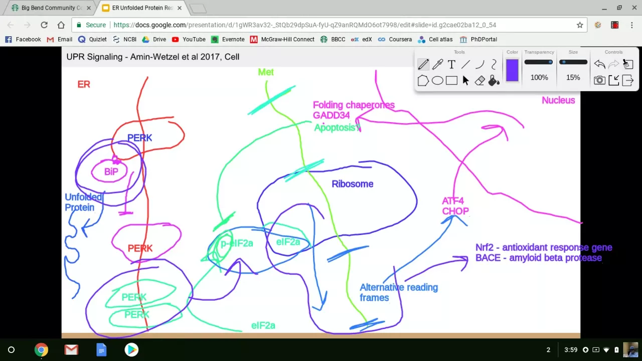
- Why this video: Fills a critical gap in stress-sensing pathways. This academic lecture details the Unfolded Protein Response (UPR) triggered by endoplasmic reticulum (ER) stress. It explains how the three transducer arms (PERK, IRE1, and ATF6) detect accumulated misfolded luminal proteins and orchestrate downstream signaling to halt translation, upregulate chaperones, and activate ER-associated degradation (ERAD).
Independent Study Gap-Fill: Heat Shock Response (HSR)
Note: While the video pool contains great coverage of structural chaperones and the ER-specific Unfolded Protein Response, it lacks a dedicated molecular video detailing the cytosolic Heat Shock Response (HSR) network.
- Independent Search Recommendation: Search YouTube for
"Heat Shock Response HSF1 molecular mechanism activation"to study how Heat Shock Factor 1 (HSF1) dissociates from Hsp90/70 during proteotoxic stress, trimerizes, translocates to the nucleus, and binds Heat Shock Elements (HSE) to induce bulk chaperone transcription.
Module 2 Knowledge Checkpoint
- Detail the cyclical step-by-step mechanism of the GroEL-GroES chaperonin system, including the role of ATP binding, chamber expansion, hydrophilic inversion, and ATP hydrolysis in substrate release.
- Distinguish between the mechanisms of Hsp70 (which binds linear hydrophobic stretches in nascent polypeptides) and Hsp60/chaperonins (which encapsulate fully synthesized client proteins).
- Map the three arms of the Unfolded Protein Response (UPR): list their transmembrane sensors, primary activation mechanisms, and distinct downstream cellular objectives.
Module 3: The Ubiquitin-Proteasome System (UPS)
When chaperones fail to fold a protein, the cell must selectively degrade it to prevent toxic accumulation. This module covers the Ubiquitin-Proteasome System (UPS), the primary pathway for targeted protein degradation in eukaryotes. You will study the E1-E2-E3 enzymatic cascade that tags substrates with polyubiquitin chains, and the structure of the 26S proteasome that acts as the cell's molecular paper shredder.
Recommended Videos

- Why this video: A clear step-by-step breakdown of the conjugation chemistry of ubiquitin. This video teaches you how a 76-amino acid ubiquitin molecule is covalently linked to a target protein. It explains the enzymatic cascade: E1 (activating), E2 (conjugating), and E3 (ligating) enzymes, culminating in the formation of an isopeptide bond with a lysine residue on the target.
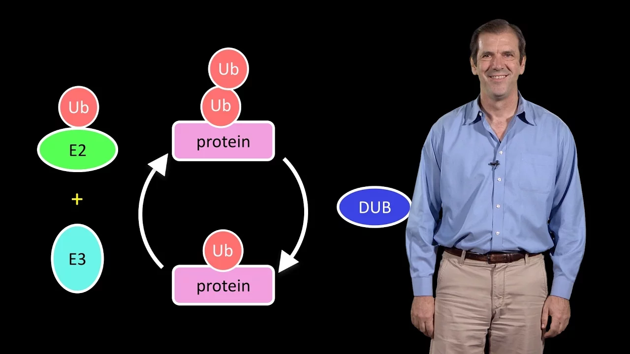
- Why this video: An excellent systemic overview of the UPS, highlighting its massive evolutionary investment—accounting for approximately 5% of the human genome. It explains how different cell types maintain proteostatic balance through highly specific E3 ubiquitin ligase networks, and introduces the functional diversity of ubiquitin signals.
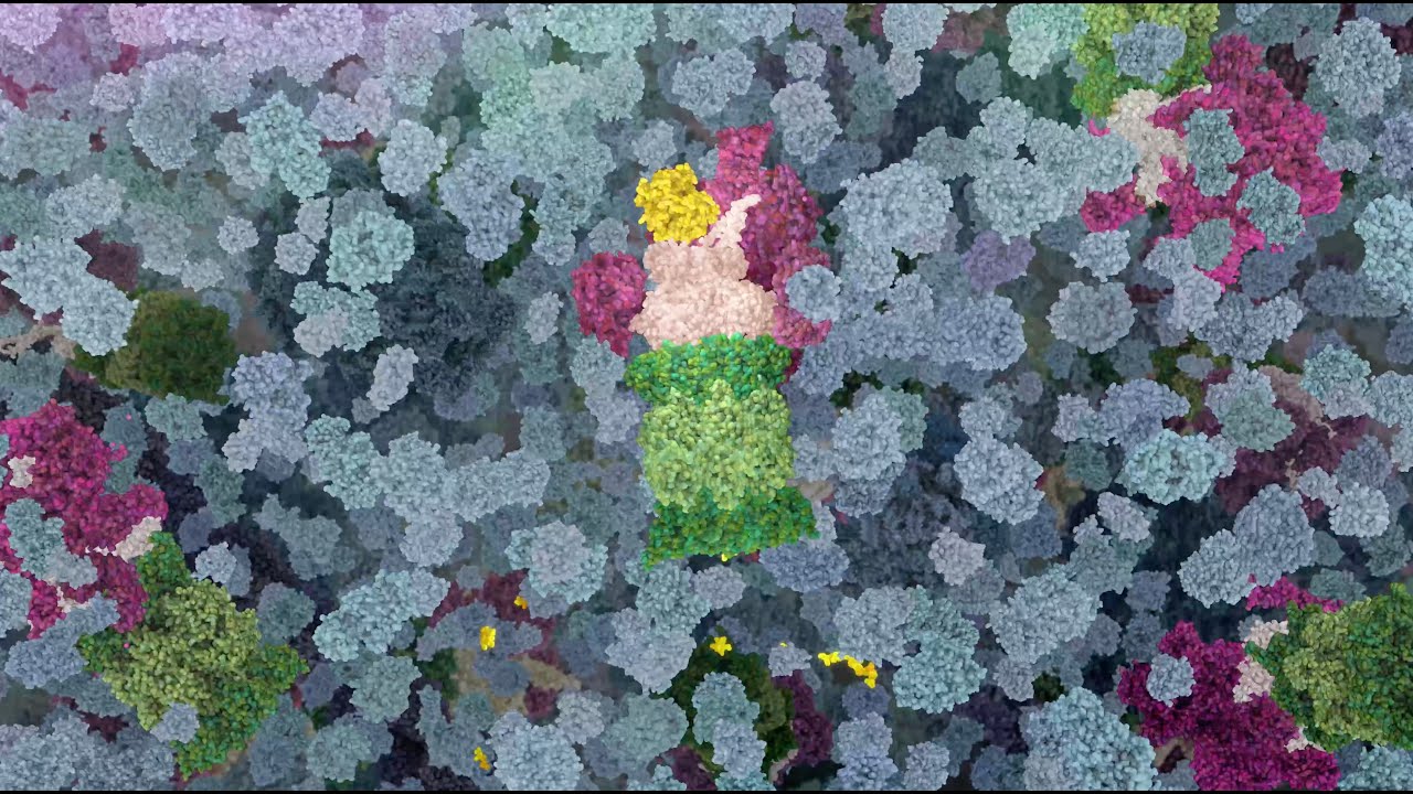
- Why this video: A state-of-the-art 3D molecular animation showing the 26S proteasome in action. It visualizes how the 19S regulatory particle recognizes the polyubiquitin tag, uses ATP-dependent motor proteins to unfold the protein substrate, cleaves off and recycles the ubiquitin tag, and translocates the unfolded polypeptide into the hollow 20S core particle for proteolytic cleavage.
Independent Study Gap-Fill: E3 Ligase Subclasses & Warning
- Terminology Warning: When searching for E1/E2 biochemistry, Google/YouTube algorithms often return organic chemistry tutorials on "E1 vs E2 elimination reactions" (such as Videos 45, 70, and 89 in our initial database). Ensure your search queries are biological, specifying "E1 E2 E3 ubiquitin cascade molecular biochemistry."
- Independent Search Recommendation: Search for
"HECT vs RING E3 ubiquitin ligase mechanism"to learn the chemical distinction between RING E3s (which act as scaffolds to transfer ubiquitin directly from E2 to the substrate) and HECT E3s (which form a covalent thioester intermediate with ubiquitin before transferring it to the substrate).
Module 3 Knowledge Checkpoint
- Draw the full E1-E2-E3 cascade, identifying where ATP is consumed and where thioester and isopeptide bonds are formed.
- Identify the structural differences between the 19S regulatory cap (ubiquitin receptors, deubiquitinases, and AAA+ ATPase unfolding ring) and the 20S catalytic core (alpha structural rings and beta catalytic rings containing protease active sites).
- Explain why a protein must be actively unfolded by the 19S cap before it can enter the 20S chamber.
Module 4: Autophagy: Lysosomal Degradation Pathways
While the UPS handles individual, soluble proteins, it cannot degrade large protein aggregates, intact damaged organelles (like mitochondria), or intracellular pathogens. These macromolecular structures are cleared by autophagy. This module focuses on the molecular pathways of macroautophagy, microautophagy, and Chaperone-Mediated Autophagy (CMA).
Recommended Videos

- Why this video: The definitive, primary-source lecture delivered by Nobel Laureate Yoshinori Ohsumi. It bypasses lifestyle clickbait (which conflates molecular autophagy with diet and weight loss) to detail his original genetic screens in yeast, the identification of ATG genes, and the molecular architecture of the double-membrane autophagosome.
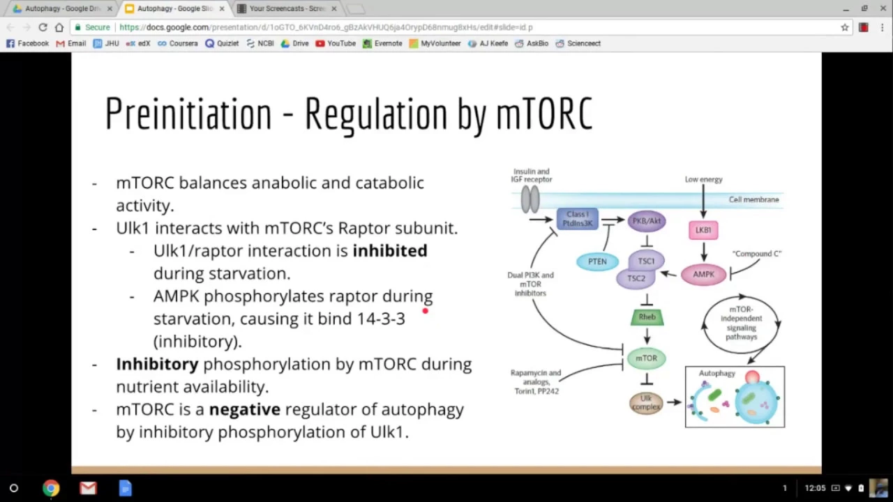
- Why this video: Highly targeted academic overview that directly addresses a major curriculum gap by contrasting Chaperone-Mediated Autophagy (CMA) with macroautophagy. It diagrams how CMA targets soluble proteins bearing a specific pentapeptide motif (KFERQ), binding them to HSC70, which then docks with the lysosomal receptor LAMP2A to form a multimeric pore for translocation.
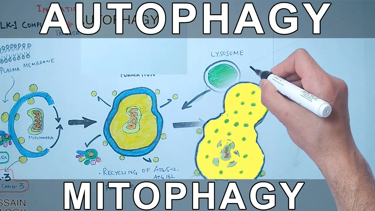
- Why this video: A clear molecular mechanism video focusing on the lipid conjugation and membrane dynamics of autophagosome biogenesis. It explains the initiation steps of the phagophore, elongation, lipid-anchoring of LC3-II, and how the mature double-membrane vesicle fuses with the lysosome via SNARE complexes.
Independent Study Gap-Fill: LC3 Lipidation Details
- Independent Search Recommendation: Search for
"LC3 conjugation cascade ATG5 ATG12 ATG16L1 biochemistry"to understand the precise enzyme cascade that converts soluble LC3-I to lipid-anchored, membrane-bound LC3-II (phosphatidylethanolamine conjugation), which is the biochemical hallmark of autophagosome membrane expansion.
Module 4 Knowledge Checkpoint
- Outline the structural and mechanical differences between macroautophagy (double-membrane autophagosome vesicle), microautophagy (direct lysosomal invagination), and chaperone-mediated autophagy (pore-based translocation).
- Describe the molecular components and chemical reaction required to link LC3 to phosphatidylethanolamine (PE) on the autophagosomal membrane.
- Detail how a CMA client protein containing the KFERQ motif is processed from cytosolic detection to degradation within the lysosome.
Module 5: Proteostasis Collapse, Aging, and Neurodegenerative Disease
As cells age, the efficiency of chaperones, the UPS, and autophagy pathways progressively declines. This systemic failure, known as proteostasis collapse, leads to the accumulation of misfolded proteins and the formation of insoluble amyloid plaques and intracellular neurofibrillary tangles. This module deconstructs the pathological mechanisms of diseases like Alzheimer's, Parkinson's, and Prion-mediated encephalopathies.
Recommended Videos

- Why this video: An interview excerpt with Nobel Laureate Venki Ramakrishnan addressing the global collapse of the proteostasis network during aging. He connects aging to the gradual decay of cellular protein stoichiometry and describes how nutrient-sensing pathways like TOR regulate proteostatic maintenance.
- Why this video: A deep dive into the unique thermodynamics of prion proteins ( to ). It explains the molecular biophysics of template-directed misfolding, demonstrating how a misfolded protein rich in beta-sheets can act as a catalyst to convert normally folded alpha-helical proteins into toxic, self-propagating amyloid fibrils.
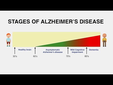
- Why this video: Provides a clear pathobiological analysis of Alzheimer's disease. It covers the biochemical differences between the two diagnostic hallmarks: extracellular amyloid-beta plaques (derived from cleavage of amyloid precursor protein by secretases) and intracellular neurofibrillary tangles (derived from hyperphosphorylated tau proteins dissociating from microtubules).
Module 5 Knowledge Checkpoint
- Explain how a single misfolded protein template () can thermodynamically drive the structural conversion of healthy, soluble proteins () without changing their genetic sequence.
- Describe the structural step-by-step formation of amyloid-beta plaques, noting the enzymatic roles of alpha-, beta-, and gamma-secretases on the amyloid precursor protein (APP).
- Formulate a hypothesis for why neurons are particularly sensitive to proteostasis collapse compared to highly mitotic somatic cells.
Course Map
Below is the recommended learning progression. Each module builds upon the structural and chemical foundations of the previous ones, culminating in the study of systemic disease mechanisms.
Key People Index
- Dr. Christian Anfinsen (Nobel Laureate, 1972): Proved that all the information necessary for a protein to fold into its native 3D conformation is encoded in its primary amino acid sequence (ribonuclease A experiments).
- Dr. Arthur Horwich (Yale / HHMI): Co-discovered the chaperonin-assisted folding machinery (GroEL/ES), establishing that active cellular systems are required to assist protein folding in vivo.
- Dr. Alexander Varshavsky (Caltech / MIT): Discovered the first biological degradation signals (the N-end rule) and identified key enzymology of the ubiquitin system (E1-E2-E3 cascade).
- Dr. Yoshinori Ohsumi (Nobel Laureate, 2016): Used yeast genetics to identify the Autophagy-Related (ATG) genes, elucidating the molecular pathways of autophagosome creation and lysosomal fusion.
- Dr. Stanley Prusiner (Nobel Laureate, 1997): Discovered prions, proving that infectious agents could consist solely of misfolded proteins without nucleic acid genomes.
Final Self-Assessment
Test your mastery of the entire Proteostasis curriculum with these comprehensive check-items:
- I can write down the chemical equation showing how ATP-dependent activation of ubiquitin occurs at the E1 enzyme via an adenylated intermediate.
- I can describe the structural transitions that occur in the GroEL chamber when ATP binds to its heptameric ring, and how this relates to hydrophobic-to-hydrophilic surface inversion.
- I can contrast the thermodynamic stability of a natively folded cellular monomer with that of an amyloid fibril, explaining why amyloid deposits are highly resistant to standard proteolysis.
- I can trace a cellular protein destined for degradation via the UPS from its initial recognition by an E3 ligase to its peptide cleavage within the 20S core of the proteasome.
- I can distinguish between the signals that direct a protein to Chaperone-Mediated Autophagy (CMA) versus the signals that target a protein for macroautophagy or the UPS.
- I can explain the biological mechanism of the Unfolded Protein Response (UPR), detailing how ER membrane receptors (PERK, IRE1, ATF6) sense luminal chaperone depletion.
- I can summarize the role of hyperphosphorylated Tau in microtubule destabilization and explain why it aggregates into intracellular paired helical filaments (PHFs).
- I can explain how the mechanistic Target of Rapamycin (mTOR) complex 1 biochemically inhibits macroautophagy under nutrient-rich conditions.