Autophagy is a cellular degradation and recycling process where cells sequester cytoplasmic components within autophagosomes (double-membrane structures) that fuse with lysosomes to form autolysosomes (single-membrane structures), breaking down damaged organelles and proteins; this process is regulated by the mTOR pathway, which inhibits autophagy under nutrient-rich conditions but activates it during starvation when amino acids and glucose levels decrease, allowing cells to recycle nutrients for survival.
Autophagy and Mitophagy: Pathway and Mechanisms Explained
Added:Deep Dive
Structure and function of eukaryotic organelles, specifically mitochondria and lysosomes.
NTSE SAT (Stage 1) Science | Cell Organelles - Mitochondria and Lysosomes | CBSE 2024-25
6.1K
This comprehensive lesson covers two essential eukaryotic cell organelles. Mitochondria are small, rod-shaped organelles found in eukaryotic cells but not prokaryotic cells. They have a double membrane with highly folded inner membranes called cristae that increase surface area for cellular respiration. The mitochondrial matrix contains enzymes, DNA, RNA, and ribosomes. Mitochondria are the 'powerhouses' of the cell, generating ATP through cellular respiration where glucose is oxidized to produce energy. Lysosomes are membrane-bound organelles formed from the Golgi apparatus, containing digestive enzymes that break down organic materials. They are not found in prokaryotic cells and are primarily present in animal cells. Lysosomes perform intracellular digestion, immune defense against pathogens, programmed cell death (apoptosis), and tissue maintenance by removing dead cells. Both organelles are essential for cellular function, energy production, and maintaining cellular homeostasis.
Click to watch →
Cell Organelles - Mitochondria & Lysosomes | Class 9 Biology Chapter 5 | CBSE 2025-26 #cbse2026
2.7K
Mitochondria are membrane-bound organelles found in eukaryotic cells (except mature RBCs and sieve tube elements) that serve as the 'powerhouse of the cell' by generating ATP through cellular respiration. They have a double-membrane structure with an outer porous membrane and a highly folded inner membrane called cristae, which increases surface area for ATP-producing reactions. The inner chamber contains mitochondrial matrix with its own DNA, RNA, and ribosomes. Lysosomes are membrane-bound vesicles containing hydrolytic enzymes that digest organic materials, foreign particles, and worn-out organelles, functioning as the cell's waste disposal system and natural defense mechanism.
Click to watch →

army nursing assistant classes 2026 | army nursing assistant model paper | top msq important que pyq
132
This section covers lysosome and mitochondria structure and function: Lysosomes are formed from Golgi body vesicles and are known as suicide bags. They contain acid hydrolase enzymes that break down carbohydrates, proteins, fatty acids, and lipids. Lysosomes are single membrane-bound organelles and part of the endomembrane system. Mitochondria are the powerhouse of the cell, generating ATP through cellular respiration. They have a double membrane structure (outer and inner mitochondrial membranes) and are semi-autonomous organelles. Mitochondria are small in size (0.2-1 micrometer diameter, 1-4 micrometer length) and cannot be seen without staining.
Click to watch →
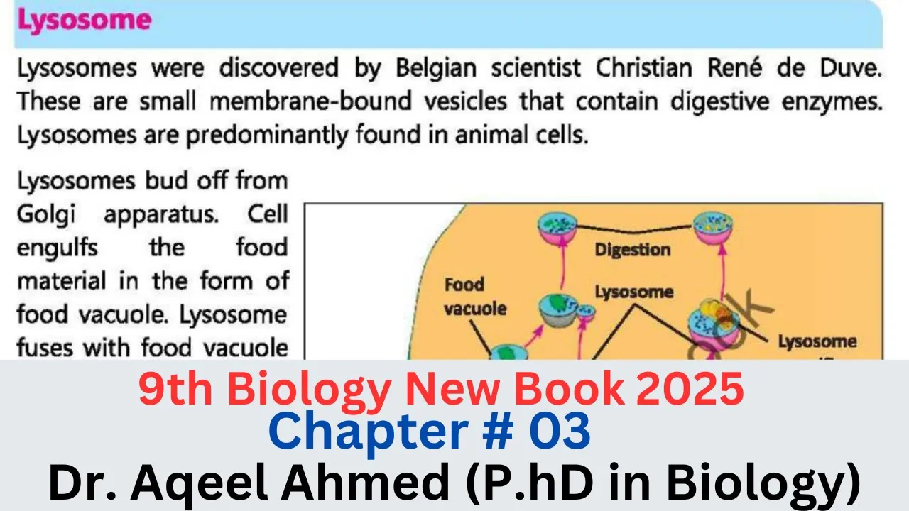
Lysosomes And Mitochondria 9th class Biology New Book Chapter 03 | 9th Biology PTTB Ch 3
776
Lysosomes are single-membrane-bound vesicles containing digestive enzymes, discovered by Belgian scientist Christian de Duve, that originate from the Golgi apparatus and perform intracellular digestion of food particles and damaged organelles through autophagy. Mitochondria are double-membrane-bound organelles found only in eukaryotic cells, known as the 'powerhouse of the cell' because they produce energy through aerobic respiration, generating ATP (adenosine triphosphate) as the cell's energy currency; their inner membrane forms infoldings called cristae that increase surface area for ATP synthesis, and they contain their own DNA and 70S ribosomes.
Click to watch →
The Fundamental Unit of Life L-4 | Cell Organelles 🧫 (Part-3) | CBSE Class 9 Biology - Umang 2021
40.8K
Lysosomes are membrane-bound organelles found in animal cells that contain digestive enzymes. They are formed in the Golgi apparatus where digestive enzymes are packaged into membrane-bound vesicles. Lysosomes are sometimes called 'suicide bags' because they can break down cellular components and trigger cell death. They protect cells by digesting waste materials, damaged organelles, and foreign particles. As cells age, lysosomes may become less effective at breaking down cellular materials, contributing to cellular aging. Dysfunction of lysosomes can lead to various cellular and tissue disorders. Mitochondria are membrane-bound organelles found in eukaryotic cells that are responsible for cellular respiration. They are often called the 'powerhouses' of the cell because they generate most of the cell's supply of adenosine triphosphate (ATP), which is used as a source of chemical energy. Mitochondria contain their own DNA and ribosomes, suggesting they evolved from free-living bacteria. They are essential for energy production in eukaryotic cells and play a crucial role in cellular metabolism.
Click to watch →
Deep Dive
The concept of cellular homeostasis and how cells respond to metabolic stress or nutrient deprivation.

Super Simplified Pathology | Cell Adaptations | Dr. Priyanka Sachdev
119.1K
Human cells are classified into three types based on division capacity: labile cells (continuously divide, e.g., skin, intestinal cells), stable cells (can divide in response to injury, e.g., liver, kidney cells), and permanent/terminal cells (never divide, e.g., heart muscle, neurons). Homeostasis is the maintenance of normal cellular structure and function where each cell type performs specific functions through specific structures. When cells face stress (physiological or pathological), they respond in a spectrum: first adaptations (reversible structural changes to achieve new homeostasis), then reversible injury, and finally irreversible cell death (apoptosis/necrosis). Cell adaptations are reversible responses to stress that achieve new homeostasis. The five types are: hypertrophy (cell size increase), hyperplasia (cell number increase), atrophy (cell size decrease), metaplasia (epithelial cell type conversion), and dysplasia (disordered development, neoplasia precursor). Hyperplasia occurs through increased growth factor production and receptor expression, shown by labile and stable cells but not permanent cells. Physiological examples include female breast enlargement during puberty/pregnancy/lactation, uterine myometrium expansion during pregnancy (combined hypertrophy/hyperplasia), and endometrial regeneration post-menstruation. Pathological examples include HPV-induced skin warts, estrogen-induced endometrial hyperplasia (causing abnormal bleeding), and compensatory hyperplasia after partial organ resection.
Click to watch →
Stressed Out Cells: What Zebrafish Are Teaching Us About Cell Survival - Danielle Hicks
148
Cell stress is defined as a disruption of homeostasis within the cell, occurring when internal balance is disturbed. Similar to human stress from deadlines and responsibilities, cells experience stress from various factors including temperature extremes, insufficient oxygen, inadequate nutrients, and excessive protein demands. The cell responds by initiating mechanisms to restore balance, analogous to how the body regulates temperature through sweating when overheated. This fundamental principle applies across all organizational levels from entire organisms down to individual organelles.
Click to watch →

Adaptación y lesión celular
279
Cells maintain homeostasis through balanced metabolic needs, waste elimination, and workload. When disrupted by harmful stimuli, cells may adapt, suffer reversible damage, or undergo irreversible damage leading to death. The determining factor is stimulus intensity and persistence. Cells have preprogrammed responses including integrated stress response, ER stress response, and autophagy. Using cardiac muscle as an example, hypertrophy initially compensates for increased workload but can become pathological by reducing ventricular cavity size and decreasing cardiac output. The hypertrophic muscle is more susceptible to ischemic damage due to higher oxygen demands and compressed coronary arteries. Cellular damage results from diverse causes: physical agents (temperature extremes, radiation, pressure changes), chemical agents (toxins, medications, heavy metals), infectious agents, immune-mediated damage, genetic abnormalities, and nutritional deficiencies.
Click to watch →

Clinical Pathology | Lab License Preparation Class | Smart Academy | NHPC
261
Homeostasis is the body's ability to maintain stable internal conditions despite external changes. Cells strive to maintain their normal state and adjust to environmental changes. When cells experience stress (such as being cut), they attempt to adapt. If adaptation succeeds, the cell returns to normal. If the stress is too severe, the cell becomes injured. Cell injury can be reversible (cell recovers and returns to normal) or irreversible (cell death).
Click to watch →
LESIÓN CELULAR (PATOLOGÍA)
1.2K
Cells require substrates (glucose, glycogen, lipids, proteins) for energy and function. Homeostasis represents equilibrium, but physiological demands fluctuate, compromising homeostasis and creating cellular stress. Stress can be physiological (exercise, pregnancy) or pathological (systemic hypertension affecting cardiac myocytes). Cells adapt through four mechanisms: (1) Hypertrophy - increase in cell size (e.g., cardiac hypertrophy in hypertension); (2) Hyperplasia - increase in cell number (e.g., uterine hyperplasia during pregnancy); (3) Atrophy - decrease in size and number of cells (e.g., muscle atrophy after nerve injury); (4) Metaplasia - change from one specialized cell type to another (e.g., squamous metaplasia in smokers' respiratory epithelium).
Click to watch →
Deep Dive
Basic mechanisms of intracellular vesicle trafficking, membrane dynamics, and fusion.

Endocytosis | Exocytosis | Vesicular Transport | Bulk Transport | Membrane Transport | Cell
64.6K
Cellular trafficking involves coordinated vesicle formation, transport, and fusion. Clathrin-mediated pathways use triskelion structures to form cages around vesicles, with dynamin facilitating pinching off. After uncoating via ATP-dependent processes, vesicles fuse with endosomes where proton pumps create acidic environments for ligand-receptor separation. Receptors recycle while ligands progress to lysosomes for degradation. Exocytosis complements this by releasing synthesized proteins, hormones, and neurotransmitters. Two pathways exist: constitutive secretion releases immediately, while regulated secretion stores vesicles until signaled. Vesicle fusion relies on v-SNARE/t-SNARE complexes and GTPases, enabling either transient openings for recycling or complete membrane integration. This bidirectional trafficking maintains cell size and membrane composition through coupled endocytic and exocytic cycles.
Click to watch →
APARATO DE GOLGI
30.1K
Vesicle trafficking relies on SNARE proteins for precise targeting: v-SNAREs on vesicles match with t-SNAREs on target membranes. This molecular recognition ensures selective fusion between compatible membranes. The mechanism explains how vesicles find their correct destinations despite the crowded cellular environment. This system is fundamental to all intracellular transport processes, demonstrating the precision of cellular logistics.
Click to watch →

Cell Biology Lecture 7 Intracellular Transport and Pathways
3.4K
Vesicles form through clathrin-mediated endocytosis where clathrin baskets shape the membrane into spheres. Adapter proteins anchor clathrin, and dynamin constricts the neck to pinch off vesicles using GTP energy. Vesicle fusion requires SNARE proteins: V-SNAREs on vesicles bind T-SNAREs on target membranes, causing membrane merger. Rab proteins bind tethering proteins to position vesicles correctly. This machinery enables precise intracellular trafficking, with different SNARE combinations directing vesicles to specific destinations.
Click to watch →

Cell and Molecular Biology: Vesicular Budding and Fusion
62.9K
Transport vesicles form through a regulated process involving cargo selection (where cargo receptors bind to specific signal sequences and adapter proteins filter cargo), coat formation (where coat proteins sculpt membrane curvature), and dynamin-mediated budding (where GTPases pinch off the vesicle); vesicles then fuse with target membranes through a highly specific process involving RAB GTPases for targeting, tethering proteins for initial contact, and V-SNARE/T-SNARE protein pairs for final membrane fusion, ensuring accurate intracellular trafficking.
Click to watch →

Células e Tecidos - aula 11 - Complexo de Golgi
27.5K
Cellular vesicle trafficking involves three sequential stages: budding, transport, and targeting/fusion. Budding requires coat proteins (clathrin, COPI, COPII) that induce membrane curvature and select cargo through adaptor proteins and GTPase recruitment. After release, vesicles lose their coats and associate with motor proteins using the cytoskeleton for directed transport. Targeting occurs via specific recognition: Rab GTPases on vesicles bind Rab effectors on target membranes, while complementary V-SNARE and T-SNARE proteins mediate membrane fusion. This precise trafficking system ensures accurate delivery of cellular components to their designated destinations.
Click to watch →
Deep Dive
The ubiquitin-proteasome system and the general concept of targeting proteins for degradation via covalent ubiquitin tagging.

Ubiquitin-Proteasome System | Targeted Protein Degradation
22.7K
The ubiquitin-proteasome system (UPS) is a cellular mechanism where E1, E2, and E3 ligases collaborate to tag proteins with polyubiquitin chains, marking them for degradation by the proteasome; this natural process has been harnessed through proteolysis targeting chimeras (PROTACs), which bring together target proteins and E3 ligases to enable selective degradation of undruggable proteins like BRD4 and CDK9, offering new therapeutic strategies for previously inaccessible targets.
Click to watch →
The Robot Repairmen Inside You (Secrets of the Cell, Ep. 9 feat. biologist Michael Behe)
434.2K
A set of micro-machines recognizes disabled proteins and places a molecular tag called ubiquitin on them. This tag serves as a signal that directs the proteasome to degrade the tagged protein. Healthy proteins are rarely consumed because they lack this destruction signal, ensuring selective protein turnover.
Click to watch →
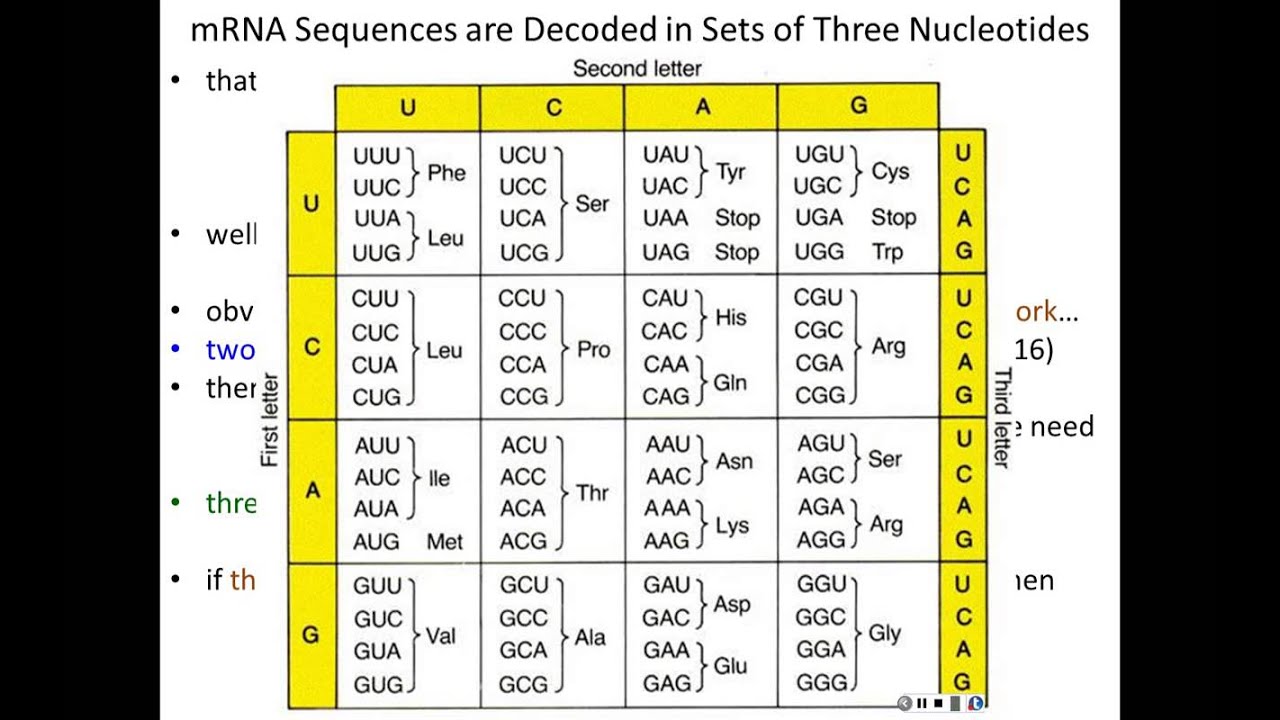
Lecture 6 - From Genes to Proteins Translation (Chapter 7, Part 2)
7K
Proteins are degraded by the proteasome, a large multi-protein complex that shreds proteins into 2-3 amino acid fragments. Only proteins tagged with ubiquitin can enter the proteasome - the ubiquitin tag serves as a molecular signal for destruction. The ubiquitin-proteasome pathway is the primary mechanism for regulated protein degradation in cells. Proteins targeted for destruction are ubiquitinated (tagged with ubiquitin molecules). The ubiquitin tag acts as a molecular signal that directs the proteasome to shred the protein. Once ubiquitinated, protein degradation is irreversible - the ubiquitin tag commits the protein to destruction. This provides precise control over protein levels in the cell.
Click to watch →
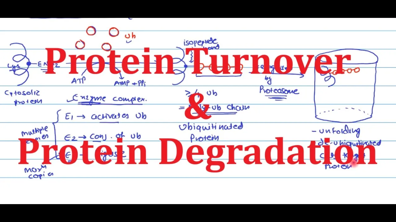
4 Protein Turnover and Protein Degradation | Biochemistry | Metabolism
4.5K
The ubiquitin-proteasome system uses a three-enzyme cascade (E1, E2, E3) to mark proteins for degradation. E1 activates ubiquitin using ATP, E2 conjugates it to lysine residues via isopeptide bonds, and E3 ligases attach multiple ubiquitin molecules. Four or more ubiquitins form a polyubiquitin chain that signals the proteasome. The proteasome unfolds the protein, removes ubiquitin, and cleaves it into fragments. These fragments are further broken down by cytosolic proteases into amino acids for recycling.
Click to watch →

UBIQUITIN - Proteasome Degradation Pathway I What is Ubiquitin? Proteasome and Ubiquitin
4.2K
Ubiquitination is a post-translational modification where a small protein called ubiquitin (approximately 76 amino acids) is attached to target proteins through isopeptide bonds; this process serves as a cellular quality control mechanism where misfolded or damaged proteins are marked for degradation by the proteasome. The degradation signal is determined by the type and linkage of ubiquitin chains: monoubiquitination (single ubiquitin) and multiubiquitination (multiple ubiquitins on different lysines) do not trigger degradation, while polyubiquitination (multiple ubiquitins linked together) does, with lysine 48 linkage specifically signaling degradation. The ubiquitin-proteasome system involves three enzymes: E1 (activates ubiquitin using ATP), E2 (transfers ubiquitin), and E3 (ligase that recognizes target proteins and facilitates ubiquitination). The proteasome complex (26S) consists of a 20S barrel-shaped core containing proteolytic subunits and a 19S regulatory cap that recognizes ubiquitinated proteins, unfolds them, removes ubiquitin, and feeds the protein into the 20S for degradation. This pathway is often referred to as the 'kiss of death' because attaching ubiquitin to a protein essentially marks it for cellular destruction.
Click to watch →
Prerequisite Knowledge
- Concept 01Structure and function of eukaryotic organelles, specifically mitochondria and lysosomes.
- Concept 02The concept of cellular homeostasis and how cells respond to metabolic stress or nutrient deprivation.
- Concept 03Basic mechanisms of intracellular vesicle trafficking, membrane dynamics, and fusion.
- Concept 04The ubiquitin-proteasome system and the general concept of targeting proteins for degradation via covalent ubiquitin tagging.
Subsequent Learning
- Step 01The pathophysiological role of defective mitophagy in neurodegenerative disorders, particularly Parkinson's disease (linked to PINK1/Parkin mutations).
- Step 02Upstream nutrient-sensing signaling pathways that regulate autophagy, such as the mTORC1 and AMPK cascades.
- Step 03Therapeutic modulation of autophagy, including the development of autophagy inhibitors and activators for cancer and anti-aging therapies.
- Step 04Other forms of selective autophagy, such as xenophagy (targeting intracellular pathogens) and pexophagy (targeting peroxisomes).
Pathway Initiation
Maturation & Fusion
Pathway Initiation
0:00Playing Section
- 1
Autophagy begins with formation of phagophore from cellular membranes.
- 2
Nutrient depletion inhibits mTORC1, triggering downstream autophagy signals.
- 3
ULK complex activates effector proteins under starvation conditions.
PINK1/Parkin-Independent Mitophagy and the Basal Turnover Debate
While mainstream models often present the PINK1/Parkin pathway as the definitive mechanism of mitophagy, a significant body of research highlights the importance of PINK1/Parkin-independent pathways, challenging their necessity for routine physiological mitochondrial clearance. In vivo studies using advanced fluorescence reporters show that basal mitophagy occurs normally in high-energy tissues like the brain, heart, and kidneys even in the complete absence of PINK1 or Parkin. Instead, receptor-mediated mitophagy—driven by outer mitochondrial membrane proteins like BNIP3, NIX, and FUNDC1—directly recruits the autophagic machinery without requiring the complex PINK1/Parkin ubiquitination cascade. This alternative perspective argues that the heavily studied PINK1/Parkin pathway is actually an emergency stress-response mechanism rather than the primary driver of daily, basal mitochondrial maintenance under normal physiological conditions.
Deep Dive
The pathophysiological role of defective mitophagy in neurodegenerative disorders, particularly Parkinson's disease (linked to PINK1/Parkin mutations).
Новое о митохондриях. Ольга Борисова.
59.4K
Basal mitophagy maintains mitochondrial quality in tissues including heart, nervous system, liver, kidneys, and skeletal muscle. The PINK1-Parkin pathway detects depolarized mitochondria and targets them for lysosomal degradation. Impaired mitophagy leads to accumulation of damaged mitochondria, contributing to Parkinson's disease pathology. Age-related decline in mitophagic efficiency represents a central mechanism linking mitochondrial dysfunction to neurodegeneration and systemic aging.
Click to watch →
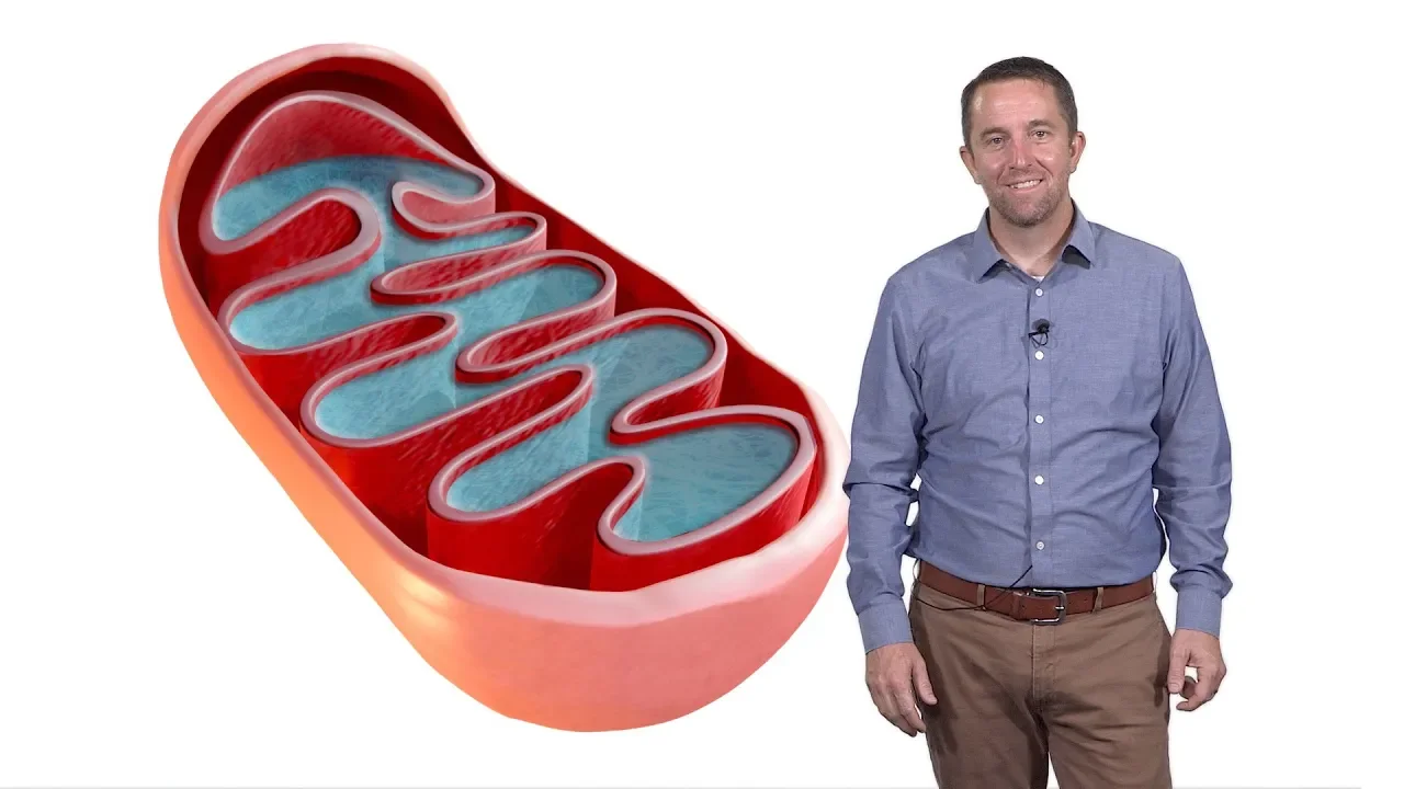
Mitochondria: Cellular Parasite Origins, Metabolism, and Signaling
191.4K
When mitochondrial quality control fails, entire organelles must be degraded through a process called mitophagy (mitochondrial autophagy). This degradation pathway is mediated by the PINK1-Parkin system, where PINK1 targets damaged mitochondria for degradation by recruiting Parkin. Mutations in the Parkin gene cause Parkinson's disease, a devastating neurodegenerative disorder. This connection highlights the critical importance of maintaining mitochondrial quality through both protein degradation within mitochondria and whole-organelle degradation mechanisms for healthy cellular function.
Click to watch →
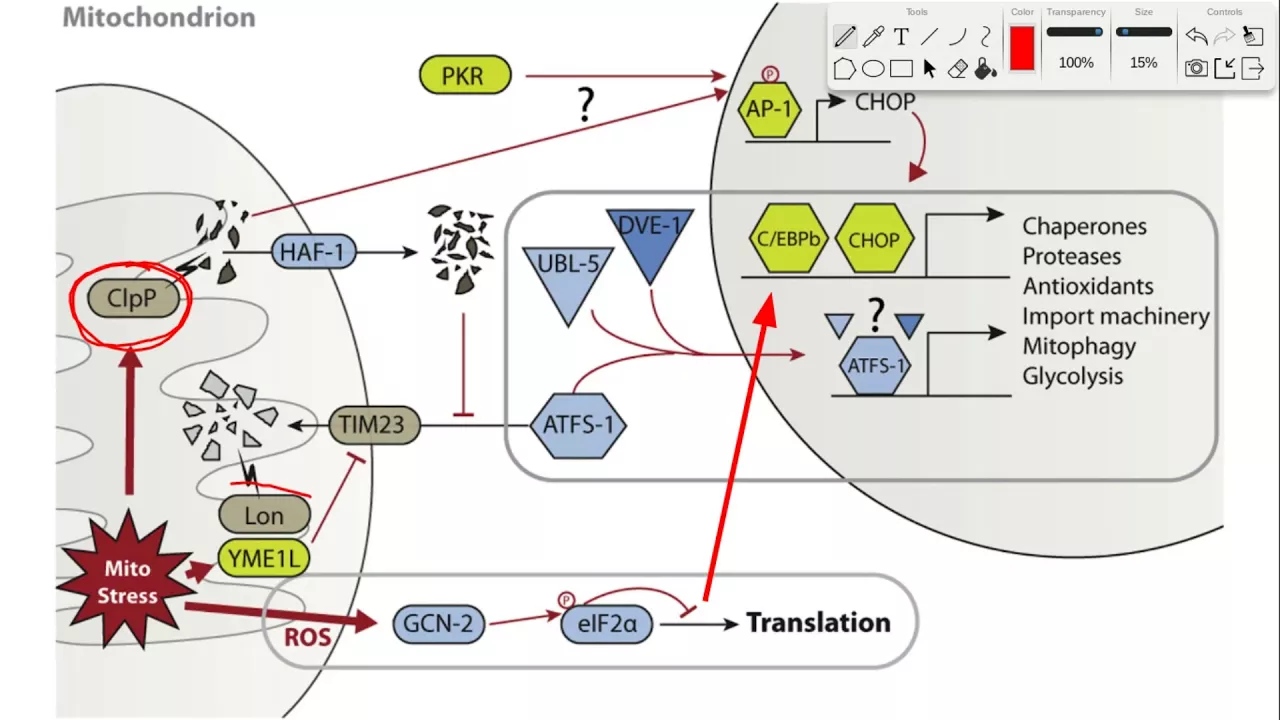
Mitophagy, mtUPR, & Translocation
6.9K
Mitophagy eliminates damaged mitochondria through selective autophagy. PINK1 kinase accumulates on depolarized mitochondria where it phosphorylates Parkin's inhibitory UBL domain, activating its E3 ubiquitin ligase activity. PINK1 also phosphorylates ubiquitin at serine 65, providing Parkin's preferred substrate. Parkin ubiquitinates outer mitochondrial membrane proteins, while USP30 counteracts this ubiquitination. Polyubiquitinated proteins are recognized by autophagy cargo adapters OPTN and P62 containing UBA domains for ubiquitin binding and LIR motifs for LC3 interaction. TBK1 phosphorylates these adapters, enhancing ubiquitin binding affinity. Mitophagy serves as negative selection pressure eliminating dysfunctional mitochondria before they damage cells through ROS release and defective DNA propagation. Parkin mutations impair activation, reducing mitophagy rate and allowing damaged mitochondria accumulation, linked to Parkinson's disease.
Click to watch →

Parkinson's Disease:- "Finding the energy: What happens to mitochondria in PD?" by Prof Sonia Gandhi
15.6K
Mitophagy is the quality control process where damaged mitochondria are decorated with ubiquitin chains by PINK1/Parkin proteins and removed by autophagosomes. Research using patient-derived neurons revealed that alpha-synuclein aggregates form on mitochondrial membranes, linking protein misfolding (the hallmark of common Parkinson's) with mitochondrial dysfunction (the hallmark of genetic forms). This integration explains how both major pathological mechanisms interact to drive neurodegeneration.
Click to watch →
Maintaining mitochondrial health – interview with Dr. Behrouz, co-founder & CEO @ Vincere Bio
588
Mitophagy represents one of the most validated therapeutic targets for Parkinson's disease due to compelling human genetic evidence. Mutations in PINK1 and Parkin genes, which directly initiate mitophagy, cause early-onset Parkinson's with high penetrance. Non-genetic Parkinson's patients also exhibit mitophagy deficits. USP30, a mitochondrial membrane enzyme, regulates the threshold for mitochondrial degradation—its inhibition shifts this threshold to promote removal of damaged mitochondria while sparing healthy ones. This selective targeting mechanism offers a potentially safe therapeutic approach by focusing on damaged components rather than indiscriminately clearing all cellular material.
Click to watch →
Deep Dive
Upstream nutrient-sensing signaling pathways that regulate autophagy, such as the mTORC1 and AMPK cascades.
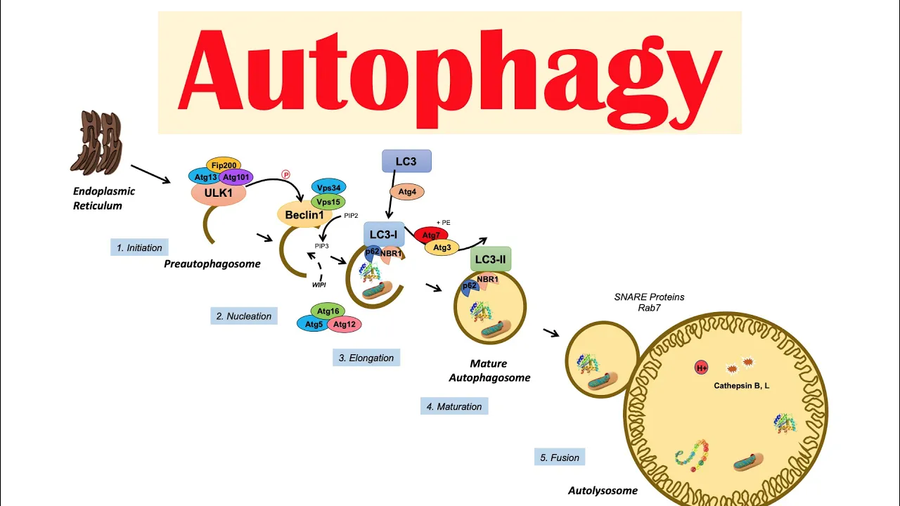
Autophagy | Macroautophagy & Importance in Health
210.6K
Autophagy is regulated by interconnected signaling networks. AMPK activates autophagy by phosphorylating ULK1; mTOR inhibits autophagy by phosphorylating ULK1 and TFEB. Bcl-2 inhibits autophagy by binding Beclin-1. The lysosomal nutrient-sensing machinery involves V-ATPase relaying amino acid signals to Rag proteins, which recruit mTORC1. When amino acids are abundant, mTORC1 phosphorylates TFEB (master lysosome biogenesis regulator), preventing its nuclear entry. Unphosphorylated TFEB binds CLEAR sequences to upregulate autophagy genes (ATG5, Beclin-1, P62, LC3). ZKSCAN3 opposes TFEB by inhibiting its transcriptional activity. This creates a nutrient-sensing feedback loop controlling autophagy flux.
Click to watch →

This Changes Everything About Autophagy WITHOUT Fasting - DR JAMNADAS
132
Two major pathways control autophagy: mTOR and AMPK. mTOR responds to nutrient abundance and promotes growth when resources are plentiful, slowing cleanup during this state. AMPK acts as an energy sensor that activates when cells use energy faster than they produce it, triggering conservation and efficiency responses. These pathways work in constant opposition throughout the day. After meals, mTOR increases; during activity or metabolic demand, AMPK rises. Both pathways can be influenced without removing food—muscle activity activates AMPK even with calorie availability, while nutrient timing affects mTOR elevation duration.
Click to watch →

mTOR Signaling Pathway: mTOR Complexes, Regulation and Downstream effects
181.3K
mTORC1 is regulated at the lysosomal membrane through a complex network involving Rheb, TSC2, AMPK, Akt, and Rag GTPases. Rheb activates mTOR while being inhibited by TSC2; AMPK inhibits mTOR through LKB1-TSC2-Rheb pathway, coupling energy status to growth. Growth factors activate Akt, which inhibits TSC2 to promote mTOR. IGF-1R and p53 also regulate TSC2. Amino acid sensing occurs through Rag GTPases regulated by GATOR complexes: arginine inhibits GATOR1 to activate Rags, while leucine inhibits sestrin2 to activate GATOR2, both ultimately activating mTORC1.
Click to watch →

Autophagy Doesn't Start When You Think (Here's When It Actually Does)
274
Autophagy is regulated by two opposing pathways. mTOR is the main growth signal that suppresses autophagy when insulin is high and amino acids (especially leucine from protein) are flooding in. This signals the body to prioritize muscle protein synthesis and storage rather than cellular cleanup. AMPK is the low fuel alarm that activates autophagy when cellular energy is running low. When AMPK rises and insulin and amino acids stay low, resources must be conserved and repurposed, making recycling a survival priority. A small frequent snack or high-protein fasting drink can keep insulin and leucine just high enough to keep mTOR active and autophagy muted.
Click to watch →
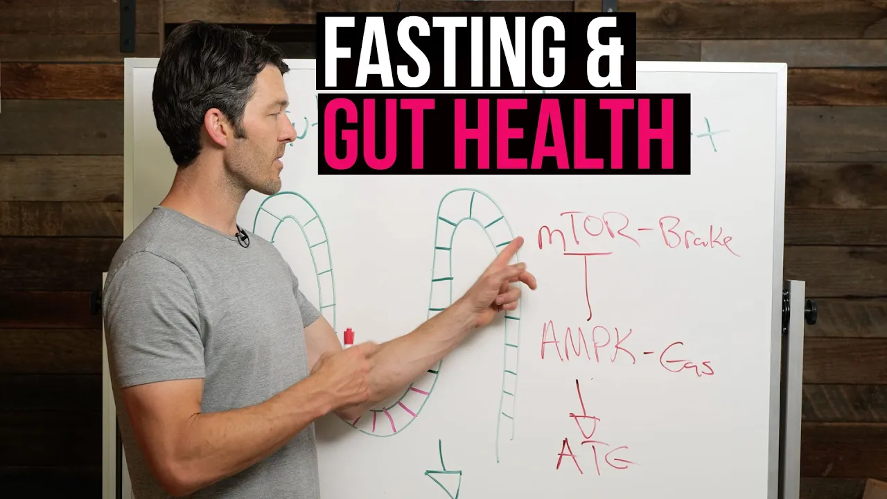
Fasting & Gut Health: Regeneration + Stem Cell Production
69.5K
Autophagy is regulated by mTOR (mechanistic target of rapamycin) and AMPK (AMP-activated protein kinase). mTOR acts as a 'gas pedal' promoting cell growth and nutrient uptake, while AMPK stimulates autophagy and also inhibits mTOR. These enzymes cannot be directly measured but can be assessed through metabolic markers: low triglycerides, low liver enzymes, low fasting insulin, and low cholesterol indicate autophagy activation. Fasting and exercise stimulate AMPK, which puts brakes on mTOR, enabling autophagy. The natural cycle requires alternating between feeding (mTOR activation for repair) and fasting (autophagy for cellular cleanup).
Click to watch →
Deep Dive
Therapeutic modulation of autophagy, including the development of autophagy inhibitors and activators for cancer and anti-aging therapies.
#114 – Eileen White, Ph.D.: Autophagy, fasting, and promising new cancer therapies
53.2K
Pharmacological agents offer promising approaches to modulate autophagy for health benefits. Rapamycin, an mTOR inhibitor, extends lifespan across multiple eukaryotic model organisms including yeast, flies, worms, and mice. While mTOR inhibition theoretically stimulates autophagy, rapamycin's longevity benefits likely involve additional mechanisms including reduced senescence cell accumulation and decreased inflammation. Metformin activates AMPK and may induce autophagy, though long-term consequences remain unclear. The 2016 Nobel Prize recognized Yoshinori Ohsumi's foundational work elucidating autophagy mechanisms in yeast, identifying essential autophagy genes and demonstrating their conservation across species. Future research directions include developing small molecule inhibitors targeting autophagy for cancer therapy, combining autophagy modulation with immune checkpoint blockade to enhance cancer immunotherapy responses, and decoding metabolism using advanced isotope tracer techniques to identify predictive biomarkers for disease prevention and intervention strategies.
Click to watch →
Autophagy | Macroautophagy & Importance in Health
210.6K
Several pharmaceutical agents modulate autophagy. Chloroquine and hydroxychloroquine inhibit autophagy by raising lysosomal pH and blocking autophagosome-lysosome fusion. Rapamycin and torin-1 inhibit mTORC1, thereby activating autophagy. TFEB activation occurs when it is not phosphorylated by mTORC1, allowing it to upregulate autophagy-related genes. The opposite effect is achieved by ZKSCAN3, which inhibits TFEB transcriptional activity and thus suppresses autophagy pathway expression.
Click to watch →

Autofagia Extrema: O Processo Proibido que Destrói Doenças em Silêncio
5.7K
A woman lying in the scanner is indistinguishable from any other 40-year-old. But when images appear on screen, researchers lean forward, fascinated. Her tissues appear to be 25 years old. She has been participating in an experimental autophagy optimization protocol for 5 years. Regular fasting, strategic exercise, precise nutrition, targeted supplementation, and results are beyond any expectation. Cellular aging markers reversed. Telomeres - the protective caps on chromosome ends that shorten with age - actually lengthened. Gene expression changed to patterns associated with youth. This is not science fiction, it is real study happening now in research institutes around the world. We are on the frontier of a revolution in understanding and manipulation of autophagy. Pharmaceutical companies are developing drugs that modulate autophagy with surgical precision - not just general activators, but compounds that can increase autophagy in specific cells, at specific times, for specific conditions. Imagine a pill that activates autophagy selectively in neurons, cleaning toxic Alzheimer's proteins without affecting other cells. Or treatment that induces intense autophagy in tumor cells while protecting healthy cells, making cancer treatable without devastating side effects of current chemotherapy.
Click to watch →

La clave de la longevidad | La Autofagia explicada
520K
Several compounds can pharmacologically stimulate autophagy: Metformin activates AMP kinase pathway; Rapamycin inhibits mTOR to promote autophagy; Resveratrol (found in grapes, blueberries, wine) activates autophagy; Spermidine (found in soybeans, green tea, mushrooms, and gut microbiota) enhances autophagy. These compounds 'hack' cellular signaling pathways to induce autophagy even under normal nutrient conditions, offering potential therapeutic applications for age-related diseases.
Click to watch →

Аутофагия. НОБЕЛЕВСКАЯ ПРЕМИЯ за СЕКРЕТ ДОЛГОЛЕТИЯ!
100K
Autophagy is activated through two primary methods: physical exercise and fasting. Exercise, particularly intense training and statodynamics (slow muscle fiber training), triggers autophagy through hydrogen ions affecting lysosomes. Fasting activates autophagy after 8-16 hours without food, serving as a disease prevention mechanism that eliminates harmful elements before they can develop into illnesses like cancer. The strongest inhibitor of autophagy is protein consumption, including both animal and plant proteins, which dramatically slows down this cellular cleaning process.
Click to watch →
Deep Dive
Other forms of selective autophagy, such as xenophagy (targeting intracellular pathogens) and pexophagy (targeting peroxisomes).
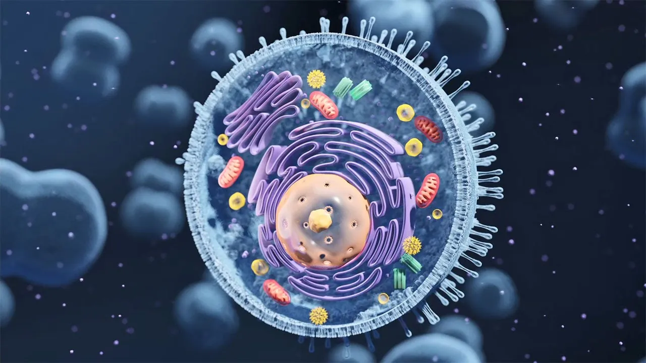
Autophagy - The orderly degradation and recycling of cellular components
7.6K
Selective autophagy is involved in regulated turnover of specific organelle portions including mitochondria (mitophagy), peroxisomes (pexophagy), and endoplasmic reticulum (reticulophagy), as well as large protein/RNA complexes such as protein aggregates (aggrephagy) and pathogens (xenophagy). Selective autophagy relies on autophagy receptors that physically link autophagosome membranes to cargo. These receptors bind cargo on one hand and LC3 proteins localized in the inner phagophore membrane on the other hand. Binding between autophagy receptors and LC3 proteins relies on the short LC3-interacting region (LIR) motif, though other motifs like ubiquitin-interacting motifs also exist.
Click to watch →

Autophagy: Intracellular Recycling System | Nobel Lecture 2016
150.9K
Beyond bulk autophagy, cells employ selective autophagy pathways to target specific substrates for degradation. These include: (1) Mitophagy for degrading damaged mitochondria, (2) Pexophagy for peroxisome degradation, (3) Ribophagy for ribosome degradation, and (4) Aggrephagy for protein aggregate clearance. Receptors on the autophagosomal membrane recognize specific cargo proteins, enabling precise targeting of damaged or unnecessary cellular components while preserving healthy structures.
Click to watch →

FBI Cyber Series - Autophagy: A Selective Eater that Protects Brain by Zhenyu Yue, PhD
1K
Autophagy is highly selective in targeting specific cellular cargo. Different selective autophagy types target different organelles: xenophagy clears invading pathogens, mitophagy degrades damaged mitochondria via PINK1-Parkin pathway, and aggrephagy clears protein aggregates. p62/SQSTM1 serves as a critical autophagy receptor containing LIR domain for LC3 binding and ubiquitin-associated domain for cargo recognition. Phosphorylation by ULK1 increases p62-cargo affinity. Autophagy deficiency causes massive protein aggregate accumulation, demonstrating that selective autophagy is essential for clearing toxic aggregates in neurodegenerative diseases.
Click to watch →

The mechanism of autophagy
41.8K
Selective autophagy enables targeted degradation of specific cellular components including mitochondria (mitophagy), peroxisomes (pexophagy), endoplasmic reticulum (reticulophagy), protein aggregates (aggrephagy), and pathogens (xenophagy). This process relies on autophagy receptors that physically link cargo to autophagosome membranes through LC3-interacting regions (LIR). Receptors are classified as ubiquitin-dependent (soluble, e.g., p62, NBR1, OPTN) or ubiquitin-independent (membrane-bound, e.g., NIX, BNIP3). Ubiquitin-dependent receptors bind ubiquitinated cargo, while membrane-bound receptors recognize organelle-specific signals. Activation involves FIP200-mediated recruitment of ULK kinase complexes, with TBK1 phosphorylation enhancing LC3 binding affinity. This ensures hermetic sequestration of targeted cargo within autophagosomes.
Click to watch →
This Fatty Nutrient Reprograms Your Immune System (and Your Gut!)
48.4K
Xenophagy differs from regular autophagy in that it specifically targets foreign invaders like bacteria that have escaped their cellular prison. When immune cells engulf pathogens through phagocytosis, some bacteria use secretion systems to escape the vacuole, creating a dangerous situation where the pathogen runs freely inside the cell. Xenophagy recaptures these escaped pathogens and destroys them, with 'xeno' meaning foreign.
Click to watch →
Pathway Initiation
Maturation & Fusion
Pathway Initiation
0:00- 1
Autophagy begins with formation of phagophore from cellular membranes.
- 2
Nutrient depletion inhibits mTORC1, triggering downstream autophagy signals.
- 3
ULK complex activates effector proteins under starvation conditions.
PINK1/Parkin-Independent Mitophagy and the Basal Turnover Debate
While mainstream models often present the PINK1/Parkin pathway as the definitive mechanism of mitophagy, a significant body of research highlights the importance of PINK1/Parkin-independent pathways, challenging their necessity for routine physiological mitochondrial clearance. In vivo studies using advanced fluorescence reporters show that basal mitophagy occurs normally in high-energy tissues like the brain, heart, and kidneys even in the complete absence of PINK1 or Parkin. Instead, receptor-mediated mitophagy—driven by outer mitochondrial membrane proteins like BNIP3, NIX, and FUNDC1—directly recruits the autophagic machinery without requiring the complex PINK1/Parkin ubiquitination cascade. This alternative perspective argues that the heavily studied PINK1/Parkin pathway is actually an emergency stress-response mechanism rather than the primary driver of daily, basal mitochondrial maintenance under normal physiological conditions.
Structure and function of eukaryotic organelles, specifically mitochondria and lysosomes.
The concept of cellular homeostasis and how cells respond to metabolic stress or nutrient deprivation.
Basic mechanisms of intracellular vesicle trafficking, membrane dynamics, and fusion.
The ubiquitin-proteasome system and the general concept of targeting proteins for degradation via covalent ubiquitin tagging.
The pathophysiological role of defective mitophagy in neurodegenerative disorders, particularly Parkinson's disease (linked to PINK1/Parkin mutations).
Upstream nutrient-sensing signaling pathways that regulate autophagy, such as the mTORC1 and AMPK cascades.
Therapeutic modulation of autophagy, including the development of autophagy inhibitors and activators for cancer and anti-aging therapies.
Other forms of selective autophagy, such as xenophagy (targeting intracellular pathogens) and pexophagy (targeting peroxisomes).
102.1K views1.6Klikes3:29@hussainbiologyOriginal Release: 2020-03-11
Oh in this video we'll be discussing about the autophagy pathway the autofill involved the sequestration of cytoplasmic components within the auto fire zones the autophagy is the intracellular degradation process rather we can say it is the celebri gradation and cytoprotective process it involves cellular cleansing and recycling of cellular nutrients as well in the autophagy pathway first there is a formation of a gopher followed by the auto phagosome and then autophagy lysosome we see the Vega for is actually a site of membrane which is derived from endoplasmic reticulum Golgi or plasma membrane now let's see this pathway in detail in a vago for formation we get the initiation signal in the form of nutrient signaling in other words we can say autophagy is initiated when there is presence of less amino acids and glucose we know am Tour c1 is the nutrient dependent M tall which gets active when there is high availability of glucose and amino acids as depicted in the diagram on the left but when the nutrient level falls extensively the mTOR pathway is inhibited we see under starvation conditions ul Kavon postulates both FIP 280 G 13 resulting in the activation of downstream autophagy effector proteins thus leading to auto fare G so when signal is mediated the Saito membrane is first selected either from Golgi plasma membrane or endoplasmic reticulum and from this the pay-go 4 is made the fog of war has LC 3 molecules on it as shown in the diagram and it also has ATG to l80 g5 ATG 16 L complex on the membrane the exact role of LC 3 is still ambiguous but there have been some researchers put downward it has been found that LC 3 plays role in Auto phagosome maturation induction of out of hedgy and the cargo selection as well as it plays role in huge n then we have a TG v 2 l6 complex on the membrane this complex is responsible for elongation pegye for and it's maturation into physical here we see the pay-go for English the mitochondria and then 82 L 80 g 5 80 g sixteen l complex completes the elongation of phago four into the vesicle and we get the fully formal out of haggis room as shown in the diagram and it must be noted that when mitochondria is taken in for degradation process its terminus might oh hey G and in that process also some LC 3 molecules leaves the membrane and 82 to l5 + 16 L complex is given off by the membrane and as shown in the diagram then in the next step we have the fusion of lysosome with Auto phagosome we see the auto phagosome pues with the lysosome and we get the auto fabulous room in that process then the lysosome releases all its hydrolytic components to degrade the cargo in the auto Faygo lysosome which concludes the auto-pay g pathway and it must be noted that auto fagala is room is the single membrane structure whereas auto phagosome is the double membrane structure so this is all about auto fare G or might of edgy I hope you liked the video if you liked it give it a thumbs up do consider supporting my work on patreon and also make sure to subscribe this channel thanks
Up Next

62:30
Why We Die: The New Science of Aging and the Quest for Immortality
@harvardsciencebooktalksand4527
117.1K views•2024-05-31

50:12
Bacterial Communication: Quorum Sensing and Biofilm Formation Explained
@JHUAAP
12.6K views•2011-06-28
4:44
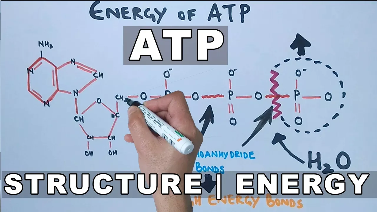
ATP Structure and Hydrolysis: Cellular Energy Release Explained
@hussainbiology
80.9K views•2018-02-18

7:08
Bacteriophages: Earth's Deadliest Killers and Future Antibiotics
@kurzgesagt
34.6M views•2018-05-13
Related Study Plans & Knowledge Roadmaps
Structured learning paths in Biology
biology
RNAi: Biogenesis, RISC & Gene Silencing
01Molecular Biology Foundations: DNA to Protein
02Introduction to RNA Interference (RNAi)
03Molecular Biogenesis of miRNA and siRNA
+ 2 more learning modules
10h5 modules
Explore Roadmap
biology
Mycorrhizae: Signaling, Biophysics & Ecology
01Foundations of Mycorrhizal Symbiosis
02Molecular Signaling and Root Colonization
03Biophysics and Bioenergetics of Nutrient Exchange
+ 1 more learning modules
11h4 modules
Explore Roadmap
biology
Circadian Clocks: TTFL, SCN & Periphery
01Foundations of Molecular Biology & Gene Expression
02Introduction to Chronobiology and Actograms
03The Core TTFL: Molecular Architecture of the Clock
+ 3 more learning modules
14h6 modules
Explore Roadmap
biology
Population Genetics: Math, Drift & Selection
01Genetic & Probability Foundations
02The Hardy-Weinberg Principle and Baseline Models
03Mathematical Modeling of Natural Selection
+ 3 more learning modules
18h6 modules
Explore Roadmap