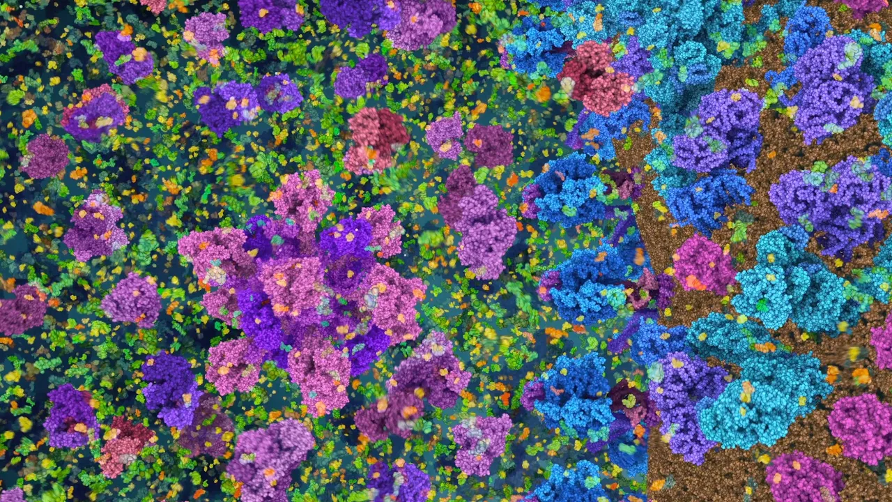
The proteasome is a large enzyme complex that breaks down old or damaged proteins into smaller amino acid peptides for cellular recycling; it consists of a regulatory particle containing ubiquitin receptors and a motor with six subunits that mechanically unfold proteins, and a core particle with six protease enzymes that cleave proteins into peptides of 2-25 amino acids, which are then repurposed for new protein synthesis or used by the immune system to detect infections.
Proteasome Function & Protein Recycling Explained | Molecular Animation
Added:Deep Dive
Basic protein structure, including the concepts of polypeptide chains, folding, and denaturation.

الحصة الثانية من المكثف -البروتينات والحموض النووية- الأستاذ محمد بطاينة 2007
115.2K
Proteins have four levels of structural organization: (1) Primary structure - the linear sequence of amino acids connected by peptide bonds, (2) Secondary structure - local folding patterns like alpha helices and beta sheets formed by hydrogen bonding, (3) Tertiary structure - the overall three-dimensional shape of a single polypeptide chain stabilized by various interactions, (4) Quaternary structure - the arrangement of multiple polypeptide chains in a protein complex. Hydrogen bonds are the primary forces that stabilize secondary, tertiary, and quaternary protein structures. In secondary structures, hydrogen bonds form between the carbonyl oxygen of one amino acid and the amide hydrogen of another amino acid four residues apart. Tertiary and quaternary structures are stabilized by multiple types of bonds: hydrogen bonds, ionic bonds (salt bridges), disulfide bridges (covalent bonds between sulfur atoms), and hydrophobic interactions. When the bonds stabilizing tertiary and quaternary protein structures are disrupted (denaturation), the protein loses its three-dimensional shape and biological function. Myoglobin is a protein found in muscle tissue that stores oxygen and has a tertiary structure. Hemoglobin is a protein found in red blood cells that transports oxygen and has a quaternary structure consisting of four polypeptide chains.
Click to watch →
Biomolecules In 2 Hours| Organic Heuristic Series| NCERT, PYQ & Concepts Covered For NEET 2025
40.6K
Proteins are polymers of alpha-amino acids linked by peptide bonds (-CONH-). Peptide bonds form through condensation reactions between the carboxyl group of one amino acid and the amino group of another. Peptides are named by number of amino acids: dipeptide (2), tripeptide (3), polypeptide (10+). Proteins are polypeptides with more than 100 amino acids and molecular mass >10,000 u (except insulin with 51 amino acids). Protein structure levels: Primary (amino acid sequence), Secondary (alpha-helix and beta-pleated sheet formed by hydrogen bonding), Tertiary (overall 3D folding stabilized by hydrogen bonds, disulfide linkages, van der Waals forces, electrostatic interactions), Quaternary (arrangement of multiple polypeptide chains, e.g., hemoglobin with 4 subunits). Denaturation is loss of secondary and tertiary structure due to temperature or pH changes, while primary structure remains intact.
Click to watch →

IMPORTANT TOPICS BIOMOLECULES ONE SHOT CLASS 12 CHEMISTRY 🔥|| BY MUNIL SIR
538.8K
Peptide bonds form when amino acid carboxyl reacts with another's amino group, releasing water. Protein structure levels: primary (amino acid sequence), secondary (alpha helix, beta sheet stabilized by hydrogen bonds), tertiary (overall 3D folding), quaternary (multiple subunits). Fiber proteins (keratin, collagen) are elongated and insoluble; globular proteins (albumin, insulin) are spherical and soluble. Denaturation destroys structure through temperature/pH changes but preserves primary structure.
Click to watch →
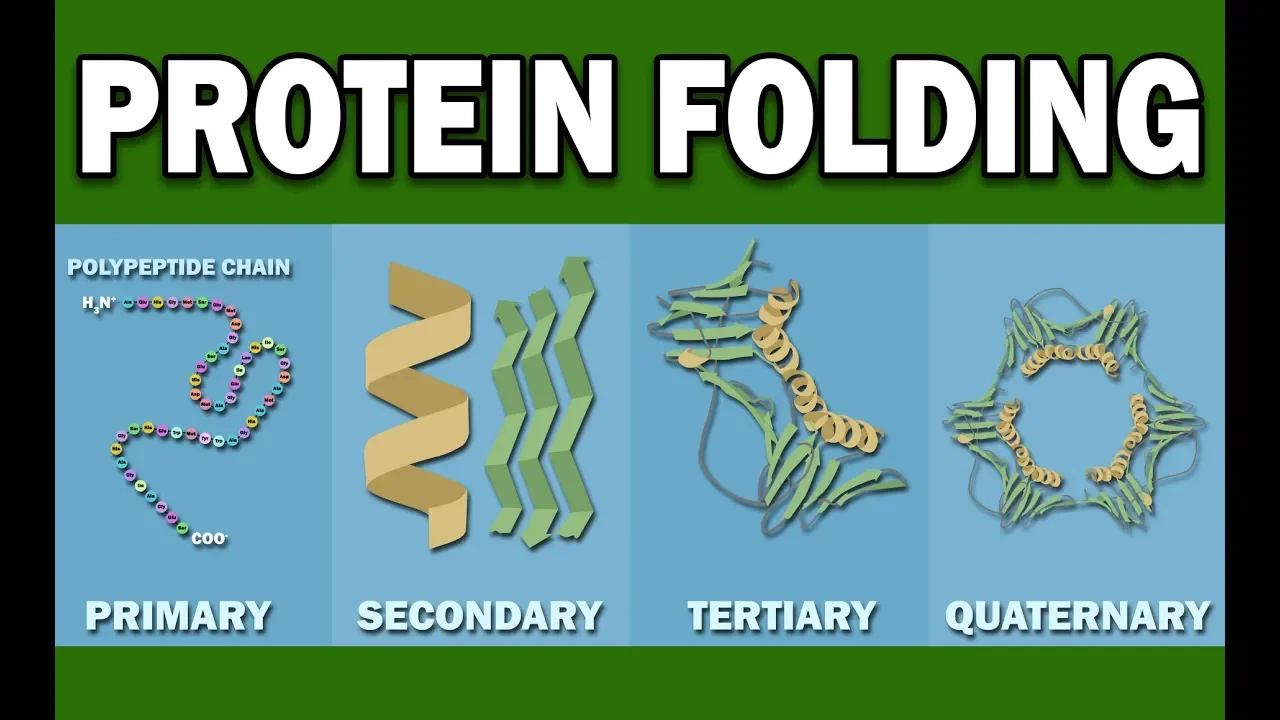
PROTEIN FOLDING
260.7K
Proteins are composed of folded polypeptide chains made from 20 different amino acids linked by peptide bonds, with their structure organized into four levels: primary (amino acid sequence), secondary (alpha helices and beta sheets formed by hydrogen bonding), tertiary (compact 3D shape determined by hydrophobic interactions), and quaternary (multiple subunits); protein folding occurs through secondary structure formation, hydrophobic collapse where nonpolar amino acids aggregate inward, and long-range interactions, ultimately determining the protein's functional conformation.
Click to watch →

Proteins: Tertiary and Quaternary Structure | A-level Biology | OCR, AQA, Edexcel
114.5K
The tertiary structure of a protein is its overall specific 3D shape formed by the integration of alpha helices and beta pleated sheets, held together by hydrogen bonds between polar R groups, ionic bonds between charged R groups, disulphide links between sulfur-containing R groups, and the arrangement of hydrophobic R groups in the center versus hydrophilic R groups on the exterior; the quaternary structure involves multiple polypeptide chains bonded together through similar intermolecular forces, and both structures are determined by the primary amino acid sequence, which can be disrupted by changes in temperature or pH causing denaturation.
Click to watch →
Deep Dive
The concept of cellular homeostasis and why cells need to regulate protein concentration and eliminate damaged proteins.

Prof. Aaron Ciechanover at ADMU, with an introduction by Uwe Morawetz
226
Cells must constantly destroy proteins because proteins are inherently unstable. At the human body temperature of 37°C, proteins continuously denature and misfold, losing their function. Every day, approximately 10% of our proteins become damaged and must be degraded. Within 15-20 days, every molecule in our body has been replaced. This represents an evolutionary trade-off: higher temperatures optimize metabolic reactions but accelerate protein degradation. Oxygen further damages proteins through oxidation. The narrow temperature range (36-42°C) that sustains human life also creates constant molecular stress requiring continuous quality control mechanisms.
Click to watch →

Proteostase: equilíbrio entre síntese e degradação de proteínas
3.6K
Proteostasis is the cellular equilibrium between protein synthesis and degradation, maintained through molecular chaperones (Hsp70, Hsp90, Hsp60, Hsp100) that assist proper protein folding and prevent aggregation, and degradation systems (ubiquitin-proteasome and autophagy-lysosome) that eliminate misfolded, damaged, or short-lived proteins; this balance is critical for cellular survival and dysfunction can lead to diseases like Alzheimer's, Parkinson's, and diabetes.
Click to watch →

Autophagy and Intracellular Aggregates - Dr. Ana Maria Cuervo
23.9K
Autophagy performs three critical functions: (1) Energy supply - under nutrient deprivation, cells activate autophagy to degrade cytoplasmic components and obtain amino acids and fatty acids for energy maintenance; (2) Quality control - elimination of damaged proteins and organelles to maintain cellular proteome integrity; (3) Cellular defense - removal of pathogens and toxic compounds. Compromised autophagy leads to inability to adapt to metabolic stress, increased pathogen susceptibility, and accumulation of damaged products causing cellular dysfunction and death. These functions make autophagy essential for cellular survival and organismal health.
Click to watch →
La célula como unidad de salud y enfermedad - Curso 2022B
13.7K
Cellular homeostasis is the state of equilibrium maintained by cells through various maintenance functions including protein production, elimination, nutrient acquisition, metabolism, and renewal of cellular components. Organelles are membrane-bound structures that perform specialized functions: the nucleus contains genetic material, mitochondria produce ATP through cellular respiration, the endoplasmic reticulum synthesizes and modifies proteins and lipids, the Golgi apparatus packages and sorts proteins, lysosomes digest waste materials, and peroxisomes detoxify harmful substances.
Click to watch →

12 Dấu Hiệu Lão Hóa - Hallmarks of Aging | Tiến Sĩ Carlos López-Otín
3.1K
Proteostasis is the cellular system ensuring proteins are properly folded and damaged proteins are removed. Proteins must fold into specific three-dimensional shapes to function correctly - errors cause loss of function or toxicity. With aging, proteostasis weakens, causing protein misfolding and accumulation that directly causes neurodegenerative diseases like Alzheimer's (amyloid beta and tau accumulation), Parkinson's (alpha-synuclein accumulation), and ALS. The body has three mechanisms to maintain proteostasis: heat shock proteins (activated by exercise and sauna bathing), the ubiquitin-proteasome system (requires adequate B vitamins and magnesium), and autophagy. Autophagy is the cellular self-cleaning mechanism discovered by Nobel laureate Yoshinori Ohsumi (2016 Nobel Prize) - cells eat their own damaged components to recycle materials and remove toxic aggregates. When autophagy fails, cells become cluttered with damaged components, contributing to aging and age-related diseases.
Click to watch →
Deep Dive
An introduction to enzymes, active sites, and how multi-subunit enzyme complexes function.

Biology - Chapter 8, Energy and Metabolism
180
Enzymes typically catalyze only one or a few similar chemical reactions due to substrate specificity determined by the active site shape. Some enzymes function as integral parts of cell structures and organelles. Multi-enzyme complexes associate several enzymes catalyzing sequential reactions in non-covalently bonded assemblies. The pyruvate dehydrogenase complex contains 60 protein subunits working in concert. These complexes offer advantages: substrates never leave the complex during sequential reactions, eliminating unwanted side reactions, and all reactions can be controlled as a unit. Fatty acid synthetase is another example with seven enzymes keeping intermediates associated throughout the reaction series.
Click to watch →
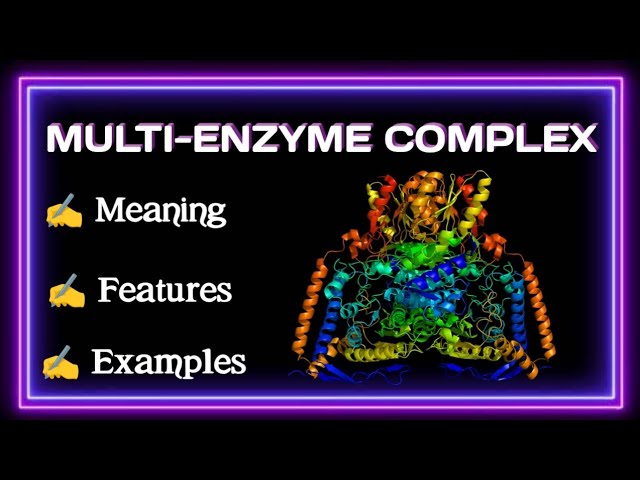
MULTI-ENZYME COMPLEX || WELL EXPLAINED
703
A multi-enzyme complex is a combination of two or more enzymes bound together that catalyze a series of sequential reactions on a single substrate. Four key features characterize these complexes: (1) Proximity and orientation ensure enzymes are positioned close to each other for efficient substrate transfer between active sites; (2) Regulation allows formation and activity to be controlled by substrate concentration, activators, and inhibitors; (3) Compartmentalization places different enzymes in specific cellular locations while maintaining functional connectivity; (4) Stability is achieved through specific enzyme interactions that hold the complex together. These features enable efficient metabolic processing through coordinated division of labor among multiple enzymes.
Click to watch →

Chp#6 Satyanarayana Biochemistry | Active Sites on Enzymes | Biochemistry Lectures
8K
Enzymes are large protein molecules that catalyze reactions on smaller substrates. The active site is the small region where substrate binding and catalysis occur. Key properties include: (1) Active sites result from tertiary structure creating 3D formations with exposed areas; (2) Active sites are formed by amino acids far apart in linear sequence (e.g., lysozyme uses amino acids 35, 52, 62, 63, and 101); (3) Active sites appear as clefts, surfaces, or pockets; (4) Active sites are flexible to accommodate substrates. The active site contains a substrate binding site and catalytic site. Substrates bind through weak non-covalent bonds, forming an enzyme-substrate complex that breaks down into enzyme and product. Enzyme specificity follows the key-lock model. Common amino acids in active sites include cysteine, histidine, and lysine.
Click to watch →

شرح المحاضرة التانية (الانزيمات) لطلاب كلية زراعة من جزء مقدمة فى التكنولوجيا الحيويه
2.2K
Enzymes are classified by structure: Monomeric enzymes consist of a single polypeptide chain with the active site (e.g., trypsin). Oligomeric enzymes consist of multiple polypeptide chains (2-60 subunits) that associate to form the functional enzyme. Multienzyme complexes are assemblies of multiple enzymes working together in coordinated sequences (e.g., pyruvate dehydrogenase complex). The active site is the specific region where substrate binding and catalysis occur.
Click to watch →
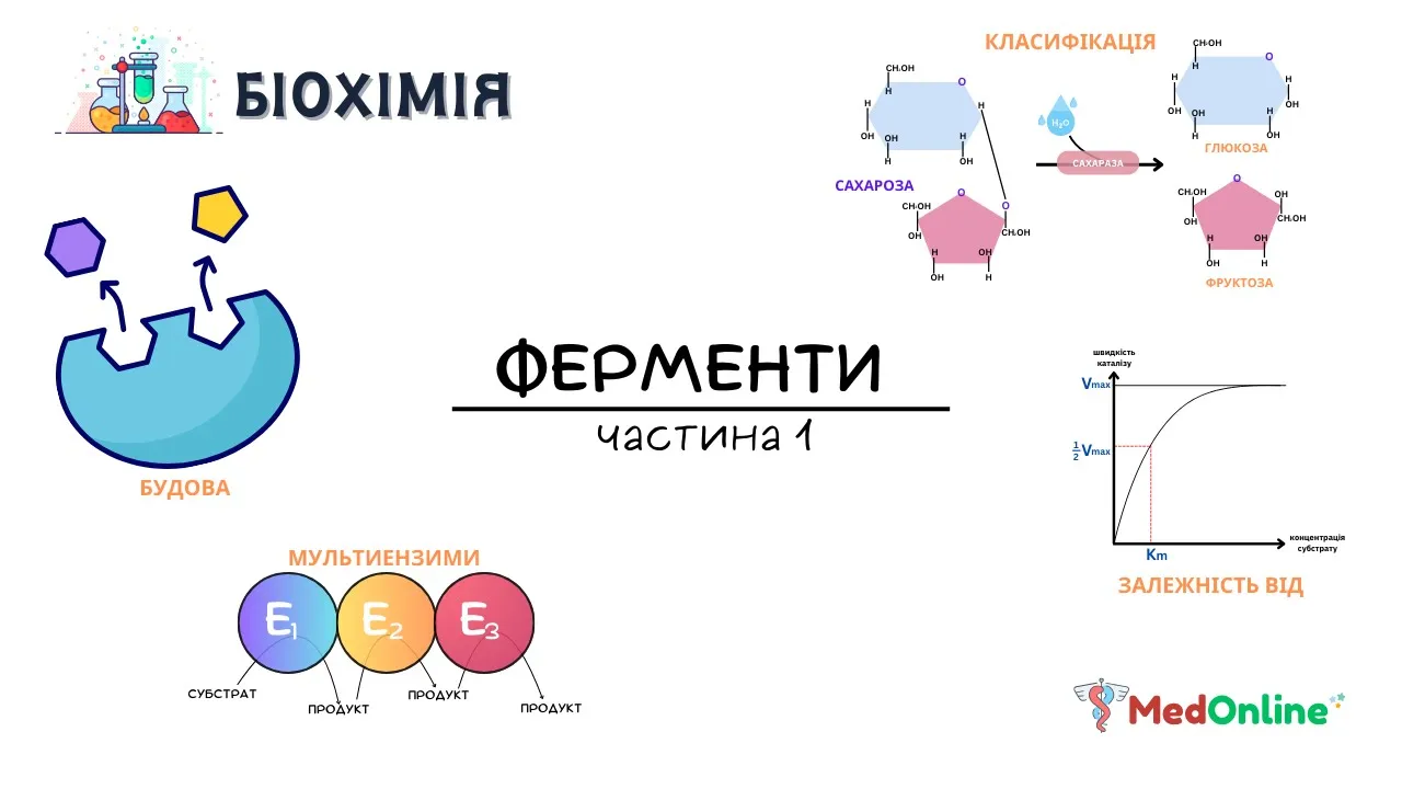
Ферменти | Будова, Класифікація, Механізм дії
2K
Enzymes are composed of subunits—autonomous structural units that perform specific functions. One subunit may catalyze a reaction while another responds to environmental changes and modulates activity. Enzymes have two functional sites: the active site (in catalytic subunits) facilitates the chemical reaction, while the allosteric site (in regulatory subunits) binds molecules that modulate enzyme activity. The active site consists of a binding site (attaches substrate) and catalytic site (facilitates conversion). The catalytic site contains amino acid functional groups (SH, OH, NH) that participate in substrate transformation.
Click to watch →
Deep Dive
The general definition of post-translational modifications, particularly how small molecules can tag proteins.
20. Cell Signaling 1 – Overview
91.8K
Ubiquitination is a post-translational modification that serves as a tagging system for proteins destined for degradation. When proteins cannot be properly folded, they undergo ubiquitination where small ubiquitin proteins are attached to them using ATP. This ubiquitin chain acts as a signal flagging the protein for destruction. The proteasome recognizes this ubiquitin tag and processes the protein for disposal.
Click to watch →
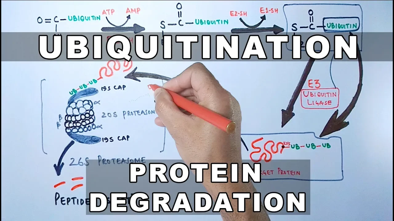
Ubiquitination of Proteins and Protein Degradation
215.4K
Ubiquitination is a post-translational modification where ubiquitin proteins are covalently attached to lysine residues of target proteins via iso peptide bonds, serving as a molecular tag that marks proteins for degradation; this process is catalyzed by a cascade of three enzymes—E1 (ubiquitin activating enzyme), E2 (ubiquitin conjugating enzyme), and E3 (ubiquitin ligase)—which sequentially transfer ubiquitin from E1 to E2 and then to the target protein, after which the ubiquitinated proteins are recognized and degraded by the 26S proteasome complex consisting of a 20S proteasome core and 19S regulatory caps.
Click to watch →

CH450 Chapter 8.2 Post-Translational Protein Modifications
127
Post-translational protein modifications are essential regulatory mechanisms that cells use to quickly fine-tune enzyme activity and protein function without waiting for new protein synthesis; these modifications include small chemical changes like phosphorylation (adding phosphate groups via kinases using ATP, with phosphatases removing them), acetylation (adding acetyl groups to histone lysines to regulate gene transcription), hydroxylation (common in collagen for structural stability), and methylation (using S-adenosyl methionine as methyl donor), as well as larger modifications such as glycosylation (adding sugars to proteins, exemplified by the SARS-CoV-2 spike protein where ~40% of the surface is covered with sugar modifications), lipidation (attaching lipids for membrane anchoring), and ubiquitination/SUMOylation (adding small peptides to target proteins for degradation or alter their function).
Click to watch →

Post-Translational Modifications in prokaryotes and eukaryotes - Types, Functions, and Examples
79.1K
Post-translational modifications (PTMs) are reversible or irreversible chemical changes to proteins after translation, primarily driven by enzymatic reactions, that significantly increase protein diversity in cells from approximately 20,000 genes to over one million functional proteins; these modifications include phosphorylation (adding phosphate groups, critical for signaling pathways like EGFR), methylation/acetylation (modifying histones to control chromatin accessibility), glycosylation (adding sugars, determining blood types), ubiquitination (tagging proteins for degradation via proteasomes), SUMOylation (directing protein transport between cellular compartments), proteolysis (cleaving precursor proteins like insulin), and protein splicing (removing inteins), with defects in these processes contributing to diseases such as cancer, diabetes, and immune disorders.
Click to watch →

Post Translational Modifications
136.8K
Post-translational modifications are biochemical processes that polypeptide chains undergo after translation to become functional proteins. These modifications include phosphorylation, methylation, glycosylation, proteolysis, acetylation, and lipidation. The purpose of these modifications is to activate proteins, direct them to their target locations within the cell, and prepare them for their specific biological functions.
Click to watch →
Prerequisite Knowledge
- Concept 01Basic protein structure, including the concepts of polypeptide chains, folding, and denaturation.
- Concept 02The concept of cellular homeostasis and why cells need to regulate protein concentration and eliminate damaged proteins.
- Concept 03An introduction to enzymes, active sites, and how multi-subunit enzyme complexes function.
- Concept 04The general definition of post-translational modifications, particularly how small molecules can tag proteins.
Subsequent Learning
- Step 01The detailed enzymatic pathway of ubiquitination, involving the sequential action of E1, E2, and E3 enzymes.
- Step 02The role of proteasome dysfunction in neurodegenerative disorders such as Alzheimer's and Parkinson's diseases, where misfolded proteins aggregate.
- Step 03Clinical applications of proteasome inhibitors (such as bortezomib) in cancer therapies, particularly for treating multiple myeloma.
- Step 04A comparative study of the Ubiquitin-Proteasome System (UPS) versus autophagy-lysosome pathways for cellular degradation.
Proteosome Role
Mechanism
Proteosome Role
0:02Playing Section
- 1
Explains proteosome as recycling machine for damaged proteins.
- 2
Details cleavage into peptides for cell maintenance and immune defense.
Ubiquitin-Independent 20S Proteasomal Degradation
While classic molecular biology emphasizes the Ubiquitin-Proteasome System (UPS)—where proteins must be tagged with ubiquitin chains before destruction—a significant alternative pathway exists: ubiquitin-independent degradation by the core 20S proteasome. Under this model, the catalytic 20S core of the proteasome can directly recognize, unfold, and degrade 'intrinsically disordered proteins' (IDPs) without any prior ubiquitin tagging or ATP consumption. This challenges the traditional dogma that ubiquitin tagging is a universal prerequisite for proteasomal recycling, demonstrating that the cell maintains a simpler, passive clearance mechanism for highly flexible or oxidatively damaged proteins.
Deep Dive
The detailed enzymatic pathway of ubiquitination, involving the sequential action of E1, E2, and E3 enzymes.

Ubiquitination of Proteins
156.1K
Ubiquitination is a cellular process where a 76-amino acid ubiquitin molecule is attached to damaged or unwanted proteins via an isopeptide bond between ubiquitin's terminal glycine and lysine residues on the target protein, marking it for degradation by the proteasome; this process requires ATP hydrolysis and involves three sequential enzymes (E1, E2, and E3) to transfer ubiquitin from E1 to E2 and finally to the target protein, with at least four ubiquitin molecules needed to effectively target a protein for breakdown.
Click to watch →
Ubiquitination of Proteins and Protein Degradation
215.4K
Ubiquitination is a post-translational modification where ubiquitin proteins are covalently attached to lysine residues of target proteins via iso peptide bonds, serving as a molecular tag that marks proteins for degradation; this process is catalyzed by a cascade of three enzymes—E1 (ubiquitin activating enzyme), E2 (ubiquitin conjugating enzyme), and E3 (ubiquitin ligase)—which sequentially transfer ubiquitin from E1 to E2 and then to the target protein, after which the ubiquitinated proteins are recognized and degraded by the 26S proteasome complex consisting of a 20S proteasome core and 19S regulatory caps.
Click to watch →
Lec 08 The Ubiquitin Proteasome system
10.8K
Ubiquitylation is a complex multi-step process requiring three main enzymes: E1 (activating enzyme), E2 (conjugating enzyme), and E3 (ligase). Precursor ubiquitin is processed by DUBs to expose the C-terminal glycine. E1 activates ubiquitin with ATP, transfers it to E2, and E3 mediates substrate selectivity by binding both E2 and target proteins. E3s are classified into RING, HECT, U-box, and PHD finger families, each with distinct mechanisms. In mammalian cells, there are ~2 E1s, ~40 E2s, and ~600 E3s, enabling recognition of diverse substrates at specific times.
Click to watch →

Ubiquitin
123
Ubiquitin is a small regulatory protein (76 amino acids) that acts as the 'fashion police' of the cell by tagging faulty or non-functional proteins with ubiquitin molecules; this tagging process involves three enzymes (E1, E2, E3) in a sequential cascade where E1 activates ubiquitin, E2 transfers it, and E3 ligates it to the target protein, and the proteasome recognizes the polyubiquitinated protein to degrade it into amino acids for recycling.
Click to watch →

UBIQUITIN - Proteasome Degradation Pathway I What is Ubiquitin? Proteasome and Ubiquitin
4.2K
The cellular outcome of ubiquitination depends on which lysine residue is used for linkage: K48 linkage between ubiquitins triggers classical protein degradation; K63 linkage signals DNA repair and endocytosis; K11 linkage regulates the cell cycle. The ubiquitination process requires a sequential enzymatic cascade: E1 activates ubiquitin using ATP, transfers it to E2, which then passes it to E3 ubiquitin ligase. E3 recognizes degradation signals (degrons) on target proteins and facilitates ubiquitin transfer. Only when E1, E2, E3, and target protein are all present can polyubiquitination occur, marking the protein for proteasomal degradation.
Click to watch →
Deep Dive
The role of proteasome dysfunction in neurodegenerative disorders such as Alzheimer's and Parkinson's diseases, where misfolded proteins aggregate.

Proteassoma e sua relação com doenças neurodegenerativas, como Alzheimer e Parkinson?
4.6K
The proteasome is a large protein complex present in all cells of humans, animals, plants, and fungi, responsible for breaking down damaged or defective proteins into reusable peptide fragments. It consists of a cylindrical 20S core unit where protein breakdown occurs, coupled with 19S regulatory units at one or both ends that recognize proteins marked with ubiquitin chains for degradation. When the proteasome functions correctly, it eliminates damaged proteins to maintain cellular health; however, when its activity decreases, toxic proteins accumulate, leading to neuronal destruction in neurodegenerative diseases such as Alzheimer's and Parkinson's. Research aims to understand how to increase proteasome activity to prevent toxic protein accumulation and potentially treat these diseases.
Click to watch →
Parkinson, Huntington kasalliklari va Demensiyalar
415
The ubiquitin-proteasome system normally degrades damaged or misfolded proteins. In neurodegenerative diseases, this system becomes dysfunctional, allowing abnormal proteins to accumulate. In Parkinson's disease, this contributes to Lewy body formation. In Huntington's disease, mutant huntingtin protein accumulates due to impaired degradation. Proteasome dysfunction represents a common mechanism in multiple neurodegenerative diseases.
Click to watch →
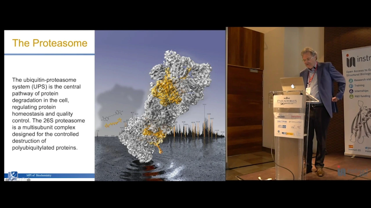
The Molecular Machinery of Protein Degradation: Structural Studies ex situ and in situ
197
In ALS models caused by glycine-alanine repeat expansions, toxic protein aggregates recruit massive numbers of proteasomes (40-fold increase) that become functionally trapped. Proteasomes attempt to degrade aggregated proteins but cannot unfold them, resulting in accumulation of proteasomes in substrate-processing states. This trapping mechanism explains why proteasome inhibition worsens ALS pathology—reducing the already overwhelmed proteasome pool impairs clearance of both normal and pathological substrates. The aggregates contain polymorphic ribbon structures with proteasomes inhabiting the material, demonstrating how misfolded proteins disrupt cellular proteostasis networks and contribute to neurodegeneration.
Click to watch →
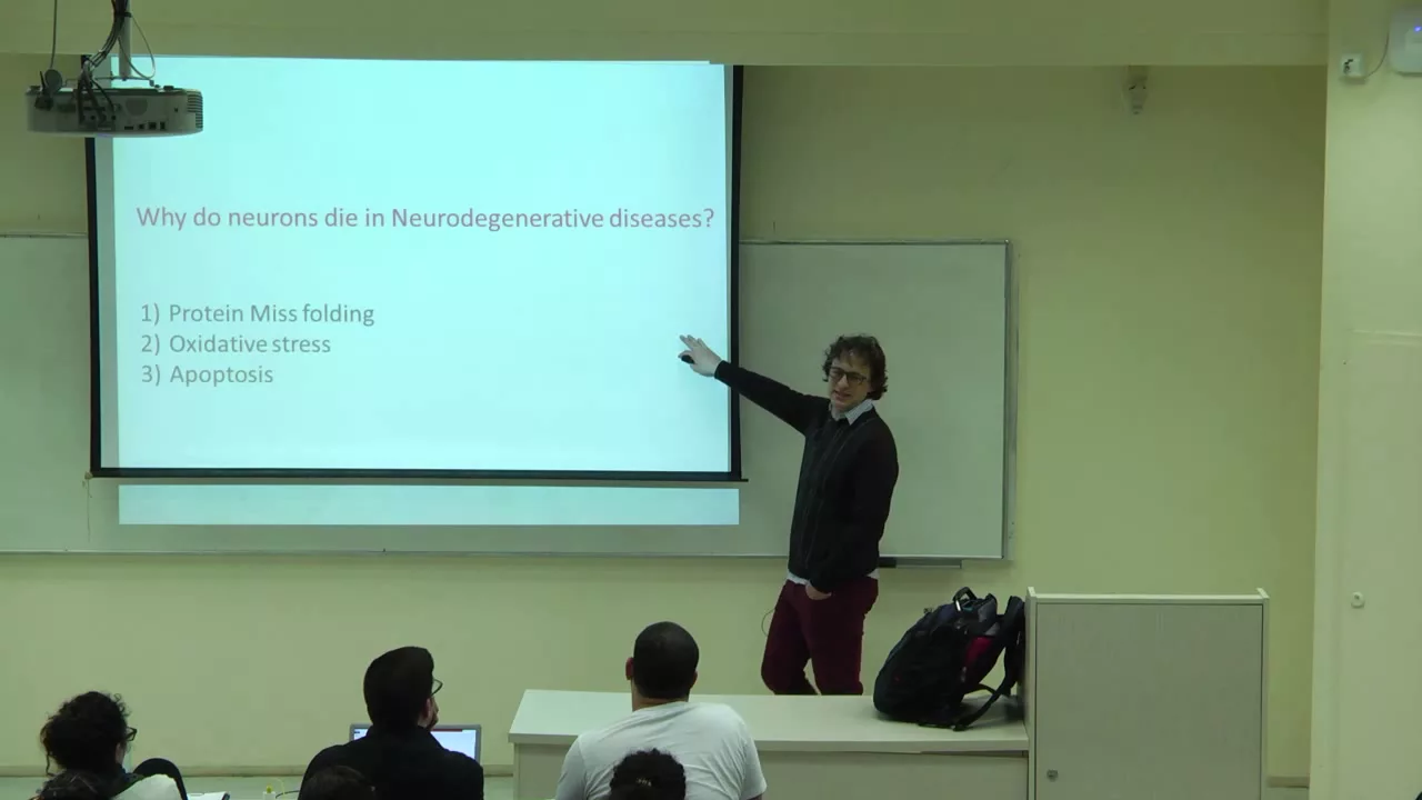
אבני פינה: המוח המתנוון - ממחקר לתקווה - שיעור מס' 1
2.4K
Proteins determine cellular function through their three-dimensional shape. Cells use chaperone proteins to ensure proper folding during synthesis and after stress-induced unfolding. When stress overwhelms chaperone capacity, misfolded proteins aggregate into insoluble fibrils. These aggregates represent quality control failure and are hallmarks of Alzheimer's, Parkinson's, Huntington's, and ALS. The ubiquitin-proteasome system normally degrades damaged proteins, but when impaired, aggregates accumulate. This protein misfolding pathway is central to understanding neurodegenerative disease mechanisms.
Click to watch →

Guest Lecture: Giuseppe Grasso - Neurodegeneration is a worldwide global health emergency
233
Neurodegenerative diseases, such as Alzheimer's and Parkinson's, result from the accumulation of misfolded proteins that overwhelm cellular quality control systems. The ubiquitin-proteasome system (UPS) is the primary cellular mechanism for degrading misfolded proteins, where proteins are tagged with ubiquitin and degraded by the proteasome. When UPS function is impaired, misfolded proteins accumulate, leading to neurodegeneration. A holistic approach to treating these diseases involves understanding the interconnected nature of cellular processes—including protein synthesis, folding, and degradation—and developing therapies that enhance proteasome activity or prevent protein misfolding, rather than targeting single pathways in isolation.
Click to watch →
Deep Dive
Clinical applications of proteasome inhibitors (such as bortezomib) in cancer therapies, particularly for treating multiple myeloma.
Functions of the Proteasome From Protein Degradation to Disease Therapy
5.5K
Proteasome inhibitors were developed by designing molecules mimicking natural substrates. Initial compounds like MG132 (three leucines) were converted into boronate derivatives, yielding bortezomib (PS-341), now used by over 700,000 cancer patients worldwide. Bortezomib selectively blocks the chymotrypsin-like site, stabilizing cell cycle regulators and inducing apoptosis through proteotoxic stress. Multiple myeloma cells are particularly vulnerable because they produce abnormal immunoglobulins overwhelming the ubiquitin-proteasome pathway. Blocking this pathway further activates the unfolded protein response and inhibits NF-kappaB signaling, making it highly effective against this disease with over 95% response rates in combination therapy. Approved inhibitors mainly block the chymotrypsin-like site, leaving other proteolytic sites functional at 50-70% normal rates, allowing cells to compensate partially.
Click to watch →

Aaron Ciechanover | Max Birnstiel Lecture
711
Bortezomib (Velcade) was the first drug targeting the ubiquitin-proteasome system, developed through serendipitous screening at NIH. It is a boronic acid derivative that specifically inhibits the chymotrypsin-like active site of the proteasome's beta subunits. Originally designed to treat pregnancy-related nausea (thalidomide), it was found to kill malignant B-cells in multiple myeloma. The drug exploits the unfolded protein response: malignant plasma cells secrete excessive immunoglobulins, some misfolded and degraded by ER-associated degradation. When proteasome inhibition blocks degradation, misfolded proteins accumulate, triggering apoptosis. Bortezomib dramatically improves outcomes in multiple myeloma, reducing tumor burden from 41% to less than 1% in bone marrow.
Click to watch →
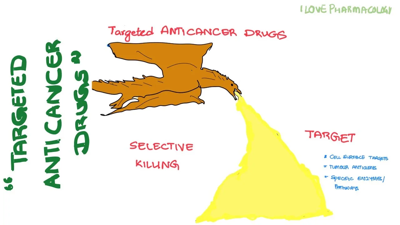
ANTICANCER DRUGS - 2 | TARGETED ANTICANCER DRUGS
1.6K
Bortezomib is a boron-containing compound that covalently binds to proteasomes and inhibits proteolytic activity. The proteasome is a complex proteolytic enzyme responsible for degradation of intracellular signaling proteins involved in cell cycle control, apoptosis, and survival responses. Mechanism: NF-kappa-B mediated signaling is interrupted by bortezomib. Under stress (hypoxia, cytotoxic drugs, DNA breaks), proteasome cleaves the Ikappa-B/NF-kappa-B complex, releasing NF-kappa-B which promotes anti-apoptosis and cell proliferation. Bortezomib prevents this cleavage, inhibiting NF-kappa-B activity and promoting apoptosis while decreasing cell proliferation. It also increases pro-apoptotic proteins. Route: IV infusion or subcutaneous injection. Uses: multiple myeloma and refractory mantle cell lymphoma. Adverse effects: peripheral neuropathy, diarrhea, fatigue, depression, thrombocytopenia.
Click to watch →

MYELOMA UNIVERSITY | Learn about steroids, IMiDs, and PIs: three classes of drugs to treat myeloma
1K
Proteasome inhibitors (bortezomib, carfilzomib, ixazomib) represent a cornerstone of modern multiple myeloma treatment, approved for newly diagnosed, relapsed, and refractory disease. These agents block proteasome function, disrupting cellular regulatory signals and inducing apoptosis in cancer cells while sparing normal cells. Bortezomib, the oldest agent, commonly causes thrombocytopenia, infections, and peripheral neuropathy, now administered subcutaneously to reduce neurotoxicity. Carfilzomib, an irreversible inhibitor given once/twice weekly, requires pre-dose blood counts and carries thrombotic and cardiac risks requiring baseline cardiac assessment. Ixazomib, an oral agent given weekly on empty stomach, offers convenient administration but requires viral prophylaxis and monitoring for cyclic thrombocytopenia. All proteasome inhibitors necessitate routine CBC monitoring and dose adjustments for renal/hepatic impairment.
Click to watch →
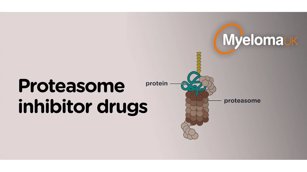
Proteasome inhibitor drugs
25.6K
Proteasome inhibitors are a class of drugs that block the proteasome, a cellular recycling system responsible for breaking down excess proteins; by inhibiting this process, these drugs cause toxic protein accumulation in myeloma cells, leading to cell death, and newer generations (carfilzomib and ixazomib) offer improved efficacy with fewer side effects, particularly reduced peripheral neuropathy, compared to the first-generation drug bortezomib.
Click to watch →
Deep Dive
A comparative study of the Ubiquitin-Proteasome System (UPS) versus autophagy-lysosome pathways for cellular degradation.

Yoshinori Ohsumi: What is autophagy? A dynamic cellular recycling process
550.9K
Autophagy differs fundamentally from the ubiquitin-proteasome system in its approach to protein degradation. While the ubiquitin-proteasome system recognizes and degrades specific target proteins through sophisticated recognition mechanisms, autophagy provides a bulk degradation pathway that can eliminate entire cellular components including organelles. This makes autophagy particularly important for eliminating damaged or excessive cellular material that cannot be targeted by specific degradation systems.
Click to watch →
Lectio magistralis del Prof. Aaron Ciechanover "Intracellular Protein Degradation"
263
Autophagy and the ubiquitin-proteasome system represent two major cellular degradation pathways that work together and regulate each other. Autophagy degrades damaged organelles and protein aggregates through autophagosome-lysosome fusion, while the ubiquitin-proteasome system degrades misfolded proteins via proteasomal degradation. Surprisingly, ubiquitination also marks proteins for autophagy, with p62 adaptors recognizing ubiquitin and LC3 to deliver cargo to autophagosomes. These pathways cross-regulate each other: when autophagy should be off, the proteasome degrades autophagy proteins; when autophagy should be on, autophagy proteins inhibit specific ubiquitin ligases. AMBRA1 serves as a central regulator, interacting with both CUL4 and CUL5 ubiquitin ligases to coordinate degradation responses based on cellular conditions.
Click to watch →
Intracellular protein degradation bsc 3rd semester zoology unit 4 🔥BSc 3rd semester zoology
2.2K
The ubiquitin-proteasome system provides precise, selective degradation of specific target proteins, while lysosomal degradation causes bulk, non-selective degradation of cellular components. In UPS, ubiquitin is specifically attached to target proteins before degradation, making it highly selective for damaged, misfolded, or unnecessary proteins. In contrast, lysosomes degrade various materials including long-lived proteins, organelles, and extracellular components without specific targeting. Lysosomes contain digestive enzymes (proteases, lipases) that work optimally at acidic pH and are responsible for breaking down cellular waste. Proteins reach lysosomes through endocytosis (external materials), phagocytosis (large particles like bacteria), and autophagy (cellular components enclosed in vesicles).
Click to watch →
Best Practices for PROTACs - UPS, MoA and Design of PROTACs (Part 1A)
2.4K
Cells employ two major protein degradation pathways: the lysosomal pathway and the ubiquitin-proteasome system (UPS). The lysosome degrades proteins non-selectively through endocytosis or autophagy in acidic compartments. The UPS performs selective degradation of ubiquitin-marked proteins, serving quality control and cell state-dependent removal. The UPS operates through a cascade of E1, E2, and E3 enzymes: E1 activates ubiquitin, E2 transfers it, and E3 ligases bring substrates close for ubiquitination. Over 600 human E3 ligases exist, with most involved in degradation. The 26S proteasome contains a regulatory cap with ubiquitin-binding domains and ATPases, plus a barrel-shaped 20S core where proteases cleave peptides.
Click to watch →

Prof XJ Wang - "Interplay between the ubiquitin-proteasome system and autophagy"
738
The ubiquitin-proteasome system (UPS) and autophagy are two major catabolic pathways that interact bidirectionally: UPS malfunction activates autophagy through P62-mediated mechanisms involving TFEB nuclear translocation, while autophagy impairment reciprocally compromises UPS function, creating a complex regulatory network essential for protein quality control and cardiac health.
Click to watch →
Proteosome Role
Mechanism
Proteosome Role
0:02- 1
Explains proteosome as recycling machine for damaged proteins.
- 2
Details cleavage into peptides for cell maintenance and immune defense.
Ubiquitin-Independent 20S Proteasomal Degradation
While classic molecular biology emphasizes the Ubiquitin-Proteasome System (UPS)—where proteins must be tagged with ubiquitin chains before destruction—a significant alternative pathway exists: ubiquitin-independent degradation by the core 20S proteasome. Under this model, the catalytic 20S core of the proteasome can directly recognize, unfold, and degrade 'intrinsically disordered proteins' (IDPs) without any prior ubiquitin tagging or ATP consumption. This challenges the traditional dogma that ubiquitin tagging is a universal prerequisite for proteasomal recycling, demonstrating that the cell maintains a simpler, passive clearance mechanism for highly flexible or oxidatively damaged proteins.
Basic protein structure, including the concepts of polypeptide chains, folding, and denaturation.
The concept of cellular homeostasis and why cells need to regulate protein concentration and eliminate damaged proteins.
An introduction to enzymes, active sites, and how multi-subunit enzyme complexes function.
The general definition of post-translational modifications, particularly how small molecules can tag proteins.
The detailed enzymatic pathway of ubiquitination, involving the sequential action of E1, E2, and E3 enzymes.
The role of proteasome dysfunction in neurodegenerative disorders such as Alzheimer's and Parkinson's diseases, where misfolded proteins aggregate.
Clinical applications of proteasome inhibitors (such as bortezomib) in cancer therapies, particularly for treating multiple myeloma.
A comparative study of the Ubiquitin-Proteasome System (UPS) versus autophagy-lysosome pathways for cellular degradation.
42.5K views2.6Klikes3:32@WEHImoviesOriginal Release: 2024-05-24
the [Applause] proteosome the proteosome is a protein recycling machine breaking down old or damaged proteins for cell maintenance this enzyme complex Cleaves proteins into short stretches of amino acids called peptides the peptides can be repurposed for new protein synthesis by the cell the proteosome is also responsible for recycling proteins from bacteria and viruses this generates peptides essential for detecting infection and developing the body's immune response the proteosome is a molecular machine it contains receptors that recognize proteins tagged for recycling and a motor which unravels the protein and threads it through to the proteome core the motor has six subunits that work together and will mechanically pull the protein through a central Channel preparing it for the proteome core the proteosome core contains a chamber that cuts the protein into peptides six cutting enzy enzymes known as proteases line the chamber receptors in the proteosome recognize ubiqutin tags marking proteins for recycling the protein is guided into the motor tethered by the ubiqutin receptor an enzyme in the proteosome removes the ubiquit and tag preparing the protein for unraveling the proteosome motor unfolds the protein delivering an amino acid strand to the core for cutting the motor subunits extend into the channel and directly Engage The Protein strand motor residues form a staircase around the Strand and mechanically unravel the protein towards the proteosome core the proteases within the proteosome core cut the protein strand into peptides of varying length typically ranging from 2 2 to 25 amino acids the peptides are released and the reclaimed amino acids will contribute to new protein synthesis completing the proteasome's central role in cellular maintenance and protein recycling
Up Next

11:35
Animal Cell Structure and Function: Crash Course Biology
@crashcourse
6M views•2012-02-20

50:12
Bacterial Communication: Quorum Sensing and Biofilm Formation Explained
@JHUAAP
12.6K views•2011-06-28
6:10
Citric Acid Cycle Explained: The Krebs Cycle in Mitochondria
@WEHImovies
140.8K views•2020-11-20

7:08
Bacteriophages: Earth's Deadliest Killers and Future Antibiotics
@kurzgesagt
34.6M views•2018-05-13
Related Study Plans & Knowledge Roadmaps
Structured learning paths in Biology
biology
RNAi: Biogenesis, RISC & Gene Silencing
01Molecular Biology Foundations: DNA to Protein
02Introduction to RNA Interference (RNAi)
03Molecular Biogenesis of miRNA and siRNA
+ 2 more learning modules
10h5 modules
Explore Roadmap
biology
Mycorrhizae: Signaling, Biophysics & Ecology
01Foundations of Mycorrhizal Symbiosis
02Molecular Signaling and Root Colonization
03Biophysics and Bioenergetics of Nutrient Exchange
+ 1 more learning modules
11h4 modules
Explore Roadmap
biology
Circadian Clocks: TTFL, SCN & Periphery
01Foundations of Molecular Biology & Gene Expression
02Introduction to Chronobiology and Actograms
03The Core TTFL: Molecular Architecture of the Clock
+ 3 more learning modules
14h6 modules
Explore Roadmap
biology
Population Genetics: Math, Drift & Selection
01Genetic & Probability Foundations
02The Hardy-Weinberg Principle and Baseline Models
03Mathematical Modeling of Natural Selection
+ 3 more learning modules
18h6 modules
Explore Roadmap