




![Traducción del ARN - Bio[ESO]sfera - Biología](https://i.ytimg.com/vi_webp/ZapoL3RuX3s/maxresdefault.webp)






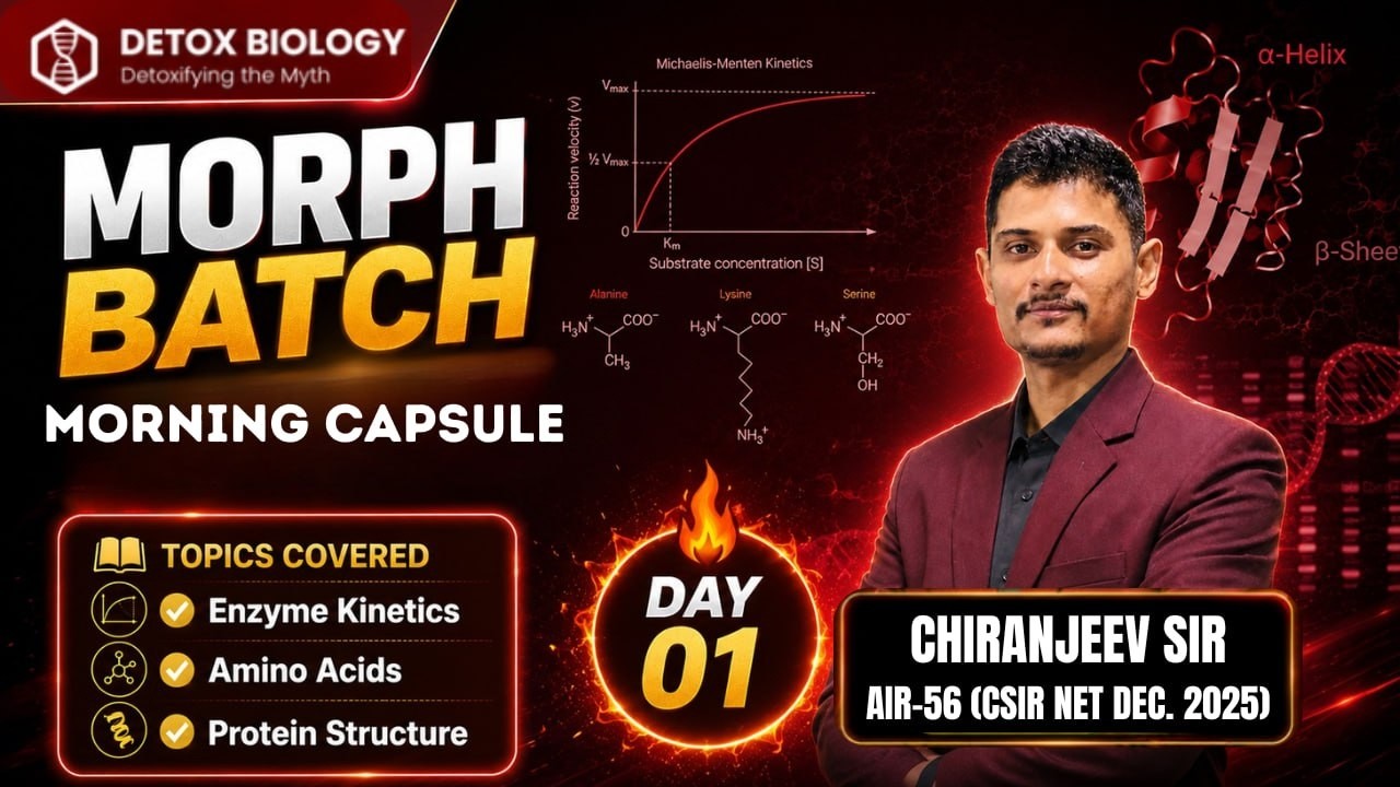


Proteins are polymers of amino acids that exist in a hierarchical structure: primary structure is the linear sequence of amino acids; secondary structure includes localized conformations like alpha helices and beta pleated sheets formed by hydrogen bonding; tertiary structure is the overall three-dimensional folding of a single polypeptide chain stabilized by hydrophobic interactions, disulfide bonds, and electrostatic forces; and quaternary structure describes how multiple polypeptide subunits assemble into a functional protein complex. Even minor changes in primary structure can dramatically alter protein function, as demonstrated by sickle cell disease where a single amino acid substitution causes abnormal hemoglobin folding.
Protein Structure Explained: Primary to Quaternary | Biochemistry
Added:Understanding of the basic chemical structure of an amino acid, including the central carbon, amino group, carboxyl group, and the variable R-group side chain.
Familiarity with peptide bond formation via dehydration synthesis to create polypeptide chains.
Knowledge of fundamental chemical bonds and intermolecular forces, such as hydrogen bonding, ionic interactions (salt bridges), hydrophobic interactions, and covalent disulfide bridges.
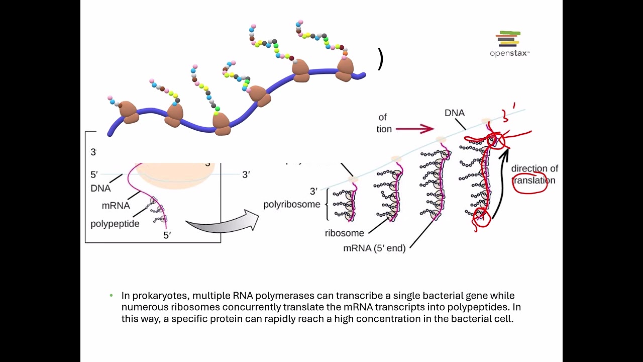
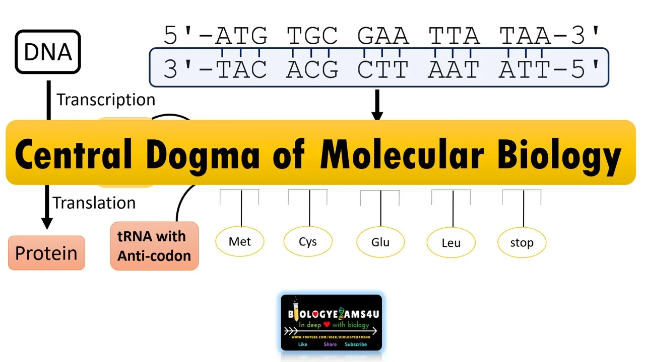
A basic grasp of the Central Dogma of Molecular Biology, specifically how mRNA codons translate into a specific sequence of amino acids.
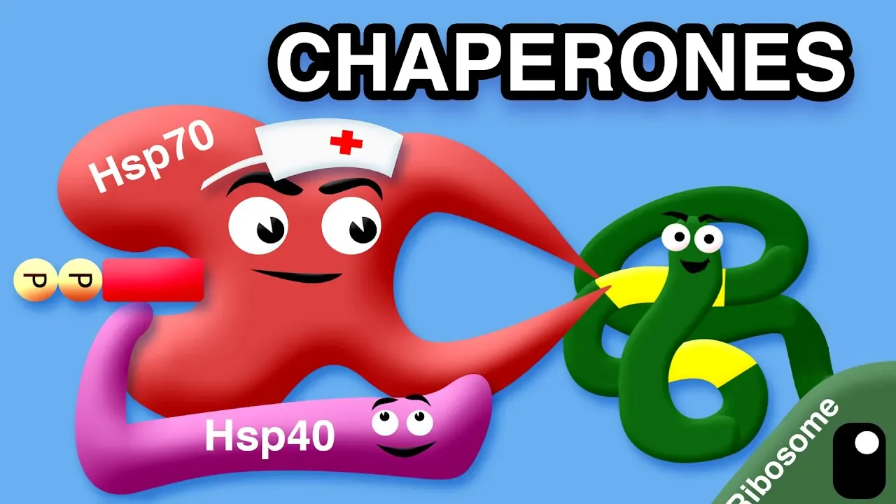
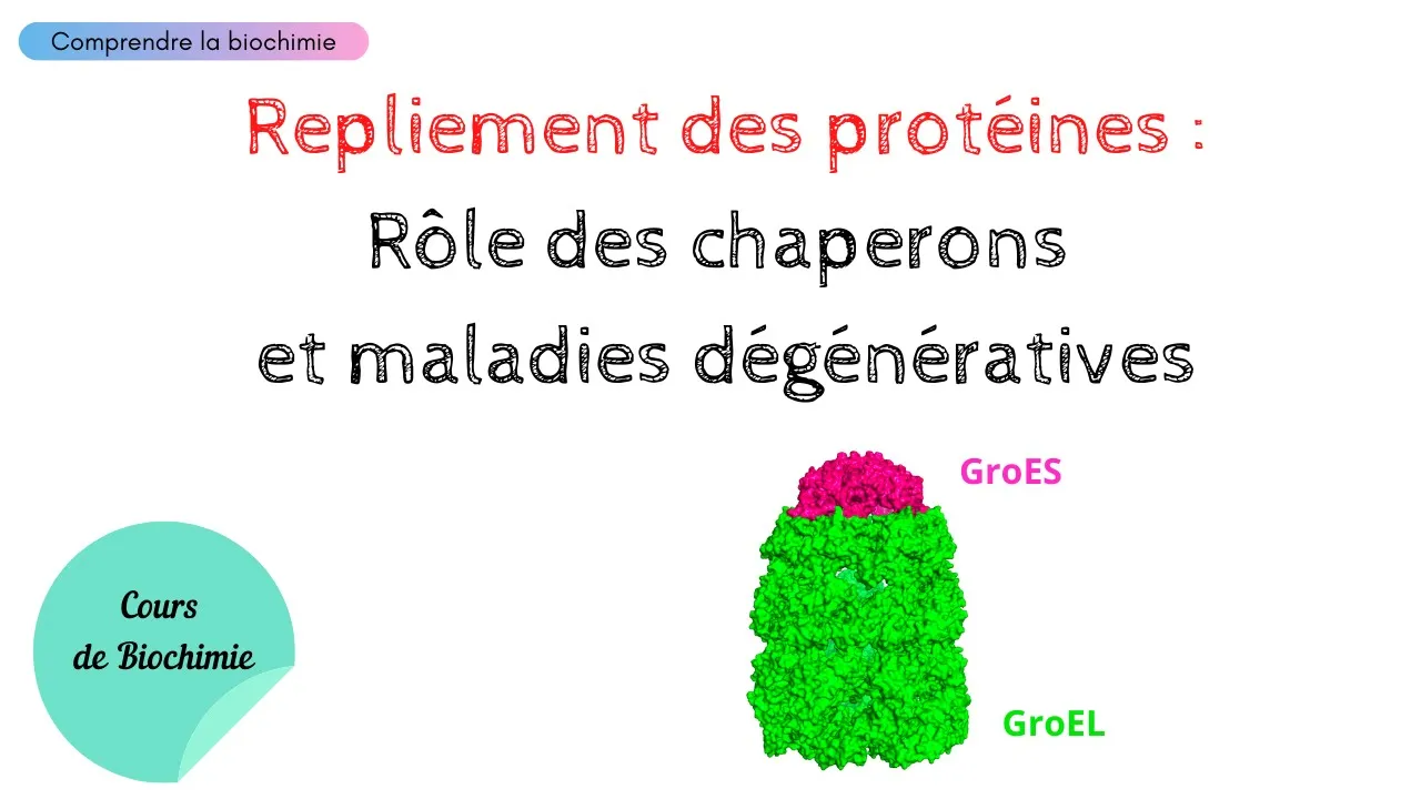
Exploration of protein folding thermodynamics and the critical role molecular chaperones play in preventing misfolding in vivo.
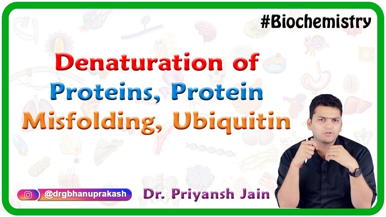
Investigation of protein denaturation and the pathological consequences of misfolding, such as amyloid plaque formation in Alzheimer's disease or infectious prions.
Study of the structure-function relationship in proteins, particularly enzyme catalysis, active site geometry, and allosteric regulation in cooperative proteins like hemoglobin.
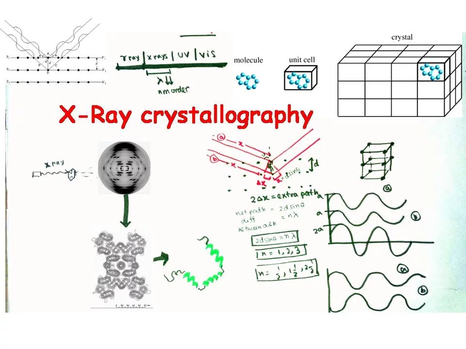
Introduction to experimental and computational biophysical methods used to determine 3D structures, including X-ray crystallography, NMR spectroscopy, Cryo-EM, and AlphaFold predictive modeling.
1.4M views27.7Klikes10:50Original Release: 2016-08-27
Hey guys, it's Professor Dave, I want to tell you about proteins.
So we know about amino acids, and these are the monomers that will form proteins, which are also known as polypeptides. Proteins are polymers of amino acids, and they are the most diverse type of biomolecule in your body.
Different kinds of proteins include enzymes that catalyze chemical reactions, receptors that control signaling in your body, hemoglobin, which carries oxygen throughout the bloodstream, muscle and organ tissue, which gives your body structure and mobility, and so many other things. So how do amino acids polymerize? This happens when amino acids form peptide bonds with one another, such as the peptide bond between these two glycine units. Peptide bond formation is an example of a dehydration reaction because the two hydrogens and the oxygen marked in blue are lost, and two hydrogens plus one oxygen equals a water molecule, so as a water molecule is lost these two amino acids come together to form a peptide bond, which results in an amide. An amide is a functional group with a nitrogen atom next to a carbonyl and this is the functional group that will connect each amino acid during polymerization. If two amino acids combine we get a dipeptide. If between three and ten come together, we would call that an oligopeptide, since oligo means just a few. And if more than ten come together we will call that a polypeptide, since poly means many and proteins are large polypeptides of around three hundred to a thousand amino acids that are folded in such a way that they have some biological activity. When we look at any peptide, we must notice that there is an N-terminus, meaning the side of the chain that ends with the amino group, and a C-terminus, the side that ends with the carboxyl group.
By convention we typically write proteins with the N-terminus on the left and the C-terminus on the right. Each monomeric unit in the polypeptide is called a residue, so on this structure we should be able to differentiate between each individual residue and locate the peptide bonds that connect them.
Proteins are very large compared to simple molecules. They contain hundreds of amino acid residues and they have very specific shapes from which their function is derived, so let's learn about protein structure.
Protein structure follows a specific hierarchy so let's start first with primary protein structure.
The primary structure of a protein is simply the sequence of amino acids with no attention paid to any aspect of three-dimensional shape.
We can abbreviate each amino acid residue with either a three-letter code or a one letter code depending on how much you feel like writing, and here are both sets of abbreviations for your reference. The shape the protein will take depends on the primary structure because it is the identity of all the side chains and the sequence they are found in that determines how a protein will fold up.
To see this in action let's look at secondary protein structure.
Secondary protein structure describes localized conformations of the polypeptide backbone, meaning the folding pattern that a protein will exhibit over a few dozen amino acid residues. There are a few different motifs that a protein can utilize so let's see what those are.
First let's understand that the backbone itself is essentially planar. The peptide bond has some pi bond character due to resonance, since there is a resonance structure where the lone pair on the nitrogen forms a pi bond, pushing the pi bond in the carbonyl up to the oxygen atom. We know that rotation is restricted around pi bonds so even though it's not a formal pi bond between the carbon and the nitrogen, the backbone is fairly rigid, while the sigma bonds to the R groups can freely rotate, so it is mainly the side chains on each residue that have a flexible conformation.
We also know that molecules with dipoles or formal charges will attempt to store energy by making electrostatic interactions, and amino acids have such features. So if this backbone can fold in such a way so as to let each residue interact with other residues, it will do so, in order to adopt the lowest-energy conformation. One way it can do this is by forming something called a beta-pleated sheet. The backbone will extend one way and then turn back to line up alongside the first part of the chain so that NH bonds from one section can form dipole-dipole interactions with the carbonyls of an adjacent section. This stores energy so it is an example of a favorable conformation and one type of secondary protein structure. Another type of structural motif is called the alpha helix. This is when the backbone forms a spiral shape with about three or four amino acids per turn, and all the R groups pointing out. Here each amide group will interact with the amide group three residues above and the one three residues below. So beta-pleated sheets and alpha helices are the most common types of secondary structure, though there are all kinds of other coils and loops that are a bit more difficult to describe. The key thing to understand is that at this level the polypeptide backbone begins to form shapes entirely dependent on how it can best store energy in dipole-dipole interactions, by adopting the lowest-energy conformation, and these structures contain so many individual atoms that we begin to depict them as colored strips rather than with conventional molecular line notation for practical purposes.
Now let's move on to tertiary protein structure. Tertiary protein structure involves the further folding of the polypeptide chain to produce its overall three-dimensional structure. This folding is not random, it is specific to the protein and will occur in that specific way every time that particular protein is formed, since it is this shape that gives the protein its function.
There are a few factors that influence tertiary structure. First, residues with hydrophobic side chains, like alkyl groups, tend to be found in the interior of the protein, so that they are not in contact with aqueous solvent. At the same time, residues with hydrophilic side chains, ones with formal charges or dipoles on them, tend to be found on the surface of a protein so that they can make dipole-dipole or ion-dipole interactions with water molecules.
All of this is just a way for the protein to maximize the electrostatic interactions it can make in solution, which is a big factor in determining the way the protein will fold.
Another motif that stabilizes tertiary structure is something called a disulfide bond. We know that cysteine has an SH group called a thiol on its side chain, which is the sulfur analog of a hydroxyl group.
If one cysteine is near another, these thiol groups can react with one another by mild oxidation to create a disulfide which causes a covalent linkage between the two sulfur atoms, and therefore can also covalently link two residues anywhere in the protein. This can help maintain the structural integrity of the protein through covalent bonds that are much stronger than the dipole-dipole interactions found in the various secondary structures. Overall, if the protein is highly folded and compact we would call it a globular protein, whereas if it is long and spindly we would call it a fibrous protein. Lastly, we can examine quaternary structure. Some proteins are just one continuous polypeptide chain, but some proteins involve multiple polypeptide subunits that can come together to form a larger protein. These units are not covalently bound, they make only electrostatic interactions with one another, but the interactions are strong enough to allow the subunits to arrange themselves in specific ways, and it is the arrangement of these subunits that determines the quaternary structure of a protein, like hemoglobin, which consists of four totally separate polypeptides arranged in a specific way. If a protein is comprised of just one polypeptide then it will not have quaternary structure. So to summarize, primary structure is just the sequence of amino acids. Secondary structure is the way the chain begins to fold on the localized level. Tertiary structure is the complete folding pattern of an entire polypeptide, and quaternary structure is the way multiple polypeptide subunits come together to form a larger protein. It is important to understand that even a tiny change in primary structure can completely change the overall protein. For example, sickle cell disease is a genetic disorder where just one amino acid residue in one of the subunits of hemoglobin is changed from glutamic acid to valine. Because the side chains of those two amino acids are so different, the mutation changes the folding pattern at that location, and the resulting hemoglobin protein, which is responsible for carrying oxygen through the bloodstream, takes on a different shape, causing the red blood cells that contain hemoglobin to look like tiny sickles, which can then clog blood vessels.
So we can already begin to see why understanding the structure and function of biomolecules is crucial if we want to understand health and disease.
Let's keep learning about different biomolecules.
Thanks for watching, guys. Subscribe to my channel for more tutorials, and as always feel free to email me:
Up Next

6:59
GroEL/GroES Chaperonin System: Protein Folding Mechanism
@AwesomeBiochemistry
8.3K views•2022-03-01

50:12
Bacterial Communication: Quorum Sensing and Biofilm Formation Explained
@JHUAAP
12.6K views•2011-06-28
13:43
Innate vs Adaptive Immunity: Immune System Explained
@ProfessorDaveExplains
599.5K views•2019-04-25

7:08
Bacteriophages: Earth's Deadliest Killers and Future Antibiotics
@kurzgesagt
34.6M views•2018-05-13
Related Study Plans & Knowledge Roadmaps
Structured learning paths in Biology
biology
RNAi: Biogenesis, RISC & Gene Silencing
01Molecular Biology Foundations: DNA to Protein
02Introduction to RNA Interference (RNAi)
03Molecular Biogenesis of miRNA and siRNA
+ 2 more learning modules
10h5 modules
Explore Roadmap
biology
Mycorrhizae: Signaling, Biophysics & Ecology
01Foundations of Mycorrhizal Symbiosis
02Molecular Signaling and Root Colonization
03Biophysics and Bioenergetics of Nutrient Exchange
+ 1 more learning modules
11h4 modules
Explore Roadmap
biology
Circadian Clocks: TTFL, SCN & Periphery
01Foundations of Molecular Biology & Gene Expression
02Introduction to Chronobiology and Actograms
03The Core TTFL: Molecular Architecture of the Clock
+ 3 more learning modules
14h6 modules
Explore Roadmap
biology
Population Genetics: Math, Drift & Selection
01Genetic & Probability Foundations
02The Hardy-Weinberg Principle and Baseline Models
03Mathematical Modeling of Natural Selection
+ 3 more learning modules
18h6 modules
Explore Roadmap