














![Lecanemab - Novo Medicamento Para Alzheimer [Guia Completo]](https://i.ytimg.com/vi/5mB1sbMmwjM/maxresdefault.jpg)








Alzheimer's disease, the most common neurodegenerative disorder worldwide, presents through four distinct clinical syndromes—memory syndrome (hippocampal involvement), visual-spatial syndrome (posterior cortical atrophy), language syndrome (logopenic variant), and frontal/executive syndrome—each reflecting different patterns of brain pathology; the disease progresses through stages from asymptomatic to mild cognitive impairment to dementia, with modern biomarkers including cerebrospinal fluid analysis and PET scans for amyloid and tau detection enabling earlier and more accurate diagnosis.
Alzheimer's Disease: Clinical Syndromes, Pathology & Biomarkers (UCSF) 59
Added:Basic neuroanatomy, particularly the structure and function of the hippocampus, cerebral cortex, and neural pathways involved in memory and cognition.
Fundamental concepts of molecular biology and genetics, including how genetic mutations and risk-associated alleles (e.g., APOE) influence protein expression.
The general mechanism of protein misfolding and aggregation, specifically the basic roles of amyloid-beta and tau proteins in neurodegeneration.
Basic principles of medical imaging modalities, such as Magnetic Resonance Imaging (MRI) and Positron Emission Tomography (PET) scanning.
Translational therapeutics and pharmacology, focusing on disease-modifying therapies for Alzheimer's, such as monoclonal antibodies (e.g., lecanemab).
Clinical trial methodology and the integration of fluid (CSF/blood) and imaging biomarkers in drug development pipelines.
The differential diagnosis of non-Alzheimer's dementias, such as Frontotemporal Lobar Degeneration (FTLD), Dementia with Lewy Bodies (DLB), and vascular dementia.
Ethical, psychological, and legal challenges associated with preclinical diagnosis and predictive genetic/biomarker testing in asymptomatic patients.
1.1M views9Klikes1:27:27Original Release: 2019-07-27
[MUSIC] Happy to see you all again.
My name is Serggio Lanata we met last week.
I'm happy to see you back to learn a little bit more about the neurodegenerative diseases.
I think we had a pretty good first session where we were able to provide you with an overview of the neurodegenerative diseases distinguish between the different diseases of the brain and the clinical syndromes they cause.
Give you a big picture view of how we think about neurodegenerative diseases.
I think we had a pretty good discussion also to kick things off.
Today, we're going to start to narrow things down a little bit.
I've invited my colleague, Dr. George Naasan.
He is the director of our clinical services in the UCSF Memory and Aging Center.
A dear colleague of mine and he's going to give you a more detailed overview of a specific disease, Alzheimer's disease, which you learned last time we met, is the most common neurodegenerative disease worldwide.
He's going to delve into the pathology and then the clinical syndrome this disease causes.
After his lecture, we're going to have an interactive question and answer session with him, as well as two invited guests from the Memory and Aging Center.
Dr. Lea Grinberg, who leads our neural pathology department on neuropathology core, and Dr. La Joie.
We know La Joie, who is a neuroscientist a researcher that is really spending his time thinking about what are the tests that we can order to diagnose Alzheimer's disease in life, specifically that PET scans.
Without further ado, I introduce my colleague, Dr. Naasan. Take it.
[APPLAUSE] All right. Thank you, everybody.
I'm really excited to be here.
I think this is the first time that I talk to a group of people that aren't medical students or physicians, so [LAUGHTER] I hope I do a good job.
We're going to talk today about Alzheimer's disease.
I'm going to start with a story.
On November 25th in 1901, so that's more than 100 years ago, Karl, who was a German office clerk, brought his wife, Auguste, who was 51 years old at the time to a mental institution in Frankfurt, Germany.
He was having difficult time taking care of her at home and said that she had started having memory difficulties a few years prior.
He also described that she was having paranoid delusions, feelings of jealousy, thinking that he is sleeping with other women.
She was convinced that there were people out there trying to kill her.
She also had difficulty speaking and verbalizing her thoughts and her ideas.
She was described as having some auditory hallucinations, hearing things when nobody was speaking, and also unpredictable behavior.
That night, the physician on call who examined her was named Alois Alzheimer.
He was 27 years old.
This is a picture of Auguste, it was taken about a year after or maybe a drawing actually, I'm not sure, but it was documented to be a year after she was admitted.
Interestingly, not necessarily with that picture, but some of the notes from Dr. Alzheimer the night that he admitted her, said that she sat on the bed with a helpless expression.
It's just interesting that this was the picture captured of her.
These are some of the notes from Alzheimer's of his conversation with this patient.
He asked her, what is your name?
She answered, Auguste.
Last name? Auguste. What is your husband's name?
Auguste, I think. Your husband?
My husband, and then she looks as if she didn't understand the question.
These are all from his notes.
Are you married?
To Auguste. Mrs. D?
Yes, yes Auguste D, which was her her name.
How long have you been here?
She seems to be trying to remember three weeks.
What is this?
I showed her a pencil and she answers a pen.
What did I show you?
I don't know, I don't know.
It's difficult, isn't it?
So anxious, so anxious.
Those were some of the notes that there was more, but this was some extracts of the notes of that first dialogue that Alzheimer's had with Auguste.
Then she passed away on April, 8th of 1906.
About 5 years after she was admitted to the institution.
Alois Alzheimer asked for her brain and described some of the findings that he found under the microscope in the brain cells.
He described what we now know, which we're going to talk about later as neurofibrillary tangles and amyloid plaques, which have become the pathological hallmark of this disease.
The actual term Alzheimer's disease wasn't coined until 1910, so a few years after, by another psychiatrist in the Handbook of psychiatry.
These are the drawings from Alzheimer's, and I will show you later what actually these look like under a microscope in reality.
We're going to cover a few things today.
We're going to talk about how does Alzheimer's disease presents itself when it affects people and what are the different symptoms that they can manifests?
How do they present to us in clinic?
We will see that there are different ways in which Alzheimer's disease can manifest itself.
These are four of the most common ways.
It could be a memory syndrome, and I'll talk about that a little bit, visual syndrome, a language syndrome, or a frontal lobe syndrome and we'll talk about that.
Then we will dive a little bit into neuropathology just to discuss these findings that Alois Alzheimer's described.
We'll talk about some of the genetic factors of the disease and then we will finish by some of the modern biomarkers or techniques to test for this disease in our current day and age.
Supposing that this is one person in the span of their life from say, when they're 30 years old and they're healthy and nothing is going on to when there are 90 and maybe they have Alzheimer's disease.
It seems that there's a gradual progression of Alzheimer's disease where it starts affecting the brain way before we actually can detect any sign.
We call this the asymptomatic phase of Alzheimer's disease.
Then they may start showing up in people's thinking, memory behavior, etc but there could be mild enough and we'll discuss what that means and we call them mild cognitive impairment.
Or there could be more progressed and cause people to be dependent on others for their well-being in their day-to-day life, and then we call that dementia.
Mild cognitive impairment, or MCI, is described really a clinical state where a person has cognitive or behavioral decline of any cause really, but it's specifically here, Alzheimer's disease, that does not significantly interfere with independent living.
These are people who maybe they forget, but they can continue to take care of themselves.
Remember to take their medications, they can shop and cook and pay bills and take care of their hygiene, etc. As opposed to this, the term dementia describes a clinical state where we do have cognitive or behavioral decline, again of any cause, but this time, it does significantly interfere with a person's independence in completing their day-to-day function.
They become progressively more reliant on family, loved ones, hired caregivers in order to provide different levels of care that they need.
Really it's important to understand that not every person who has dementia or MCI necessarily has Alzheimer's disease.
Both these terms MCI and dementia are just clinical terms and a lot of things can cause them.
Also, not every person with mild cognitive impairment necessarily will progress to have dementia.
We have all of the possibilities.
A person can be clinically asymptomatic, they can have mild cognitive impairment or they can have dementia.
One may progress to the other but not necessarily.
All right. So we're going to start diving a little bit more into Alzheimer's disease and how it manifests itself.
I'll talk in a little bit about the pathology of Alzheimer's disease that you see on the left side but let us focus first on the clinical syndromes, which means the way that the disease manifests itself in humans.
These are, like I said earlier, the four most common syndromes.
The first one which we're going to start talking about, the memory syndrome is probably the one that is considered the most typical or classical Alzheimer's disease manifestation.
When people in the community say Alzheimer disease, usually this is what they think of.
They think of memory loss. I would like to start.
This is a poem that I love by Emily Dickinson.
This is even before Alois Alzheimer described this finding.
It's in the 1800s, but it's really interesting to me how she describes what we could feel when we have memory problems.
I'm just going to read it real quick.
A thought went up my mind today that I have had before, but did not finish.
Some way back I could not fix the year, nor where it went, nor why it came, the second time to me, nor definitely what it was, have I the art to say but somewhere in my soul, I know I've met the thing before.
It just reminded me 'twas all and came my way no more.
Let me start talking about memory syndrome.
People who have this typical or classical, what we call amnestic manifestation of Alzheimer's disease.
The first sign usually is a sign of short term memory loss, meaning that they start forgetting things that happened recently, not long ago, last week, three weeks ago, this morning.
As the disease progress, that time delay might become shorter and shorter, where they might forget a conversation that just happened an hour ago, or something that someone said 10 minutes ago.
Typically, the memories for things that occurred long time ago remain preserved sometimes to even at later stage of the disease.
However, we do know that the past can start marching backwards in the sense that people start recalling more vividly things progressively from earlier period of their times.
In fact, some patients, when they progress, they might start recalling very vivid memories from when they were at school or children, etc. The way that it manifests itself is that people may forget events that they went to, they may forget conversations, they may repeat stories, tell you something that they have already just told you or that they told you three days ago.
They can misplace their belongings because they forget where they put something down and they go to look for it.
Over time, other symptoms might arise that are not necessarily related to memory.
They might start having difficulty navigating and getting lost.
This is a combination of not recalling where things are in their internal map, but also having some visual problems.
We'll talk a little bit about visual spatial difficulties in Alzheimer's disease in a little bit.
It can also progress to include language difficulties, forgetting words that they used to be able to say.
It may affect behavior.
Some people may become anxious, other people might become more detached.
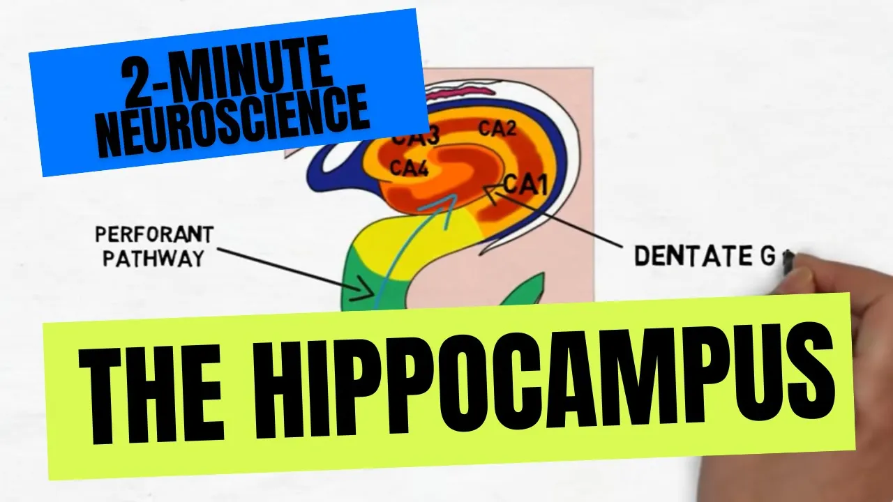
Late in the disease, other cognitive domain might be affected as well, such as exact to functions, problem solving, etc. When the classical or typical Alzheimer's disease occurs, we believe that the memory is happening because the disease is affecting a very particular part of the brain called the hippocampus, which on this cartoon you can see it right here.
This is an MRI of a patient that doesn't have the disease.
You can see the hippocampus is nice and plum.
It's that gray rounded area and it's quite big and bulky.
I'll show you in a little bit what it looks like in disease.
I really like the word hippocampus, is a little bit interesting how it's named.
It's named after a seahorse.
If you take this out, it looks like that.
Because it looks so much like a seahorse, hippocampus is seahorse in Greek.
In patients who have Alzheimer's disease, the hippocampus might really lose a lot of volume, because brain cells are dying as time goes by, and so instead of having that sort of really nice and plump hippocampus, you might have a space and the place where it's supposed to be, and you may not see a hippocampus any longer.
This was in summary sort of the presentation of somebody with primary memory problem.
Now people with Alzheimer's disease may also present with primary difficulties visual processing, not necessarily affecting their memory.
This is a syndrome that is currently known as posterior cortical atrophy.
Some people refer to it as the posterior variant of Alzheimer's disease.
In these people, usually the disease presents earlier for reasons we don't understand, they are younger, maybe in their early 60s or late 50s.
The first sign and symptom of this is impairment and visual processing, they start having difficulty locating things in space.
They may open a fridge to look for the milk, and there's a lot of things in the fridge, and they're not able to visually process where the carton of milk is located in the fridge.
They may have difficulty with sizes of things, go to fetch a big glass or mug and they fetch a smaller one or placement of how to put the table placement.
They also have difficulty navigating their own space.
A room like this would be very difficult to navigate for somebody with this type of Alzheimer's disease, because to be able to go through where is the door and on which side I have to go, and how do I not bump into people.
It's very difficult.
They may also have difficulty recognizing faces or objects.
These patients may earlier in the course of the disease, have difficulty recognizing family members or famous people on television.
Over time the disease may progress to then include memory problems and then it can later on start look the same like the amnestic form of Alzheimer's disease but most of the time in the first two or three years, it's primarily a visual problem for these patients.
These patients are the least likely to not have insight of their disease, which means out of all the people with Alzheimer's disease, they are the ones that recognize the most that there is a problem going on, and that they are unable to manage their space.
Some people believe that because of that, but also maybe other factors related to where the illnesses is attacking, they may be very depressed or very anxious.
This is a picture that we often show our patients in order to determine how well they are processing visual cues and stimuli.
We ask them to describe to us what's going on in the picture and as you can see, there's a lot going on in this picture, right there is water running.
The mom has her back turned to her kids who are maybe stealing cookies from the cookie jar.
This would be the way that you would want somebody to describe that picture but people with this type of Alzheimer's disease may actually not be able to do that because they may not be able to tend to all of the visual stimuli going on at the same time.
They may be able to describe one thing, I've had patient just describe what's outside the window and not talk at all about what is happening inside the kitchen.
This is one way in which we can test this at the backside.
In people with this type of falls and disease, it affects a very different part of the brain, which is the posterior part of the brain.
Let me orient you a little bit.
This is an MRI of a person with this type of Alzheimer's disease and this is the front of the head and this is the back, imagining that there are laying down on the scanner on the table and you are looking through their feet, not through the top of the head but through their feet.
This is the right side.
This is the left side, this is the front, and this is the back.
We are starting at the very top of the brain, the very top of the head and you see here, if you just compare the front to the back, you see that there's a lot of these black spaces in the back that don't exist in the front.
The front is mostly comprised of brain that is nice and full and plump and gray and the back is a lot of deep spaces that are black like the one that I'm showing here, or here, or here.
This is a part of the brain that's called the parietal lobe and it is responsible for a lot of our visual processing, but also some other things like math and calculation.
It plays a role in language, and it plays some other roles and sensory how we feel things, etc. You notice how much volume loss there is in the back part of the brain, whereas the remaining brain looks more or less okay.
This is the hippocampus here, it's a different cut than the one I showed you earlier, but it is relatively slightly more plump than what we expect for somebody with Alzheimer disease and so the main problem is here in the posterior part.
So far, two different ways that presents and two different parts of the brain that are being affected.
The language syndrome, what is referred to as Logopenic variant of Alzheimer's disease is a third way in which Alzheimer's disease can present itself.
It also tends to occur mostly in younger people.
The first sign is a language difficulty and specifically, people have a really hard time coming up with words and spontaneous speech.
They would be talking and then all of a sudden they blank on a word.
They may try to talk around it.
Like if they were to say a hanger, they might say the thing that it goes in the closet and then you put your clothes on it and they just, they blank on the word and they say all of these things around the word.
The exact process that is being impaired in these patients is that they have a really hard time with anything that comprises the sounds of language.
To explain that to you a little bit, let me invite you to think a little bit about language with me.
Languages are very abstract concept, who decided that if I say the sound door, that means something to us, the door that we open to enter a place and if I say the sound, [NOISE] that's nothing, it doesn't mean anything, but that's still a sound, it's a very abstract concept we have learned since we were young that these particular sounds are associated with a particular meaning or concept.
There's a part in our brain that is responsible at all times to sift through all the sounds that you get from your environment and allow you to differentiate between ambient sound and language sounds and then once you are able to process all of that, you then attach the meaning to each of the sounds and that's how language even starts to be processed in the brain.
There are other parts sort of take over for you to add the richness and texture of what it is that you are being told how you're going to respond to it, etc. These are other things that we're not going to cover today but that initial process of understanding the sounds of language is what's being affected in these patients and to show that's why they start blanking on words because they can't hear them, they cannot hear what they sound like and also they start having difficulty listening to people talk, especially if they are being told something that's really long because its way more sounds, if I were to tell you, "Get out," these are two sounds, two syllables.
It's way easier to process than if I say, "Make sure to get ready we are going now for dinner.
We're going to be late. It starts at 08:00 PM."
These is a lot of sounds that the brain now has to process, for people who have this variant of Alzheimer's disease, will have a really hard time with these long sentences as opposed to shorter sentences.
[NOISE] As the disease progresses, they may have more and more difficulty with language so that communication might be really impaired.
Some patients do become sort of close to mute or are unable to really provide any meaningful conversation.
Early on it might also affect calculation faculty because we're going to see this is the part of the brain that's close to where we process sounds of language and then they may start developing more visual problems similar to the first group that I showed you right before this one.
These patients probably also have a lot of memory problems that might be hard to detect because if you cannot speak, you may not have an opportunity to know that you forgot something because it may not come up in a conversation.
Let me show you just a video of a patient that has this and this is a test that we give to this patient in order to see how they're processing the sounds of language.
The test is really simple.
We ask them to repeat a sentence and repeating a sentence does not require you to really understand it, or that it requires you to do is to be able to process the sounds of that sentence and reproduce these sounds.
People may not be able to understand what they're saying, but they may still be able to repeat it.
However, people with this Alzheimer's disease have a really hard time.
Let me just play this real quick.
Can you repeat this phrase?
Today was a warm and sunny day.
Today was a wonder and fundy day [inaudible] Today was a warm and sunny day.
Can you repeat that again?
Yes, today was a sun was, oh God.
Summer and...I'm trying to go too fast.
It's tricky, isn't it? Let's start with something easier.
How about today is Monday?
Today is Monday.
How about the flowers were in the park?
The flowers were in the block.
You notice how he's really struggling with these sounds even when those sentences were relatively simple.
What's interesting, and if you notice, there were some errors in the words that he made, they sounded like what you're saying but they were not the right words.
He said summer as opposed to sunny day.
This is an error that often these patients make because as they are searching for the sounds, other sounds are being grasped and the brain, and they're going there.
This is to show you where in the brain this is.
Again I'm going to reorient it like we did the first time.
The front of the head or actually you can see here, these are the top of the eyes.
That's the face here, that's the back, this is your left side, this is your right side.
I'm going to bring your attention to this part of the brain where you see that there is a lot of spaces like some of these spaces here as compared to that side of the brain.
This is the right, this is the left.
This is part of the parietal lobe which I showed you earlier, but it's a little bit lower.
It is where we are processing some of these sounds of language.
This is the reason why these patients struggle.
The last variant we're going to talk about today, but not the least is the executive syndrome or the frontal syndrome.
This is a variant that also occurs at earlier age and it affects the front part of the brain, which we haven't talked about yet.
The front part of the brain is responsible for a lot of really important day-to-day things.
I think of it as the CEO of our brain.
It is little guy sitting there making decisions allowing us to know what we want to do today, plan our day, execute that plan.
Motivate us in order to go ahead with our plan.
It motivated all of you to come here today and listen to this lecture and helps us organize, etc. When it is affected by Alzheimer's disease, and then people start losing this capacity and they might start having difficulty with problem-solving.
Maybe they will always go at mechanics and they stop knowing how to do that.
Maybe they tended to their garden and knew how to organize it and stop doing that.
A lot of people, it can show up at work because remember, these are people that could be younger in their '50s.
Oftentimes they are still working and they may start making errors or judgment problems at work. Yes, we have a question.
It's also referred to as frontotemporal dementia.
The question is whether this is referred to as frontotemporal dementia?
Not necessarily.
It can depending on how it presents itself.
Frontotemporal dementia is a another syndrome that describes, it has a particular criteria about what patients present with.
I think it's going to be talked about in the next lecture after this one but this may be called frontotemporal dementia as a syndrome if the primary presentation as that of a behavioral change, but then the the cause of it would still be Alzheimer's disease.
That's a great question to segue into.
The next thought, which is because the front part of the brain, in addition to being the decision-maker and the executive processor, it is also the part of the brain that is helping us select the best behavior and any particular social setting that we are in.
If you think about social situations, you could be in a very professional situation or in a family situation, or you could be out with friends having drinks.
If the same scenario or the same conversation happen in each one of these situations, you might respond differently because you're in a different space.
Maybe you're having fun at night with your friends, but with your boss you have to take things seriously.
The front of the brain is what is allowing us to process all the time what's coming at us from the environment and be able to select what is the best way to behave right now given the setting that I'm in.
Patients may start having difficulty with knowing what to do from a social perspective.
A lot of them become either an appropriate and social setting or disinhibited, etc. Again, over time this condition progresses to involve also other cognitive domains like memory, language, and visual, as we saw previously.
I'm going to move next to talk a little bit about the neuropathology of Alzheimer disease or what is happening inside the brain.
We talked about how it manifests itself.
Now we're going to try and take a dive into the pathology.
The hallmark as we saw in that first story I told you about Alzheimer, is the presence of two main findings.
One called amyloid plaques, and the other called neurofibrillary tangles that are made of a protein called tau.
Let's take a deep dive into the brain to understand better what that means, on our brain cells we all have this molecule called the amyloid precursor protein and the amyloid precursor protein, we're not quite sure what these do, but we know that when it is snipped in ways to cause what we call Beta amyloid, which is the smaller yellow portion that you see on the screen here.
These Beta amyloids can aggregate together and form what we call a Beta amyloid plaque.
The Beta amyloid plaque can come in two forms.
Either what we call diffuse plaques, like what you see on here, all of these spots or what we call a neuritic plaque.
Both of these are outside a brain cell, this is the brain cell, it's being clipped.
Outside of it, we are having all of these aggregate together.
I really want to thank Dr. Legrum who is going to be joining us later today as a moderator.
She provides us with a lot of these nice pictures and images in order to talk about pathology.
She knows way more than I do about this.
If I make an error I'm sure that she was able to clarify that afterwards.
This is the amyloid protein part of Alzheimer's disease.
Then there's another part of Alzheimer's disease which is related to another protein called tau.
Tau is a protein that is usually part of the architecture of a cell.
Any cell in our body and specifically our brain cells are made of a skeleton.
That skeleton that holds the cell together is made of proteins that are called tau.
They like come together in order to make these tubules like here to make the skeleton of the cell.
However, at times when somethings going wrong inside the cell, which we're not quite sure what that something is.
The tau proteins may become hype-phosphorylated, meaning they have too much phosphorus attached to them and they are unable to form this really nice tubules.
Then instead they aggregate together and form this unwanted part of the cell.
I will show you here on this scan.
This is neurofibrillary tangle here or here you can see it in black.
These are actually inside a brain cell because the tau protein is supposed to be inside to form the skeleton.
Then when it's unable to do that, it aggregates inside the cell and contributes to causing brain cells to die as time goes by.
Alzheimer's disease is the term that we use to describe the specific neurodegenerative disease of the brain that is associated with progressive accumulation of these amyloid plaques and tau tangles that over time they lead to irreversible degeneration of neurons, meaning that neurons die as time goes by.
There are different ways in which we can stage Alzheimer's disease from a pathology perspective.
Let me actually start with the Beta amyloid staging.
We had talked about these two different types of amyloid, the diffused deposits and then neuritic plaques.
Then there are two different ways in which pathologists look at a brain and decide as to how much of amyloid plaque there is or what is the distribution of it.
Down here you see different ways of staging by distribution of the diffuse a neuritic plaques inside the brain.
Phases 1-5 inform us about where in the brain these amyloid plaques are being found.
This is another scheme by which we can quantify, it's by looking just at the neuritic plaques and see how dense they are on anyone microscope slides.
You can see, for example, here there's nothing, but also I think I'm cheating.
This is just a blank square.
This is not actually [LAUGHTER] from a patient, but then this is when they are sparse.
You see a few of them, but they're not too many.
This is moderate where you see a little bit more and this is frequently, you see a lot more in a slide.
These are two ways in which we quantify the amyloid presence in the brain. Yes.
This is an autopsy?
Correct. This is all on autopsy.
Occasionally, very rarely patients get a biopsy of their brain because people aren't quite sure what's going on and they think this might be a good diagnostic way, and we sometimes find that on biopsy, but most of this is on autopsy. Correct.
Regarding the tau staging, tau seems to have a much more predictable way in which it spreads in the brain, and it seems to always start close to the hippocampus and this region called transentorhinal region.
You don't need to remember that.
Some of us don't even remember that at times.
Then it starts spreading from there to the hippocampus.
Then involves more of the hippocampus and of that part of the brain called the temporal lobe.
Then as it spreads more to the cortex and involve different cortices, it becomes more and more widespread in the brain.
Every one of these has a stage which was first described by Braak and Braak, and so we call this a Braak staging of the tau presence in the brain.
The more the Braak staging in the brain, the more diffuse the tau tangles are, and usually this correlates with the severity of the symptoms.
So the more tau you have, the more symptoms you have but the accumulation of amyloid does not always necessarily correlate with the severity of the symptoms.
Yes. [BACKGROUND] The question was, as this moves from one stage to the other, is there anything that is being done for patients that have this?
Unfortunately, we have not yet scientifically found how we can stop this disease from spreading, and we do not currently have medications that have been shown to hold it in its place.
There are several experimental medications, some of which have been tested for the past decade to try and clear the amyloid plaques from the brain, and so far we have gotten non-favorable results.
In some patients, we were able to demonstrate that the amyloid plaque has been removed, but that wasn't paralleled by an improvement in disease or a stopping of the progression.
There may be other factor that we still don't understand or know about this.
There are other current medications also that are trying to clear the tau tangle from the cells and see if we can sort that from continuing to form.
But this is also still under experimentation and we're not quite sure where we're going to go.
But what we're hoping that as we learn more and more about the biology of this and how it spreads from cell to cell and how it forms in the first place, that we'll be able to have more targeted medications to try and hold this process or even reverse it if we can.
We talked about the pathology, we talked about the manifestation, and let me talk a little bit about the genetics of Alzheimer's disease before we dive in into ways in which we can detect it in our current day and age.
Alzheimer's disease is actually rarely caused by a single gene variant.
This is what we call a pie chart.
It's like a pie and we're slicing it.
What you see in orange here is all the cases of people with Alzheimer's disease that we call sporadic, meaning that they presented to us with no family history of Alzheimer's disease whatsoever, and this is the majority, 75 percent of people.
Then in purple, you see the part of the slice of patients with Alzheimer's disease that presented to us with familial Alzheimer's disease.
But this is also defined as awesome disease in people who have two or three relatives with Alzheimer's disease, but they don't necessarily have a dominant gene, meaning a gene that if you were to have it, you have the disease, no question.
These are a very small portion of the pie in green here you can barely see it.
They are less than one percent, and here you can see better the number.
Seventy five percent of patients with sporadic, no family history of Alzheimer's disease, 24 percent of patients with familial, having two or more relatives with Alzheimer's disease, and less than one percent of patients have an autosomal dominant genetically transmitted Alzheimer's disease.
Out of these less than one percent, the majority have a gene called presenilin 1, 65 percent, the second group have a gene called APP, it stands for amyloid precursor protein, and a rare group have a gene called presenilin 2.
I'll talk in detail about some of these genes.
Presenilin 1 occurs on chromosome number 14.
All of us have 23 pairs of chromosomes in every cell of our body.
These chromosomes really contain in them the entire codes of who we are.
Internally, on the outside, everything, it is the genetics that dictates everything about us, and every different cell in our body express sometimes different parts of these 23 chromosomes.
You can imagine a skin cell is very different than a cell in the heart than a cell in the brain.
So these three cells have the same 23 chromosomes with all the codes.
However, they are expressing different parts of them to become these different cells in the body.
Presenilin 1 is on chromosome 14.
When patients have it, they can present with Alzheimer's disease at a fairly young age.
Usually, the onset is between the age of 25 and 60, with the average age around the age of 14.
The symptoms can be quite different than what we just talked about.
It's not just memory problems, but people can have Parkinson-like movement problems, ataxia, which means incoordination in their movement.
They can have behavioral changes.
Of course the mutation causes buildup of amyloid Beta in our brain cells, and this is what causes the disease.
There are a few founder variance, meaning that these are the places in the world where we have found families that have this gene.
You see that it's really in very specific parts of the world that this gene is present.
The presenilin 2 remember, is extremely rare.
So the majority of cases are presenilin 1 or APP.
This is extremely rare and it is present on chromosome 1.
Again, the onset is young between 40-75 years of age with a mean of 50, and it mostly occurs in people with German, Italian, and Spanish descent.
Again, leads to build up of amyloid protein.
The amyloid precursor protein, which is the second most common gene, is on chromosome 21.
For those of you who know this, people with Down syndrome who have trisomy 21, this is that chromosome.
You'll have three copies of that chromosome instead of the regular too that all of us have.
Some because of the three copies, they are at a very highly increased risk of developing Alzheimer's disease.
In fact, people with trisomy 21, the majority of them, if not all of them, if they live long enough, they will develop Alzheimer's disease.
Again, this is caused by buildup of amyloid Beta as well.
Yes, sorry, I can't see you. Go ahead.
The question was, what is being done about the families that have this in terms of trying to understand better this disease?
There are studies that are looking at understanding better the biology of course, but also trying to look at treatment in this population.
Because what is unique about this population is that we know they're going to get Alzheimer's disease if they have the gene, so even when they are young and have no symptoms and have no buildup in their brain, we already know that this is going to lead to that.
This is a great group of people in whom we can, if we find a good drug that can stop or even prevent Alzheimer's disease from happening in the first place, this is a good group of people in whom to test this.
I am aware of a couple of studies that are looking at that.
I don't think that there is any results yet that are translatable to our community, but hopefully we will have more information as time goes by.
Thank you.
Of course. Before we move on from this, I would like to talk a little bit about Apolipoprotein E or APOE, which some of you might have heard of and a lot of people now and the day and age of 23 and me, they are getting their genetic testing done on their own with various private companies, and they are getting results of whether or not they are "at risk" for things like Alzheimer's disease.
Apolipoprotein E is a gene that has three major different forms.
You have two copies of it because we all have like I said, a pair of chromosomes.
You can have two of any of these three forms, the E2, E3, and E4.
E2 is considered to be protective against Alzheimer's disease.
E3 is considered to be neutral, it doesn't necessarily do anything, and E4 is considered to confer risk on patients who have it to develop Alzheimer's disease.
None of these is causative.
You do not need apoE4 to have Alzheimer's disease and also having apoE4 does not mean that you will develop Alzheimer's disease.
So this is not a cause, it just increases the likelihood or the risks that you may develop it given maybe other factor.
Having E4 may mean that you develop Alzheimer's disease earlier in life than people who don't have the apoE4.
But like I said, it's not necessary or sufficient for the disease and we actually medically speaking, do not recommend for people to get their apoE4 gene testing and know what their status is because we may not have any recommendations to give you one way or another.
This is just showing what the majority of people have.
As you can see, most everybody in the world has a E3, E3, so two E3s 61 percent of people.
About 25 percent have at least one apoE4 or two and then the rest, they have like E2 or E2, E3.
This is just to show you the risk of Alzheimer's disease.
This is the risk if we don't look at genotyping whatsoever.
It's about 10-11 percent in men and 14-17 percent in women.
This is lifetime risk at all ages.
Having E2, as you can see, can be protective.
So the risk becomes less, 4-5 percent or 6-8 percent in women.
Having E3 is neutral, so it doesn't really change the percentage that much but when you start having E4, that's when the percentages increase, both in men and women.
In general, one E4 copy is associated with an 18-35 percent lifetime risk of Alzheimer's disease, and two E4 copies are associated with 31-40 percent of lifetime risk of Alzheimer's disease.
Before I move on from this, I just want to thank our genetic counselor, Jamie Fong, who does a lot of great work with us and she actually provided me with most of the information on these slides.
I just want to say thank you to her.
We talked about how it presents, we talked about what it looks like in the brain, we talked about some genetic factors that can affect it.
Let's finish by talking about, well, how can we detect or be more confident about Alzheimer's disease when we are still alive and we're not doing an autopsy?
In our day and age, we have biomarkers of Alzheimer's disease, which are like a proof of evidence, if how I like to think about it.
Having a biomarker that is "positive" increase the certainty that whatever syndrome you're seeing, so the memory problem or the visual problem or the language problem is indeed being caused by Alzheimer's disease.
Biomarkers fall into three categories.
There are biomarkers for amyloid deposition, where we can detect whether there is amyloid pathology.
These come in two forms which I'm going to talk about.
One is measuring the amyloid protein levels in the fluid that is around the brain and our spine.
If you can imagine, at all times our brain and our spine are actually floating inside a cavity and our brain that is filled with a fluid.
This fluid is called cerebrospinal fluid, because we're not very creative.
That cavity extends from the skull all the way down to the spine.
People here who have gotten or have seen people got an epidural, know, this is what a spinal tap is and I'll show you in a little bit some images about that.
But this is where we get that fluid from and we can test it for amyloid markers, and I'll show you a little bit what the findings might be.
Or another technique for amyloid is doing a PET scan, which I'll describe in a little bit and checking whether there are amyloid plaques or amyloid depositions in the brain.
The second biomarker, which is really state of the art and is still under investigation, but it's really promising, is a PET scan for tau protein.
It's a PET scan that can allow us to visualize or to see a biomarker of tau depositions in the brains of people who have Alzheimer's disease.
Then the third type of biomarker is really biomarker of neuronal injury, meaning that it may not necessarily be specific to Alzheimer's disease in the sense that it's not showing us the amyloid plaques evidence for that or the tangles, but it is telling us that brain cells are dying.
We can see this again in cerebrospinal fluid or what we call CSF.
I'll show you in a little bit or on an MRI, like what I show you earlier, a scan of the brain or on a PET scan that is being done with just sugar to see how the brain is eating and utilizing sugar.
We'll talk about all of these things.
Let's start with the cerebrospinal fluid of truth is as what I call it.
This is how a lumbar puncture is done.
We go with a needle in the lower back of a person after, of course, we've cleaned it really well and we've given them an anesthetic so that they don't feel the pain and we go in the space.
This is your vertebral bones.
Then they are there to protect that cavity that is filled with that fluid and that the spine is in.
Now, where we go in the needle, the spine has already ended, but there are nerves going down, so we're not at risk to injure the spinal cord that is extending from the brain.
But we just want to get to that cavity in order to get the fluid.
That fluid is in continuum with the fluid around the brain. It's really the same.
It's one pool and the fluid is all the time passing through this entire pool.
It contains information that are also important to the brain.
Once we get this fluid, we send it to the lab and it comes back with a report where they show us the levels of the amyloid protein in that fluid and the top protein in that fluid.
Now, this may not make a lot of sense, but bear with me if the amyloid protein is low and the CSF fluid, this is the marker for Alzheimer's disease.
We don't quite understand this biology to you well, but the way that I tried to think about it to make sense of it is that if the amyloid is forming the plaque, then not enough of it is circulating in the fluid for us to detect it, so keep that in mind.
If the amyloid level is low, then this is a biomarker of Alzheimer's disease.
If the tau level is high, this is a biomarker of neuronal loss.
It's not a biomarker of tau deposition necessarily.
It doesn't necessarily tell you that there is tau tangles in the brain, but it tells you that brain cells are dying, and as they're dying, they are exploding and putting all that tau protein in the fluid.
This is what we are picking up.
What's really nice about doing a spinal tap or a lumbar puncture is that we can also measure for other things that sometimes maybe causing memory problems and aren't Alzheimer's disease, like inflammations in the brain, infections in the brain, tumors, lymphomas, things like that.
Getting a sample of the fluid allows us to test for other things that aren't Alzheimer's disease, and that's always nice and it complements the work-up.
Now, let's talk a little about the PET scan.
I want to really thank Renaud La Joie, who you're going to meet in a little bit.
He's going to be part of our panel as well because he gave me all of these slides.
He's way more eloquent than I am, but I'm going to try to do a good job explaining this.
A PET scan stands for positron emission topography.
It's a complicated process that we're going to try to simplify.
Basically, we have a certain material, whatever it is that we want, that we tag with a substance that's radioactive.
Something that's radioactive is all the time emitting a positron, which are just little like nuclear molecules.
Then we inject the blood with that substance.
Then that substance goes through the bloodstream and reaches our brain because it's in the blood and our brain is receiving blood all the time from the heart.
Then depending on what we targeted with, it will go to places in the brain.
For example, if we put a piece that fits onto the amyloid plaque and then we tag that piece was something radioactive, the piece will go to the amyloid plaque and then will emit the positron.
and then that complicated machine will detect the positron and say, aha, I see that this molecule that you tagged is hanging out in the brain.
Whereas if there's nothing in the brain, it's going to pass through the blood, go to the brain, fight nothing to latch onto, then be cleared and then we take an image and nothing's going to happen.
We're not going to get any positron.
Does that make sense? Great.
It really depends on what you injected with.
We call the thing we injected with a tracer cause it's really like going and trying to latch onto the thing that's looks like it.
You can have an amyloid tracer or a tau tracer.
Or we see in a little bit we could just have sugar that is being traced and tag with a radioactive material.
When we use an amyloid tracer, here's what happens.
It goes to the brain.
If there is amyloid, it will latch on to the amyloid and they will able to detect that.
On the top part here you see an amyloid PET scan of a patient that doesn't have Alzheimer's disease.
If you look at the colors, anything that's dark or blue means that nothing was taken up.
There's a really low uptake of the tracer.
Then the more you go and the colors of yellow, orange, and red, then there's really high uptake.
This is a patient that does not have Alzheimer's disease.
Notice that there's a little bit of uptake here and what we call the white matter of the brain.
The white matter of the brain is usually on the inside, not on the surface.
The best way to think about it it's all the wires that is connecting different brain cells to each other as opposed to the surface of the brain while most of the brain cells live.
Then down here you see the scan of a person who has Alzheimer's disease.
You can notice the huge difference between the two where now there's a lot of uptake.
You don't see any differentiation anymore between the white matter that's on the inside and the gray matter that's on the outside.
The entire scan is what we call hot, there's really, really high signal.
This is because the tracer went and latched onto all the amyloid plaques and is giving out all these signals.
This is just to show you the correlation of the amyloid PET scan with actual autopsy results.
This has been done on a 179 patients who died three years after they got a PET scan.
Then we did autopsy on their brains.
Notice how the PET scan starts detecting a signal.
Remember earlier I showed you this different phases.
For the Thal phase, as early as Phase 3, maybe even with two, we start seeing a signal but really 3,4,5.
Then for the CERAD score when it's moderate and frequent the PET scan as you did detecting it.
It's really faithful and correlates really well with autopsy.
It's a very specific way to detect amyloid plaques in the brain.
Now the tau PET is a similar concept where instead of the tracer latching onto the amyloid plaque, it's going to latch onto the tau protein.
This is still not as well refined as the amyloid PET scan.
It's still being under investigation because we are finding other molecules in the brain what we're calling the tau tracer is latching onto.
The tau tracer doesn't seem to be right now as specific to the tau protein, but it's latching on to other things.
However, in Alzheimer's disease, it is actually incredibly specific.
That's primarily because there's a really high signal of tau in the parts of the brain that are affected by Alzheimer's disease that I showed you earlier.
The hippocampus, the seahorse, the parietal lobe, the temporal lobe, etc. It's still being refined, but I hope it's a very promising technique.
What's really nice about it is that tau does correlate with symptoms as we talked about earlier.
I think it's going to provide a really great insight once it has very well refined about where we are in the disease stages.
Finally, this is a PET scan.
That is when people just say PET scan, usually that's what they mean.
A lot of people might be familiar with PET scan from like the cancer world or other medical fields.
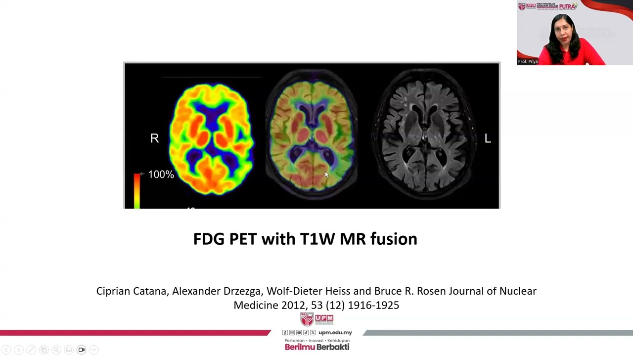
This is a PET scan that we call FDG-PET scan.
It's basically molecules of sugars that are tagged radioactively.
What we are looking for is how different cells in the body and in the situation brain cells are eating up the sugar.
Healthy cells should be eating the sugar really well because they're taking blood and they are feeding of the blood.
We are supposed to see high signal, as you see here in a clinically normal person, you see high signal all throughout and it's hot all around because we want it that way.
We want the sugar to be utilized in the entire brain.
Then for somebody who has Alzheimer's disease that may have lost brain cells in these areas.
That's the temporal lobe here, parietal lobe there.
You see that you start losing that nice signal here.
There is no uptake as much as in the other parts of the brain.
That's because the cells are not functioning well enough to take up the sugar.
That's another way in which we can detect injury but this is not specific to either amyloid protein or the tau protein.
To summarize, so here are the few things to hopefully remember from all of this if you don't remember anything else.
The neuropathological hallmark of Alzheimer's disease is accumulation of amyloid and tau protein inside the brain.
Alzheimer's disease can lead to significantly different clinical syndromes, and patient can go through distinct stages from silent disease or asymptomatic to mild cognitive impairment to dementia.
Not all patients with mild cognitive impairment or dementia necessarily have Alzheimer's disease.
Less than one percent of the cases really are genetically dominant.
Then there are modern biomarkers that can improve the accuracy of our diagnosis, but currently the autopsy remains the gold standard of confirming the disease.
Thank you so much everyone, I congratulate all these people who helped me put this talk together.
Thank you. [APPLAUSE] All right. Thank you very much.
Now we're going to open up the space for some questions.
Today we have two guests.
We have Dr. Lea Grinberg, who as I said before is co-leads our neuropathology course.
She would be the person to ask questions around neuropathology.
We also have Renaud La Joie, is that correct, La Joie?
[LAUGHTER] Good.
I think I didn't say it correctly last time.
Who is a neuroscientists in [inaudible] Center and he's focused on doing research on the biomarkers, especially the PET biomarkers.
He would be the right person to ask all the questions that are on PETs.
Then clinical questions to our colleague, Dr. Naasan.
The last time I started with questions in the front row and went back row-by-row maybe today we can start with the back row and go to the front and to kickoff the questions, maybe I have two questions to get the engine's going.
Maybe the first one would be for Renaud.
So maybe you can help us understand the difference between the sensitivity and the specificity of tests and how that pertains to PET scans.
Within the context of AD and then what are the limitations basically of these PET scans?
Thank you for the question, and thank you for inviting me.
I'm very happy to be speaking in front of you today and addressing some questions.
You saw nice images and I'm trying to talk a little bit about what can be complicated about using these images.
So you mentioned sensitivity and specificities.
These are two terms that we use a lot in the biomedical field.
They are like basically complementary measures to measure how good a test can be to detect a disease.
An ideal test would always detect the disease when it's present.
It would always tell you, oh, there's no disease when the disease is not there but we know that no tests in medicine is like this.
So the sensitivity is very important.
A sensitive test is a test that's going to actually tell you when the disease is there.
When a patient has a disease, the test is going to be positive.
It's going to be, oh, there is something going on with this patient.
So you want a test to be very sensitive but also you want your test to be very specific.
Because the specificity is about being able to rule out the disease when there's no disease.
So you also don't want a test that's going to be positive in everyone, you want it to only be positive when there's a disease.
So this is like the trade-off of any biomarker in medicine.
That's really a struggle for us to find tests that are both sensitive and specific.
I think George showed nice data on amyloid PET and comparing amyloid PET results to autopsy data, which is our gold standard.
What I can tell you is that amyloid PET, amyloid imaging is a pretty sensitive measure.
It's also very specific, meaning that when we have the signal on images, almost all the time, it means there's amyloid in the brain but amyloid PET is a great measure of amyloid plaques.
I think George made a very good point in explaining that Alzheimer's disease is not just amyloid, its amyloid and tau.
So actually we're having issues of a lot of people who can be cognitively normal and actually have amyloid in their brain.
We've known that from pathology studies.
We can see that now with imaging, but amyloid is not Alzheimer's disease.
So the test itself, amyloid PET, is not very specific to Alzheimer's disease.
It's specific to amyloid and I hope this distinction is very clear.
Good. Great. I have a question for Dr. Grinberg also to get us thinking about other questions that may come up.
We've presented so far a purposefully way of thinking of these diseases in a structured simple way but as you and we know there's far more complexities to this.
So you, that are seeing the brains.
Sometimes stumble on a patient that has reached autopsy, for example, that has been characterized clinically as having one of the variance of Alzheimer's disease but when you look inside of the brain, you may find that there isn't real evidence of Alzheimer's disease.
So I want you to talk a little bit about this dissociation that sometimes happens where a clinical syndrome looks to be pointing towards Alzheimer's disease, but we find a different pathology and that's because that's something that my colleague next week is going to expand a little bit more about.
What does that mean to you and how much do the history matter in your evaluation as a neuropathologist?
I will start by saying, good evening to everybody and thank you for coming here.
It's really a pleasure to be talking to you.
This is a very relevant question.
We, pathologists, we have this joke that we are always right, but we are always late.
Most of our researcher revolves around of not only learning what is the process going on in the brain when someone develops dementia but what we can do to be better in predicting what is causing dementia in living patients.
So we will be able to treat.
Because of this kind of studies, we also cause a lot of confusion because the more we learn, we change the naming the nomenclature.
So if you had been here three years ago, probably you are hearing something slightly different this time because we are evolving very quickly.
One of the things that it's very clear when we do this kind of studies in which we have the opportunity to contrast what we see in the brain with the clinical history is that we have a lot of patients that will come to our clinics and they will say they fit the bill for Alzheimer disease.
They start having memory problems and they evolve in the way we associate with Alzheimer's disease but actually when we look at the brain, they have something different.
So what are these things that can cause something that's very similar to Alzheimer's disease?
The first one of them and I think it's very important because it's very prevalent and we don't talk a lot about it, is what we call vascular dementia.
So in the same way that when our vessels are not good and we have heart problems, we also have brain problems.
These brain problems, they look a lot like Alzheimer's disease.
In most of the people, they will have a combination of plaques and tangles.
These vascular changes in the brain and the more we put these things together the worse it is.
So everything we can do to prevent blood vessel problems, it will help the brain, so this is the first thing.
The second thing that's becoming more evident from us and we have been learning.
There's a lot in the news about these athletes, especially football players, they have what they call these repetitive brain injury.
This leads many years later to a disease that we call chronic traumatic encephalopathy, have you ever heard about it?
A very complicated name.
CTE.
We have been learning that people that had brain trauma, not as severe as these football players, maybe because they were in car accidents in the time we did the new safety belt, or they were even in amateur sports, that they would have some kicks in their head.
It might predispose to dementia later on in a way that looks very much like Alzheimer's disease clinically.
So it opens to us a lot of opportunities because both of the conditions I told you about, they are preventable in a way.
So we feel that only by preventing these two diseases, we can lower the incidence of dementia by a lot. I hope it does.
Thank you so much for these insightful responses.
Also one of the sessions that we have in the future is going to touch on prevention as well.
So I thought maybe we can start with the last row and go in order.
Some questions in the last row.
Yes, sir. Pretty question.
The question is, what is it about these regions of the brain that makes them have a predisposition for Alzheimer's pathology?
Who wants to take that question?
It's an area of research, I will say that.
I can take that question.
This is, I think one of the most important questions that we are trying to solve nowadays, we call this phenomenon selective vulnerability.
The question is why some neurons in the brain, they are more vulnerable to a disease and other's they are resistant to the disease.
The truth is that we don't know very well.
So what are we doing to find out?
So the first step is really to map, to understand what's going on, to identify which are the neurons that are more vulnerable and which are the neurons that are less vulnerable.
This seems relatively simple, but it's not.
The reason for this is because one of the only ways we can do it is by doing postmortem studies.
Because imaging, although it's getting better and better each day, it doesn't have the resolution yet to see single cells.
Most of the brains that come to our hands to research, they are donated by research participants that we enrolled in a memory clinic.
So they already are very sick by the time the brain gets to us.
So it's very difficult to really understand early stages of the disease but we do have some of these brains and then with these brains we've mapped which cells go first.
In the past, we used to do biochemical studies, immunohistochemical studies to understand what they have but now we have much better tools.
For instance, one thing we are using nowadays, it's called single nucleus RNA sequencing.
So we can nowadays put barcodes in each one of the cells we are studying, and then the genetic studies or any other kind of biochemical studies and understand what is coming from each one of these cells.
So by doing this, we are comparing what are the difference from these vulnerable cells to these not vulnerable cells because the idea is to create treatments that can create a shield in the vulnerable cells so they won't be susceptible to the disease.
Great.
I will just add briefly, and I learn something new every time I listen to Lea talks, but thank you. I will just add briefly that there's progressively more evidence which is still at the infancy stage.
Like I think there's more work to do to demonstrate that but people are starting to see that sometimes when patients develop the syndromes that aren't the memory syndromes like say the language syndrome.
There is a portion of these people that have all their life had some sort of a language problem.
Maybe they had dyslexia when they were younger, maybe not.
That doesn't mean that people with dyslexia will develop on these but it seems like they are represented in that population in a much higher frequency than what you would expect just by chance.
Same for people who have the visual syndrome, there has been some mild evidence that sort of remains [NOISE] to be proven that they may have had some like eye injury or some eyesight problem or something that sort of lead to that region of the brain being weaker or more vulnerable.
So when the disease does hurt for whatever reason that we don't understand, it is hitting these parts of the brain first, as opposed to the hippocampus, which is what we think of as maybe the default part that should be hit by Alzheimer's disease.
Yes. Oh, go ahead.
Actually, just one last word about this, I didn't think about it until you start to talk about differences between people and some factors that can make some people more vulnerable.
I think George mentioned this gene that is a risk factor for Alzheimer's disease, APOE, especially the APOE4 form of the gene.
And we know that it's not just a random risk factor for the disease.
It's a risk factor specifically, or more strongly for the amnestic form of the disease.
When you look at patients with the language variant or the visual spatial variant, actually they don't have that gene so much.
We think that the gene APOE4, it really makes your brain more vulnerable to Alzheimer's disease, but especially just like hippocampus system and these amnestic form of the disease.
I think there's a mix of genetic factors in the process.
I think we have a question in the back.
Lady in the back. Yes. Great question.
The question is, if at the very last stage of dementia, do all syndromes develop?
Maybe Dr. Naasan can address how the disease changes as dementia advances?
Yeah, this is a very important question and I think a really difficult one to answer for many reasons and I'll say why but long time ago and I was a medical student and learning about this, I think people thought that, yeah, as dementia progresses, all dementia will eventually look the same because they're going to affect the entire part of the brain.
But I don't know necessarily that we see this and Lea can talk more about sort of the pathology, but I think what happens is that the parts of the brain that are the most affected become affected enough that you might not be able to see anymore if the other areas are still active or not.
Let me give you an example, which I think we mentioned briefly for the language.
If somebody is having a language problem and that gets advanced enough that they are mute or unable to communicate, we're never going to know whether this disease progressed to affect memory or not because there's no way that we're going to be able to ask them to remember something because they can't communicate.
Same if somebody can't see things, can't leave the room, can do something, it might be difficult to think, well, gee, are they doing this because they cannot find their way, or is this a behavioral problem?
I think the symptoms start clouding each other as the disease progress and I do think that as we become more specific about diagnosing these, we need to be careful to provide the correct matched support for patients having in mind what is the predominant symptoms that they have.
Taking that in mind, and I guess, do you want to talk about pathology also?
Thank you for your question.
Even at the early stages of the disease, it's not that the whole brain is affected.
We still have many areas of the brain, they are preserved.
The case of Alzheimer disease, it's a very long disease because the areas of the brain, they are important for our survival get affected very late in the disease but then even at this point, we don't see these areas affected.
Just to make a contrast, this is the complete opposed to the Lou Gehrig disease, amyotrophic lateral sclerosis, because in this disease, the areas that we need to survive for breathing, and motor control, they are affected very early.
Partly the person will pass because of this, even with all the cognition still doing very well, so depends on the area and even at late stages it won't take the whole brain.
Let me just add to this and summarize the concept which I think is an important concept we have to keep in mind throughout this series that, these diseases like I explained in the first lecture, our focal onset.
Meaning that there's certain like Dr. Grinberg just explained certain areas that are affected earlier and therefore, we if we see patients early, we can pick up on the signs and symptoms that take us to that region of the brain and then make us think of what is the pathology that maybe getting there.
But as the patient's advance in their disease, these substrates, these proteinopathies, they will travel to different regions of the brain or expand and therefore the clinical syndrome will change.
Therefore a person that begins with just an amnestic presentation over time will have language problems, will have visual problems, and if they live long enough with this disease, then as you just heard, that disease is going to start affecting those vital centers of the brain that control things like walking, swallowing, breathing.
Unfortunately, this is when things get complicated.
Next row, so yes, sir.
Vascular dementia is a different cause of dementia.
Think what we were hearing about earlier is that what I was trying to introduce is the concept that oftentimes, especially the older the patient is, it's not always very clean, meaning that it's not like you only have Alzheimer's disease in your brain.
Alzheimer's disease as we advance in age tends to be accompanied by vascular changes as well.
We would say this person had both Alzheimer's disease and vascular disease causing that person's dementia.
Does that make sense?
In other words ED has linked to vascular dementia? Is that what you're saying?
I would say that's a fair statement for especially for patients over what age?
You would say rough.
I think maybe the right statement based on evidence we have right now is that they are so prevalent, both of them, they often they happen together.
There are some questions if they will feed each other.
I know synergistically.
I don't think we have super strong evidence to say yes or no, but certainly they are very prevalent they work together.
So we know that this is for postmortem studies.
That's very possible for someone to pass away with a lot of amyloid and tau in the brain without having any symptoms.
However, it's not possible to see the same situation if the person on the top of it has also this micro vascular changes in the brain.
If the brain structure is not doing well to start with because there is lack of oxygen and no, it's the same situation for the heart.
The brain has more difficulties to overcome this accumulation of proteins there, and again this is preventable in a way.
Maybe over here. And then we'll move forward.
Yes. We'll discuss more in a subsequent session, but I can say that diabetes is recognized as one of the risk factors of cognitive impairment late in life.
It's an association.
We have some hypotheses as to why one would be that diabetes leads to vascular changes in the brain just like it does in the heart and other parts of the body.
Yes, sir. Then we'll move this way.
That's a great question. The question is, how do we think about cerebrospinal fluid and why does it contain proteins in the first place that we are measuring?
It's a fluid that like any fluid in our body, is composed of different types of molecule and things.
For example, if you take a sample of your blood, in your blood you can measure various different things, sodium level, potassium level, enzymes, etc. The cerebrospinal fluid is another type of fluid.
It's not blood, but it does contain a lot of information about the cells that are in the brain and in the spine.
It can contain, first of all, cells, like cells that are there to protect the brain.
If there's an infection, sometimes the number of cells would be really high in the fluid because they're all be coming to the rescue of the brain.
It can contain blood.
If you're bleeding somewhere it will appear in the cerebrospinal fluid, and it can contain different types of proteins that if you're not measuring them, you won't know that they're there, but if you measure them then you can know the level of them.
One of them is amyloid, and normally all of us, if we were to take a sample of everybody here from their cerebrospinal fluid, we will have an amyloid protein level.
It's usually pretty high and like the 800 is sort of the number, and then the more that level decreases, the more the likelihood that we have Alzheimer's disease going on.
Or I guess I should correct and say amyloidosis going on, meaning that there's some amyloid protein process happening in the brain.
Does that help frame that a little bit?
[inaudible] [inaudible] it doesn't have the chance to go to the spinal fluid anymore.
This is how we understand.
It is amyloid starts to get solid in a way and it deposits in these blocks.
So there is less amyloid to go to the spinal fluid, but tau is the opposite.
Tau is inside the cell and starts killing the cell.
When the cell is killed, this tau is released, so you have an increase of tau in the spinal fluid.
One question here and then we'll move to the next row.
When we talk about Alzheimer's in other primates.
Oh, maybe for Lea.
[LAUGHTER] Alzheimer is a human disease.
What we have in primates, in some of them we can have accumulation of amyloid.
We've not necessarily cognitive decline.
I had the opportunity once to examine a collection of very old chimpanzees.
They were almost over 40 years of age, which is very old for chimpanzees, and they only have tau in very specific structures of the brain stem, which is this part of the brain that's in our neck.
They don't accumulate it.
They don't accumulate also in rodents, even the amyloid itself.
When we produce animal models to study Alzheimer's disease, we have to in our artificial way add these proteins there because they don't occur naturally.
But finally, dogs, they accumulate amyloid and it cause them to get blind when they get older, and some dogs they also can get agitated because they are not seeing very well and they get anxious and they can bite.
This happens to dogs a lot.
Is it due to amyloid accumulating in the cortical amyloid or [OVERLAPPING] It's amyloid accumulating especially in the areas that control vision, and actually, in humans, we know that amyloid has this tendency to accumulate in vessels individual area too.
So it's a vulnerable area for amyloid in dogs and humans.
We'll go down this way.
Is there a synergistic effect between Macular Degeneration and Alzheimer's?
I don't think so. Not that we have a very solid scientific evidence for, however, that being said, there is more and more studies being done on the retina and changes that happen in the retina that might reflect changes that are happening in the brain.
In many ways, the eyes are just a part of the brain.
So it's just the direct extension of the brain.
There's a lot of theories that changes that are occurring in the brain could be displayed or could affect the eyes, and there are other neurodegenerative disease in which I think we are starting to have more evidence.
I don't know if you guys will talk about Lewy body disease in another session.
Briefly, yes.
Yeah, but Lewy body disease is another type of neurodegenerative disease that causes brain cells to die as well and can look like Alzheimer's disease at times, and there's progressively some evidence that there are changes in the retina that are very similar to the changes that we see in the brain in Lewy body disease and so I think that this [BACKGROUND] is something that hopefully we will learn more about as time goes by and more scientists can look at that.
I think I've been alerted that we have time for one more question.
Maybe going down the road to be democratic.
The question is, what's the association between hearing loss in the brain and why are we treating hearing loss in the first place if that doesn't treat the brain?
I think having hearing loss may predispose some parts of the brain to be vulnerable to disease because they're not being stimulated by sound when you don't receive enough sound, to process it inside the brain, and so in the same way that becoming blind, might predispose the visual parts of the brain to degenerate because they're not working.
Not hearing might predispose the hearing part of the brain to also degenerate.
But that being said, if you do have a few cells that are still working to try and understand language or what's happening in the environment.
It will be great to help them by making sure that they are receiving clear sound and clear speech.
I think the hearing aid is important in a therapeutic way, and I do spend a lot of time in the clinic trying to convince my patients why this is important.
I think it's exactly for that, even if it's not necessarily treating the disease itself, whatever remaining brain cells you have that are really doing as much as they can to support your cognitive function, it will be great to not make them work even harder to hear, but allow them to receive the sound as clear as possible.
Does that answer your question?
[inaudible] What do you think Lea, do you want to?
I doubt neurogenesis or neuroplasticity is something that's still very controversial.
We don't know exactly how it happens and if it happens, but what we know is that the neurons have the capacity to try to work harder, at least for a certain time, to overcome the disease, at least in early stages of the disease.
What we see biologically in Alzheimer's disease, for instance, is when a region of the brain gets affected instead of going down in terms of function, it goes up at least for some times, and then it cannot cope with this anymore.
In a way, it's plasticity, not more neurons, but certainly a higher functioning of these neurons.
But I think we have still a lot to learn about it, and again, one of the issues in studying this has to do with this difficulties in getting to research individuals that don't have dementia, but they want to participate in this kind of risk.
With that hint that we need volunteers.
[APPLAUSE] [LAUGHTER] It was great to have you.
[MUSIC]
Up Next
6:18
Brain Anatomy: A Comprehensive Guide to the Lobes and Structures
@neurotransmissions
194.3K views•2015-11-29

21:55
Deciphering Neural Circuits Controlling Anorexia in Mice | Richard Palmiter
@AllenInstitute
2.3K views•2011-10-10

4:10
Vagus Nerve (CN X): Anatomy, Nuclei & Functions Explained
@Alilamedicalmedia
305.2K views•2022-10-31

13:03
How Exercise Benefits Your Brain: Science Explained
@TED
11.4M views•2018-03-21
Related Study Plans & Knowledge Roadmaps
Structured learning paths in Neuroscience
neuroscience
Appetite Regulation: Brain Circuits & Hormones
01Foundations of Endocrinology and Brain Anatomy
02Introduction to Energy Homeostasis
03Peripheral Signals: Leptin, Ghrelin, and Insulin
+ 2 more learning modules
14h5 modules
Explore Roadmap
neuroscience
Neuroeconomics: Value, Risk & Brain Mechanisms
01Foundations of Neuroscience and Brain Anatomy
02Introduction to Neuroeconomics and Subjective Value
03Dopaminergic Prediction Errors & Reinforcement Learning
+ 3 more learning modules
18h6 modules
Explore Roadmap
neuroscience
Spatial Navigation: Cells, Grids & Brain Maps
01Foundations of Brain Anatomy and Memory
02Place Cells and Head-Direction Cells
03Grid Cells: The Brain's Coordinate System
+ 2 more learning modules
12h5 modules
Explore Roadmap
neuroscience
BCIs: Neural Decoding & Prosthetic Control
01Neuroscience Foundations: How the Brain Signals Intent
02Signal Acquisition: Recording the Brain's Activity
03Neural Decoding: Translating Brain Waves into Data
+ 2 more learning modules
25h5 modules
Explore Roadmap