The hippocampus, located in the temporal lobe of each cerebral hemisphere and named for its seahorse-like shape, is a critical brain structure primarily responsible for memory formation; it is part of the hippocampal formation along with the parahippocampal gyrus and dentate gyrus, and processes information through a sequential pathway from the entorhinal cortex to the dentate gyrus, through CA3 and CA1 regions, to the subiculum, which serves as the main output region projecting to various cortical areas and subcortical structures via the fornix.
Hippocampus Anatomy & Memory Function: 2-Minute Neuroscience
Added:Deep Dive
Basic neuroanatomy, specifically the location of the temporal lobe and the general organization of the cerebral cortex.
Introduction to Neuroanatomy: Brain Structures & Landmarks
117.5K
This extensive section covers the cerebral cortex organization into gyri (hills) and sulci (valleys) to maximize surface area. Four undisputed lobes are defined: frontal (motor), parietal (sensory), temporal (auditory/limbic), and occipital (visual). The central sulcus separates frontal from parietal lobes, with the precentral gyrus containing primary motor cortex and postcentral gyrus containing primary somatosensory cortex. The paracentral lobule represents the leg in the somatosensory homunculus. The frontal lobe contains superior/middle/inferior frontal gyri divided by superior and inferior frontal sulci. The temporal lobe contains superior/middle/inferior temporal gyri. The parietal lobe contains superior and inferior parietal lobules divided by the intraparietal sulcus. Medial structures include the cingulate sulcus, cingulate gyrus, and precuneus (bounded by parieto-occipital and calcarine fissures). Individual variation in sulcal patterns requires familiarity with idealized models.
Click to watch →

Cerebral cortex | Organ Systems | MCAT | Khan Academy
752.4K
The cerebral cortex is the outer layer of gray matter covering the cerebrum, characterized by its wrinkled appearance with ridges (gyri) and grooves (sulci). Large grooves called fissures divide the cortex into four major lobes named after corresponding skull bones: frontal lobe (frontal bone), parietal lobe (parietal bone), temporal lobe (temporal bone), and occipital lobe (occipital bone). This structural organization maximizes surface area within the skull, enabling the complex information processing capabilities of the cerebral cortex.
Click to watch →
Neurology | Cerebral Cortex Anatomy & Function: Overview
833.5K
The cerebral cortex is organized into four main lobes (frontal, temporal, parietal, and occipital) separated by specific sulci, with each lobe containing primary sensory/motor areas and association areas that work together for complex neural processing; the frontal lobe contains the primary motor cortex (voluntary movement, contralateral control), motor association cortex (planning/sequencing), frontal eye fields (eye movements), prefrontal cortex (memory, learning, personality, behavior, judgment), and Broca's area (speech production); the temporal lobe contains the primary auditory cortex (sound perception: pitch, frequency, location), auditory association cortex (sound analysis and recognition), Wernicke's area (language comprehension), and primary olfactory cortex (smell awareness); the parietal lobe contains the primary somatosensory cortex (touch, proprioception, vibration, pressure awareness) and somatosensory association cortex (pattern recognition), plus a multimodal posterior association area integrating visual, auditory, and somatosensory information; the occipital lobe contains the primary visual cortex (visual awareness) and visual association cortex (color, movement, angle analysis for object recognition); and the insula (not covered in detail here) contains gustation (taste), visceral sensation, and vestibular cortex (movement and balance).
Click to watch →
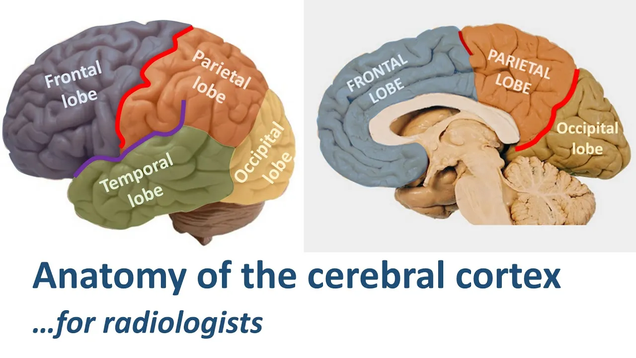
Radiological anatomy of the cerebral cortex... made easy.
71.3K
The temporal lobe contains superior, middle, and inferior temporal gyri separated by superior and middle temporal sulci, with the basal surface containing lateral occipital temporal gyrus (fusiform gyrus), medial occipital temporal gyrus (para hippocampal gyrus), and hippocampus. Heschl's gyrus contains the primary auditory cortex and appears as a small knob-like structure on top of the superior temporal gyrus, resembling a mushroom on coronal images or an A-bomb cloud on sagittal images. The collateral sulcus separates the lateral occipital temporal gyrus from the para hippocampal gyrus. These structures enable localization of auditory and visual processing areas.
Click to watch →

Neuro series: Cerebral hemisphere 2 || External morphology and divisions of the cerebral cortex
374
The parietal lobe contains the postcentral gyrus (primary somatosensory cortex) and posterior parietal cortex divided by the intraparietal sulcus into superior and inferior lobules for spatial awareness and motor planning. The temporal lobe is bounded by the lateral sulcus superiorly and temporal stem inferiorly, containing three gyri: superior temporal gyrus (primary auditory cortex in Heschl's gyri), middle temporal gyrus, and inferior temporal gyrus. The fusiform gyrus on the medial surface is involved in face recognition. The superior temporal sulcus is a key landmark for identifying the temporal lobe.
Click to watch →
Deep Dive
The structure of a neuron and the basics of synaptic transmission and neural communication.

واخيرا🔥مراجعة للاختبار⭐اقوى ملخص التنظيم العصبي| ثانية ثانوي ⭐ راجع بذكاء
351.5K
Neurons (العصبونات) are the basic functional units of the nervous system, coming in multiple shapes including star-shaped, pyramidal, unipolar, and bipolar. A neuron consists of a cell body (جسم خلوي) containing the nucleus, from which a long axon (ليف عصبي) emerges. The axon is surrounded by myelin sheath (غمض النخاعين), a fatty substance that insulates and speeds up signal transmission. Short dendrites (زوائد شجرية) receive signals. The axon hillock (القطعة الابتدائية) integrates incoming signals and generates action potentials when threshold is reached. Synaptic vesicles (الحويصلات المشبكية) in axon terminals store neurotransmitters. Neural signals are encoded electrically as action potentials (كمونات عمل) in neurons. At synapses, electrical signals are converted to chemical signals through neurotransmitter release. The synaptic cleft (الشق المشبكي) is the gap between neurons where neurotransmitters diffuse. Synapses connect neurons to neurons (neuro-neuronal), neurons to muscles (neuro-muscular), or neurons to glands (neuro-glandular).
Click to watch →

Os NEURÔNIOS são fantásticos! | Biologia com Samuel Cunha
105.9K
Neurons have three main structural parts: the cell body (soma) containing the nucleus and organelles, dendrites that receive information from other neurons, and the axon that transmits information away from the cell body. The space between the axon terminal of one neuron and the dendrites of another is called the synapse. When electrical impulses reach the axon terminal, they trigger the release of neurotransmitters that cross the synaptic gap and bind to receptors on the receiving neuron. The myelin sheath, formed by oligodendrocytes in the central nervous system and Schwann cells in the peripheral nervous system, provides electrical insulation that enables rapid signal transmission through saltatory conduction.
Click to watch →

Plus One Biology Public Exam | Chapters 14,15, 16,18 | Exam Winner
103.7K
Neurons have three main parts: (1) Cell body (soma) - contains nucleus and organelles, integrates incoming signals, (2) Dendrites - receive signals from other neurons, (3) Axon - transmits signals away from the cell body. Synaptic transmission involves: (1) Action potential arrives at axon terminal, (2) Calcium enters cell, (3) Synaptic vesicles fuse with presynaptic membrane, (4) Neurotransmitters released into synaptic cleft, (5) Neurotransmitters bind to postsynaptic receptors. Synapses can be electrical (direct transmission) or chemical (neurotransmitter-mediated).
Click to watch →
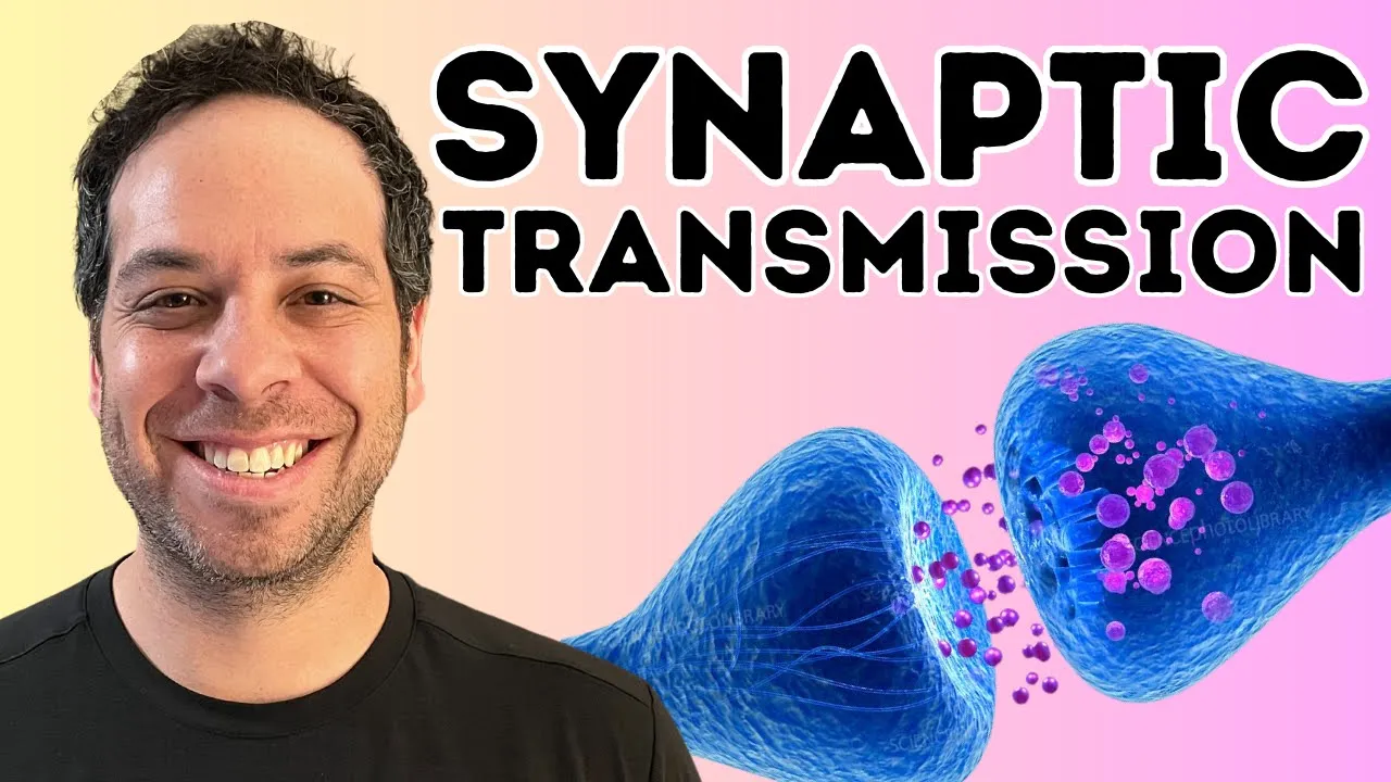
Synaptic Transmission Explained: How Neurons Communicate
147.6K
Neurons consist of three main parts: dendrites (tree branch-like structures that receive messages), the soma (cell body containing the nucleus), and the axon (which transmits signals away from the cell body). The point where two neurons communicate is called the synaptic cleft. The neuron sending the signal is called the presynaptic neuron, while the receiving neuron is called the postsynaptic neuron. The end of the axon, called the axon terminal or synaptic bulb, contains specialized structures for transmitting signals across the synapse.
Click to watch →

Nervous System | Railway Biology Classes | Railway Rankers Batch | Biology By Radhika Mam
18K
A neuron consists of three main parts: (1) Dendrites - receive incoming signals from other neurons; (2) Cell body (soma) - contains the nucleus and integrates signals; (3) Axon - transmits signals away from the cell body. The gap between neurons is called the synapse or synaptic cleft. Neurotransmitters (chemical messengers like acetylcholine) are released at the synapse to transmit signals between neurons. The myelin sheath covers the axon for faster signal transmission.
Click to watch →
Deep Dive
The concept of the limbic system and its broad association with emotion and memory.

How To CLAIM Your VISION with CREATOR CONSCIOUSNESS | Lukis Mac
142.6K
The limbic system is the part of the brain that processes memory and emotion. It exists beyond time and operates in a nonlinear space, experiencing the present, past, and future happening simultaneously. This can work both towards and against us. When we have experienced trauma and haven't processed those emotions, the energy becomes frozen and stuck in the body, causing the limbic system to loop past experiences into the present moment.
Click to watch →

Phoenix 2.0: Biology Most Important Video for NEET 2025 | Unacademy NEET Toppers | Udaan
157.8K
The limbic system includes structures involved in emotion and memory: (1) Amygdala - controls anger and rage, (2) Hippocampus - converts short-term memory to long-term memory (especially during sleep), (3) Hypothalamus - controls sexual behavior and basic drives. The limbic system works with the hypothalamus to regulate emotional responses.
Click to watch →

Il Sistema Nervoso (Riassunto) | Il Corpo Umano
334.3K
The limbic system represents the brain's emotional and memory processing center, comprising deep structures including the hippocampus and amygdala. The hippocampus converts short-term memories into long-term memories and enables spatial navigation, while the amygdala processes emotional experiences and assigns emotional significance to memories. The diencephalon contains the thalamus (sensory relay station) and hypothalamus (regulator of homeostasis, temperature, sleep, hunger, and pituitary control). The cerebellum, located at the brain base, coordinates motor precision, timing, and learning of skilled movements. It enables automation of learned skills—once mastered, movements become unconscious. Together, these systems integrate sensory input, emotional processing, memory formation, and motor control to produce coherent behavior and adaptive responses to environmental challenges.
Click to watch →

Станислав Дробышевский — о том, как древние предки влияют на современных детей
172.6K
The limbic system, located between the intermediate and cerebral cortex, is responsible for smell, emotions, and memory. It evolved at least in reptiles and is present in all mammals. Memory is divided into short-term (hippocampus) and long-term (neocortex) storage. The limbic system connects emotions to memory, which is why emotional experiences are remembered better. This is why the speaker uses emotional content in lectures - it helps students remember information. The brain evolved this system because animals need to remember dangerous places (where they were scared) and rewarding places (where they were happy).
Click to watch →
![The Human Brain (part 2): Explaining ASMR [science, psychology, anatomy]](https://i.ytimg.com/vi_webp/Pg0TCbW70Uk/maxresdefault.webp)
The Human Brain (part 2): Explaining ASMR [science, psychology, anatomy]
122.3K
The limbic system produces emotions and responses that go beyond basic grab-or-avoid reactions in the vertebrate brain, producing subtle and complex actions that are not always predictable. It also contains structures that encode experiences as memories to be recalled for guiding future action. Together, the emotional and memory faculties increase the range and complexity of behavior that mammals display, allowing behavior governed not purely by instinct.
Click to watch →
Deep Dive
An introductory distinction between different memory systems, such as working memory versus long-term storage.

Lecture 5.1 - Acquisition, Storage, Retrieval and the Route
465
Working memory and long-term memory differ fundamentally in several ways. First, working memory has limited capacity—research shows we can typically hold only a small number of items at once, like items on a grocery list. In contrast, long-term memory has enormous capacity, allowing storage of vast amounts of information including facts, concepts, and experiences spanning decades. Second, moving information into working memory is easy (e.g., repeating a phone number), but encoding into long-term memory is difficult. Third, accessing information in working memory is straightforward since it's the information we're actively thinking about, while retrieving from long-term memory requires effort. Fourth, working memory contents are fragile and easily disrupted (e.g., by distraction), whereas long-term memories are stable and resistant to change.
Click to watch →
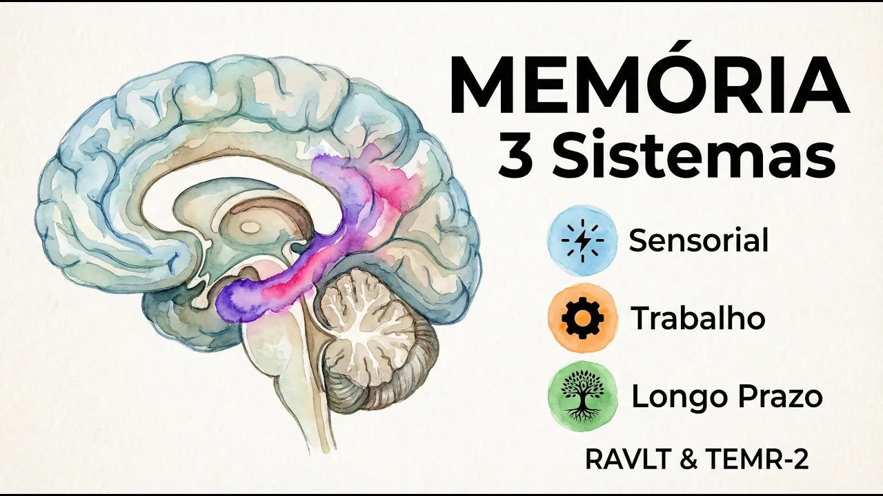
AVALIAÇÃO DE MEMÓRIA: Tipos, Testes e Interpretação (RAVLT e TEMR-2)
245
A fundamental distinction must be made between working memory and long-term memory: working memory is a temporary workspace (like a desk) for active processing, while long-term memory is permanent storage (like a library archive). Confusing these two systems is a fundamental error in understanding memory function. This distinction is vital for both academic understanding and clinical assessment, as different interventions may be needed for each type.
Click to watch →

Anthony Wagner presents Intro to Science of Memory. Vanderbilt Judicial Colloquium, Feb. 2014
939
Memory has traditionally been divided into two main classes: working memory (or short-term memory) and long-term memory. Working memory involves systems that allow us to interact with the world at a given moment, building internal representations of external information and maintaining it briefly in mind, often at the focus of attention, so we can operate on that information over small time gaps between encountering information and needing it. Long-term memory is plastic, meaning it gives rise to durable changes resulting in structural changes in the brain, allowing encoded information from one point in time to be reinstated and brought back to mind at some future point—minutes, hours, days, weeks, or even years later—given the right cues.
Click to watch →

Capítulo 2 - Evolução das teorias do conhecimento
142
Human memory consists of working memory and long-term memory subsystems. Working memory contains three complexes: phonological loop (storing and converting sounds to acoustic images), visuospatial sketchpad (interpreting visual images and their positioning), and central executive (intermediating between working memory and long-term memory rules). Non-declarative memory stores skills and procedures without consciousness (procedural memory), while declarative memory enables conscious recall of facts and events. Episodic memory recalls specific events with space and time context; semantic memory stores general knowledge about facts. Memory encoding in working memory is acoustic, visual, and semantic, while long-term memory encoding is only semantic.
Click to watch →

Curso - Aprendendo a aprender - Como aprender a estudar - Aula 07
5.4K
The brain has two major memory systems for learning: working memory and long-term memory. Working memory handles immediate, conscious processing of information, while long-term memory stores information for extended periods. These two systems are related, as information can be transferred from long-term memory into working memory for active thinking.
Click to watch →
Prerequisite Knowledge
- Concept 01Basic neuroanatomy, specifically the location of the temporal lobe and the general organization of the cerebral cortex.
- Concept 02The structure of a neuron and the basics of synaptic transmission and neural communication.
- Concept 03The concept of the limbic system and its broad association with emotion and memory.
- Concept 04An introductory distinction between different memory systems, such as working memory versus long-term storage.
Subsequent Learning
- Step 01The cellular and molecular mechanisms of memory consolidation, including Long-Term Potentiation (LTP).
- Step 02The specific neural pathways of the hippocampal formation, such as the trisynaptic loop (dentate gyrus, CA3, and CA1).
- Step 03The role of the hippocampus in spatial navigation, cognitive mapping, and the function of place cells.
- Step 04Clinical pathologies related to hippocampal dysfunction, such as anterograde amnesia (e.g., the case of Patient H.M.) and Alzheimer's disease.
Hippocampus
Hippocampus
0:00Playing Section
- 1
Located in temporal lobe; named for seahorse resemblance.
- 2
Key for memory; part of hippocampal formation with dentate gyrus.
- 3
Info flows via entorhinal cortex to CA regions, then subiculum.
Multiple Trace Theory of Memory Consolidation
While traditional neuroscience often teaches the Standard Consolidation Theory—where the hippocampus temporarily stores memories before transferring them permanently to the neocortex—the Multiple Trace Theory (MTT) offers a major counterpoint. MTT argues that the hippocampus is never truly bypassed for detailed, episodic memories. Instead, it posits that every time an episodic memory is retrieved, a new trace is created within the hippocampus. Consequently, older memories are more resilient because they have multiple traces, but they still rely on the hippocampus for retrieval. This challenges the classic view of the hippocampus as merely a temporary transit station.
Deep Dive
The cellular and molecular mechanisms of memory consolidation, including Long-Term Potentiation (LTP).
Нейробиология и психофизиология памяти - Шульговский Валерий
19.2K
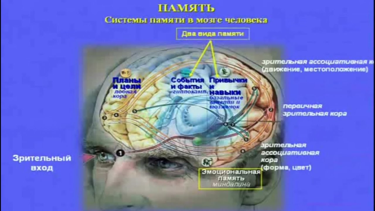
Long-term potentiation (LTP) is a persistent strengthening of synapses based on recent patterns of activity, considered a cellular mechanism underlying memory formation. In the hippocampus, stimulation of the perforant path at theta rhythm frequencies produces lasting synaptic strengthening. This phenomenon was first described in 1975 by Bliss and Lomo. Eric Kandel received the 2000 Nobel Prize for studying LTP in mollusks, demonstrating that memory involves specific molecular changes including activation of growth hormone-producing cells. LTP has become the primary model for studying memory at the cellular level, bridging the gap between behavioral memory phenomena and their underlying neural mechanisms.
Click to watch →

Биохимия памяти. Долговременная потенциация
59.9K
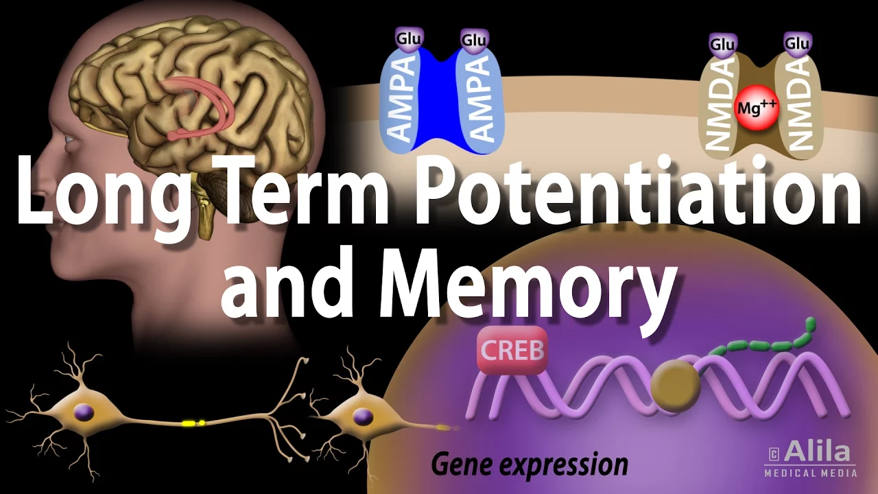
LTP involves two types of glutamate receptors: AMPA and NMDA. Under weak stimulation, only AMPA receptors are activated, causing slight depolarization. Strong stimulation removes magnesium block from NMDA receptors, allowing calcium influx. Calcium binds to calmodulin, activating protein kinases like CaMKII. This kinase enhances synaptic transmission through two mechanisms: phosphorylating existing AMPA receptors to improve their function, and promoting insertion of new AMPA receptors into the postsynaptic membrane. Additionally, calcium enhances presynaptic glutamate release. This bidirectional strengthening creates lasting changes in synaptic strength, forming the cellular basis for memory formation.
Click to watch →
Long-Term Potentiation and Memory Formation: Cellular Basis
433K
Long-term potentiation (LTP) is the cellular mechanism underlying memory formation, where high-frequency neural signals strengthen synaptic connections through a two-phase process: the early phase involves calcium-triggered phosphorylation of existing AMPA receptors and receptor insertion, supporting short-term memory; the late phase involves new protein synthesis and gene expression that enhances synaptic strength and correlates with long-term memory consolidation.
Click to watch →
learning and memory
60.4K
This section explains long-term potentiation (LTP), discovered in 1966, as the cellular mechanism underlying memory formation. LTP occurs when intense stimulation of the perforant pathway causes long-term increases in excitatory postsynaptic potential, making synapses more responsive. The mechanism involves glutamate release, AMPA receptor activation causing sodium influx and depolarization, which removes magnesium from NMDA receptors. This allows calcium influx through NMDA receptors, triggering protein kinase cascades and nitric oxide release. Calcium is the key signal that activates CREB and other transcription factors, leading to gene expression changes, new protein synthesis, and structural changes including dendritic spine growth and receptor addition. This bidirectional communication between pre- and postsynaptic cells strengthens the synapse for lasting memory storage.
Click to watch →

روانشناسی فیزیولوژیک پارت 8 دکتر پیری
226
Memory consolidation follows a cascade: (1) Increased glutamate in hippocampus, (2) Long-term potentiation (LTP) formation lasting 30-40 seconds, (3) Protein synthesis for memory consolidation, (4) Structural changes including dendritic spine formation. Glutamate (excitatory) promotes memory through LTP and spine formation. GABA (inhibitory) promotes forgetting through long-term depression (LTD) and spine removal. Alcohol and sedatives are GABA agonists, enhancing forgetting and impairing memory.
Click to watch →
Deep Dive
The specific neural pathways of the hippocampal formation, such as the trisynaptic loop (dentate gyrus, CA3, and CA1).
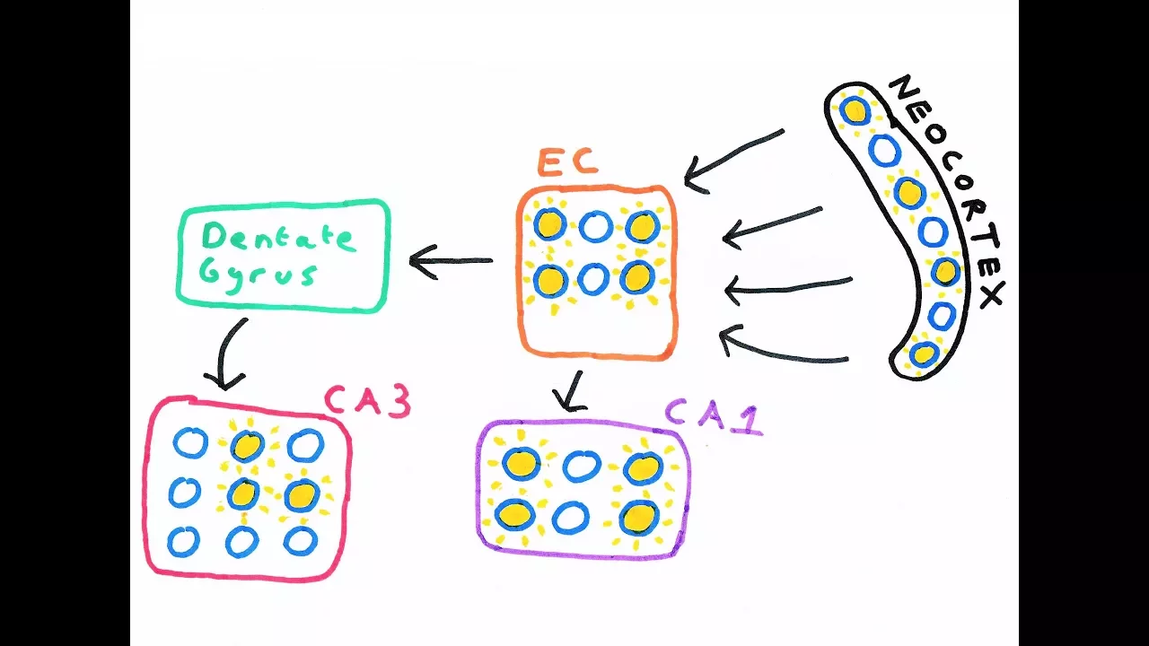
The Hippocampus and episodic memory
125.6K
The hippocampus, named for its seahorse shape, is a paired medial temporal lobe structure consisting of four CA areas (CA4, CA3, CA2, CA1) containing pyramidal cells, plus the dentate gyrus (granule cells), subiculum, and entorhinal cortex. All input enters through the entorhinal cortex via the perforant pathway to dentate gyrus and CA3. Mossy fibers connect dentate to CA3, while Schaffer collaterals connect CA3 to CA1. CA3 has dense recurrent connections. CA1 projects to subiculum, which returns to entorhinal cortex, completing the loop. This circuitry enables the hippocampus's role in episodic memory and navigation.
Click to watch →
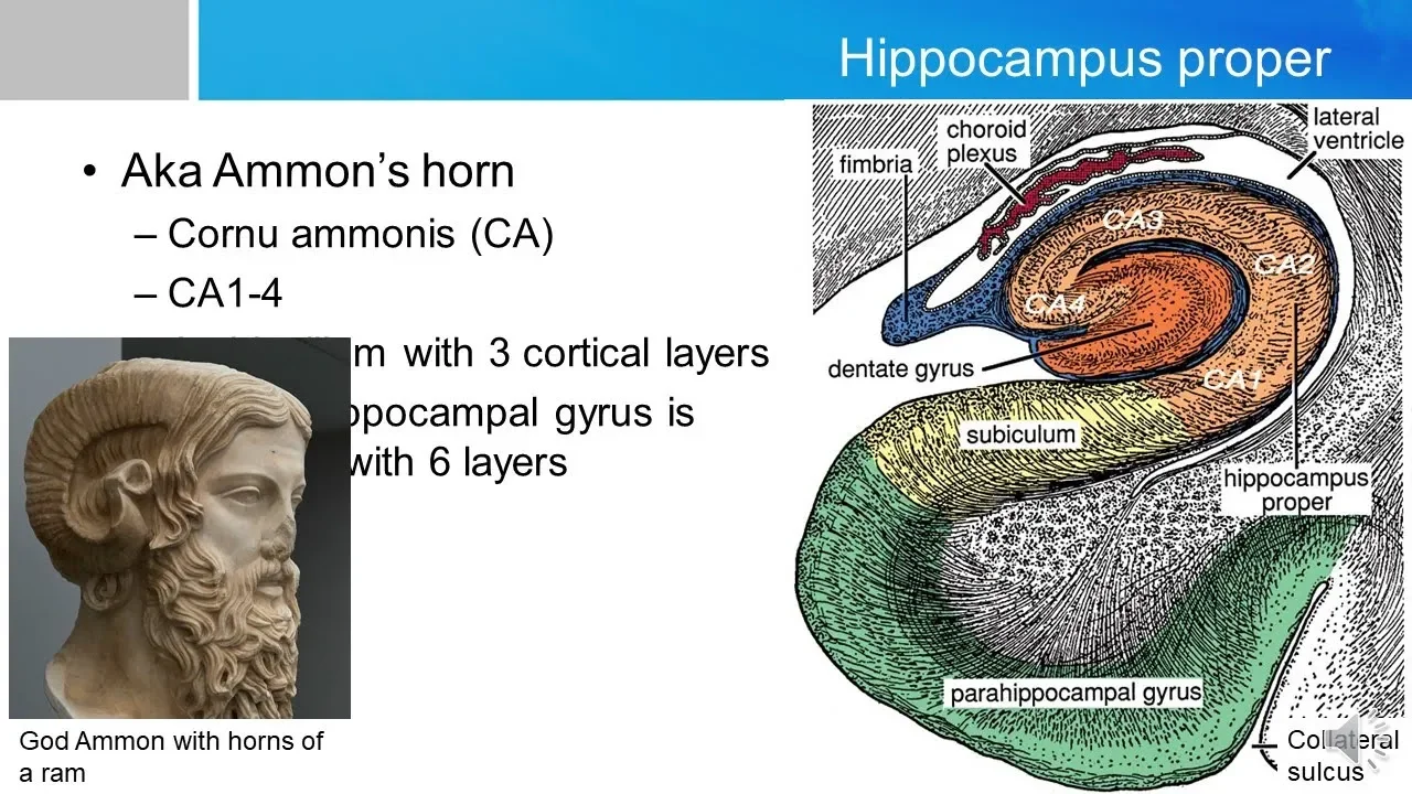
Neurosurgery written board crash course - Hippocampus
19.6K
The hippocampal formation consists of the hippocampus proper (CA1-CA4 regions named for their sequential arrangement), dentate gyrus, and subiculum, forming a trisynaptic loop where information flows from the entorhinal cortex through the dentate gyrus to CA3, then to CA1, and exits via the subiculum; the major output pathways include the fornix projecting to mammillary bodies and the subiculum projecting to the entorhinal cortex, with the CA1 region being particularly susceptible to hypoxic and excitotoxic damage, and the hippocampus playing a critical role in recent memory formation and manifesting as psychogenic seizures with automatic behaviors and psychic symptoms.
Click to watch →

Khazar Ahmadi: Deep Dive into hippocampus from a laminar perspective
249
The hippocampus receives major input from the entorhinal cortex through the trisynaptic pathway: entorhinal superficial layers send input to dentate gyrus (perforant path), CA3, and CA1. Dentate gyrus connects to CA3 via mossy fiber collaterals, CA3 projects to CA1 via Schaffer collaterals, and CA3 neurons form recurrent collateral connections. CA1 and subiculum then project outputs to deeper entorhinal layers. This fundamentally differs from neocortical feed-forward/feedback models. The dentate gyrus and CA4 lack true laminar structures, containing only granular cell layers.
Click to watch →

Deep Cerebral Structures & Cerebellum | Samra Mirza | NEU 241
135
The hippocampal formation consists of hippocampus, dentate gyrus, and subiculum, all with three layers. The perforant pathway perforates through the sulcus separating entorhinal cortex from dentate gyrus, involving GABA release from mossy fibers to dentate granule cells. The mossy fiber pathway goes directly from entorhinal cortex to CA3, involving excitatory transmission through AMPA and NMDA receptors. The subiculum, CA3, and CA1 contain pyramidal cells (excitatory, glutamate release); dentate gyrus contains only granule cells (inhibitory, GABA release).
Click to watch →

Dietmar Schmitz: Interneuron Diversity and Circuit Mechanisms of Brain Rhythms in Memory and Recall
274
The hippocampus has a characteristic trisynaptic loop circuit where information flows from the dentate gyrus through CA3 to CA1. The dentate gyrus granule cells project to CA3 via mossy fiber synapses, which have relatively few inputs per CA3 neuron (approximately a dozen). CA3 neurons then connect recurrently among themselves and project to CA1. This convergence-divergence pattern creates a bottleneck at the dentate gyrus-CA3 interface. CA3 cells can receive input from a single granule cell firing at different frequencies, enabling frequency coding of information. This architecture allows the hippocampus to integrate multiple sensory inputs into coherent memory representations.
Click to watch →
Deep Dive
The role of the hippocampus in spatial navigation, cognitive mapping, and the function of place cells.
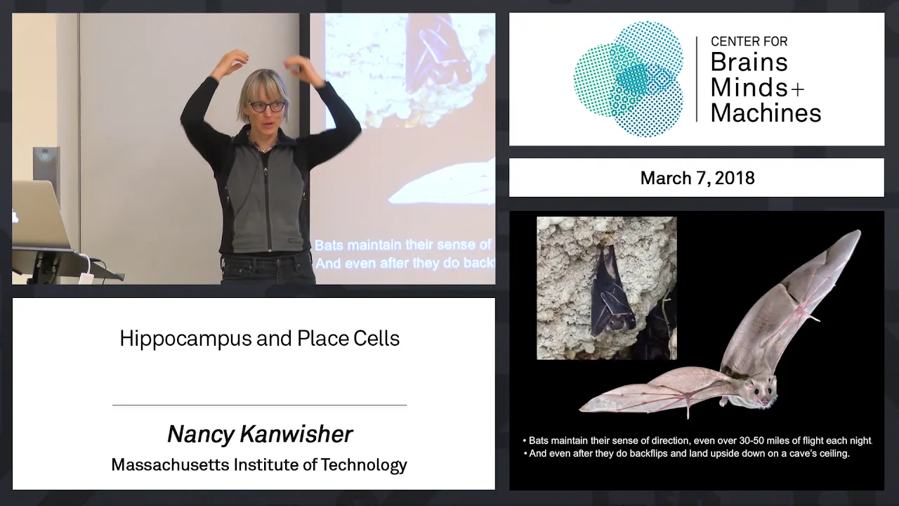
6.3 - Hippocampus and Place Cells
19.3K
The hippocampus contains specialized neurons called place cells that create a cognitive map of the environment, firing selectively when an organism is in specific locations; these cells were first discovered in rats through maze experiments, have been observed in bats as 3D place fields, and have been recorded in humans using intracranial electrodes during virtual navigation tasks, demonstrating that the brain maintains a continuous sense of spatial location through distributed neural representations.
Click to watch →
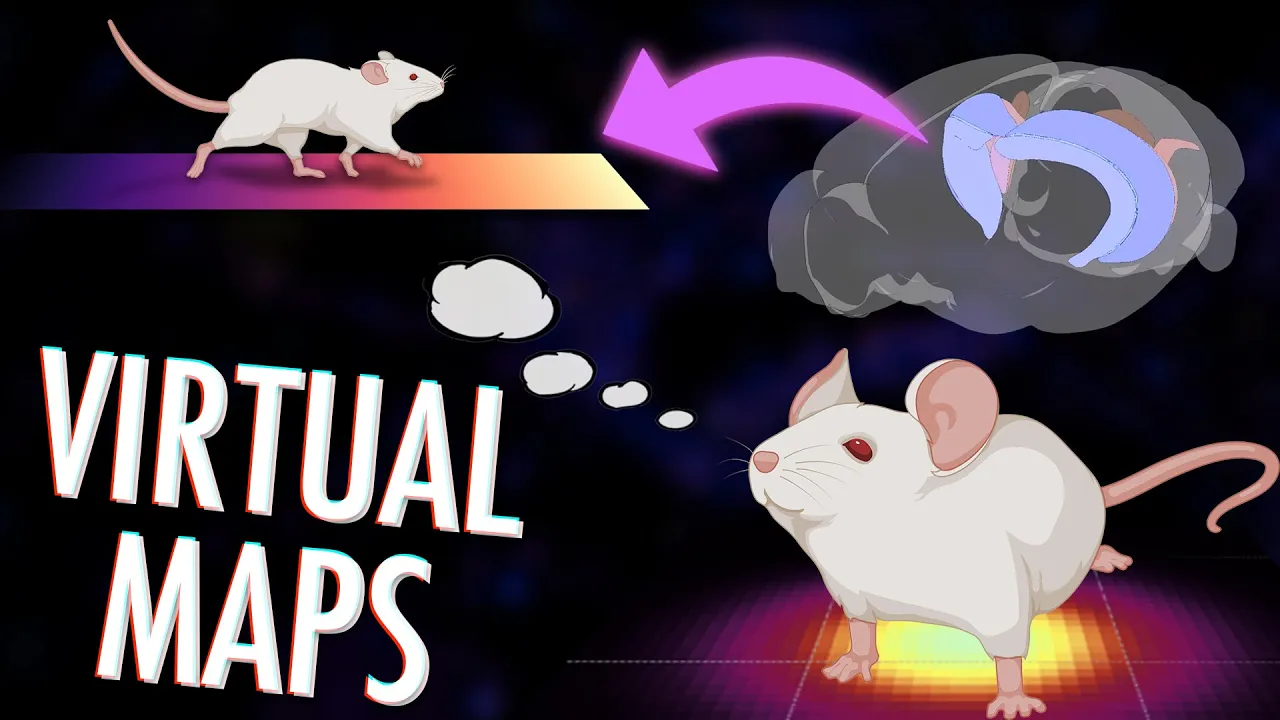
Place cells: How your brain creates maps of abstract spaces
102.2K
The hippocampus contains specialized neurons called place cells that create cognitive maps of physical and abstract spaces; these cells fire selectively when an organism occupies specific locations (place fields), and their activity patterns can be modified by environmental changes (remapping) or non-spatial contextual factors like fear conditioning, demonstrating that the hippocampus constructs internal representations of space that integrate spatial navigation with contextual and episodic memory.
Click to watch →
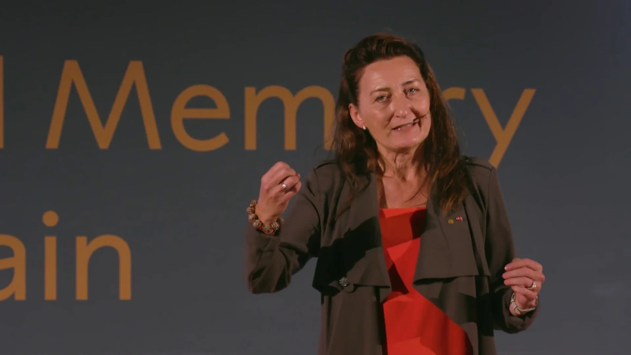
‘Space, time and memory in the brain’ May-Britt Moser, Nobel Prize in Physiology or Medicine
19.2K
The hippocampus, a seahorse-shaped structure in the temporal lobe, is essential for episodic memory encoding. Patient HM, who had both hippocampi removed for epilepsy treatment, could learn new motor skills but couldn't form new episodic memories—he couldn't recognize people he'd met minutes before or navigate familiar spaces. This established the hippocampus as critical for spatial and temporal memory. John O'Keefe then discovered place cells: individual hippocampal neurons that fire only when an animal occupies specific locations. By recording from approximately 100 place cells simultaneously, researchers can predict an animal's position with 5 cm precision. Each place cell has its own preferred location, creating a cognitive map of the environment. This work earned O'Keefe the Nobel Prize and laid the foundation for understanding how the brain represents space.
Click to watch →

May-Britt Moser | The Brain’s GPS: Mapping How We Navigate the World
300.5K
The hippocampus, a seahorse-shaped structure behind each ear, is essential for spatial navigation and memory formation. Patient HM's bilateral hippocampus removal demonstrated that without this structure, individuals cannot form new episodic memories, recognize previously encountered people, or navigate familiar environments. John O'Keefe's pioneering work revealed that individual hippocampal neurons (place cells) fire selectively when an animal occupies specific locations, creating a cognitive map of the environment.
Click to watch →

How Your Brain Maps Your Location and Self in Space
267.3K
Place cells are neurons in the hippocampus that fire specifically when an animal occupies a particular location, creating internal GPS-like maps. John O'Keefe discovered these cells in rats by recording individual neurons during spatial navigation. Remarkably, place cells maintain their firing patterns even when environmental cues are removed, indicating they encode spatial location independently of external landmarks. When rooms are stretched, place cell firing fields scale proportionally, showing that the brain treats spaces like elastic structures. This research revealed that navigation requires both landmark-based memory and an internal coordinate system, with the hippocampus serving as the brain's spatial memory center.
Click to watch →
Deep Dive
Clinical pathologies related to hippocampal dysfunction, such as anterograde amnesia (e.g., the case of Patient H.M.) and Alzheimer's disease.

«Почему наша память несовершенна и что нам с этим делать?» Лекция Полины Кривых
6.4K
Amnesia can be classified into two types: retrograde amnesia (forgetting events before onset) and anterograde amnesia (inability to form new memories after onset). Patient HM had anterograde amnesia while retaining his long-term memories. Alzheimer's disease primarily affects memory, and examination of Alzheimer's patients' brains shows that hippocampal cells are destroyed. The hippocampus shrinks and cannot function properly, explaining the memory impairment characteristic of Alzheimer's disease.
Click to watch →

Brain and Behavior - Introduction to Brain and Behavior
188.7K
Patient HM (Henry Molaison) had both hippocampi surgically removed at age 27 to treat severe epilepsy. While his epilepsy was alleviated, he developed anterograde amnesia—losing the ability to form new long-term memories. Study of his brain revealed the critical role of the hippocampus in memory formation. MRI imaging later showed the precise extent of his hippocampal damage.
Click to watch →
Where Are Memories Stored?
158.6K
Patient HM (Henry Molaison) had bilateral removal of his hippocampus and amygdala in the 1950s to treat severe epilepsy. After surgery, he developed anterograde amnesia—unable to form new declarative memories—yet retained old memories and could still learn new skills (non-declarative). This case proved the hippocampus is critical for forming new declarative memories, while other brain systems handle non-declarative memory.
Click to watch →
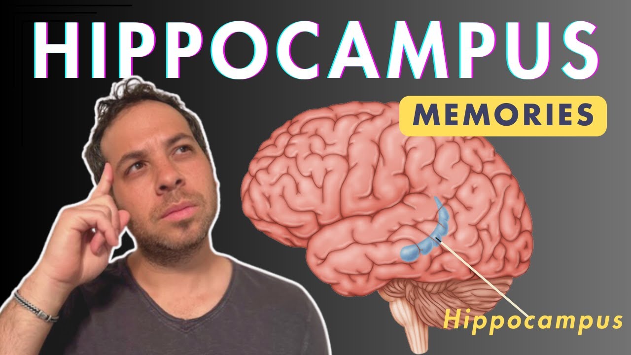
Hippocampus and Memories
143.9K
Damage to the hippocampus results in two types of amnesia. Anterograde amnesia is the inability to form new memories after the injury, while retrograde amnesia is the inability to recall old memories from before the injury. The term 'retro' relates to backward or past, helping distinguish between these conditions. Henry Molaison (HM) demonstrated this after bilateral medial temporal lobe removal, retaining childhood memories but unable to form new episodic memories.
Click to watch →

Clase Nº 3: Neurociencia cognitiva
182
HM is a famous case of anterograde amnesia caused by brain surgery to treat epilepsy. He cannot form new memories but retains memories from before his surgery. This case demonstrated that the hippocampus is critical for forming new memories.
Click to watch →
Hippocampus
Hippocampus
0:00- 1
Located in temporal lobe; named for seahorse resemblance.
- 2
Key for memory; part of hippocampal formation with dentate gyrus.
- 3
Info flows via entorhinal cortex to CA regions, then subiculum.
Multiple Trace Theory of Memory Consolidation
While traditional neuroscience often teaches the Standard Consolidation Theory—where the hippocampus temporarily stores memories before transferring them permanently to the neocortex—the Multiple Trace Theory (MTT) offers a major counterpoint. MTT argues that the hippocampus is never truly bypassed for detailed, episodic memories. Instead, it posits that every time an episodic memory is retrieved, a new trace is created within the hippocampus. Consequently, older memories are more resilient because they have multiple traces, but they still rely on the hippocampus for retrieval. This challenges the classic view of the hippocampus as merely a temporary transit station.
Basic neuroanatomy, specifically the location of the temporal lobe and the general organization of the cerebral cortex.
The structure of a neuron and the basics of synaptic transmission and neural communication.
The concept of the limbic system and its broad association with emotion and memory.
An introductory distinction between different memory systems, such as working memory versus long-term storage.
The cellular and molecular mechanisms of memory consolidation, including Long-Term Potentiation (LTP).
The specific neural pathways of the hippocampal formation, such as the trisynaptic loop (dentate gyrus, CA3, and CA1).
The role of the hippocampus in spatial navigation, cognitive mapping, and the function of place cells.
Clinical pathologies related to hippocampal dysfunction, such as anterograde amnesia (e.g., the case of Patient H.M.) and Alzheimer's disease.
425.2K views5Klikes2:00@neurochallengedOriginal Release: 2015-06-02
Welcome to 2 minute neuroscience, where I simplistically explain neuroscience topics in 2 minutes or less.
In this installment I will discuss the hippocampus.
There is a hippocampus in the temporal lobe of each cerebral hemisphere.
The name “hippocampus” comes from the Greek for "seahorse" because when it is removed from the brain, it vaguely resembles a seahorse.
Although it has many functions, the hippocampus is best known for its role in memory.
The hippocampus is part of a larger structure in the temporal lobe called the hippocampal formation.
Definitions of what structures are included in the hippocampal formation vary, but generally it is considered to at least include the hippocampus, the adjacent cortex which is called the hippocampal or parahippocampal gyrus, and a strip of grey matter in between the two called the dentate gyrus.
The hippocampal gyrus contains areas called the entorhinal cortex and subiculum, which are both involved in the flow of information through the hippocampus.
In addition to being compared to a seahorse, the hippocampus has also been likened to the curved horn of a ram or the horns of the ancient Egyptian god ammon and thus has been called Ammon’s horn or cornu ammonis.
Accordingly, the hippocampus has been subdivided anatomically into 4 regions designated CA1 through CA4; the CA stands for cornu ammonis.
The hippocampus receives information from the rest of the cerebral cortex primarily via the perforant pathway, which originates in the entorhinal cortex and projects to the dentate gyrus.
Fibers then leave the dentate gyrus and project to neurons in the CA3 region of the hippocampus; neurons in CA3 then send axons to neurons in the CA1 region, which projects to neurons in the subiculum.
The subiculum can be considered the main output region of the hippocampal formation; fibers from the subiculum project back upon neurons in the entorhinal cortex and then fibers from the entorhinal cortex travel out to a variety of areas in the cerebrum.
Output fibers also leave the subiculum and hippocampus and enter the fornix, a fiber bundle that connects the hippocampus with a variety of subcortical areas like the thalamus and hypothalamus.
Up Next
58:43
Adult Hippocampal Neurogenesis: Regulation & Function | Neuroscience Lecture
@Labroots
9.7K views•2016-03-16

21:55
Deciphering Neural Circuits Controlling Anorexia in Mice | Richard Palmiter
@AllenInstitute
2.3K views•2011-10-10
1:59
How Smell Works: Olfaction Explained in 2 Minutes | Neuroscience
@neurochallenged
303.7K views•2018-01-31

13:03
How Exercise Benefits Your Brain: Science Explained
@TED
11.4M views•2018-03-21
Related Study Plans & Knowledge Roadmaps
Structured learning paths in Neuroscience
neuroscience
Appetite Regulation: Brain Circuits & Hormones
01Foundations of Endocrinology and Brain Anatomy
02Introduction to Energy Homeostasis
03Peripheral Signals: Leptin, Ghrelin, and Insulin
+ 2 more learning modules
14h5 modules
Explore Roadmap
neuroscience
Neuroeconomics: Value, Risk & Brain Mechanisms
01Foundations of Neuroscience and Brain Anatomy
02Introduction to Neuroeconomics and Subjective Value
03Dopaminergic Prediction Errors & Reinforcement Learning
+ 3 more learning modules
18h6 modules
Explore Roadmap
neuroscience
Spatial Navigation: Cells, Grids & Brain Maps
01Foundations of Brain Anatomy and Memory
02Place Cells and Head-Direction Cells
03Grid Cells: The Brain's Coordinate System
+ 2 more learning modules
12h5 modules
Explore Roadmap
neuroscience
BCIs: Neural Decoding & Prosthetic Control
01Neuroscience Foundations: How the Brain Signals Intent
02Signal Acquisition: Recording the Brain's Activity
03Neural Decoding: Translating Brain Waves into Data
+ 2 more learning modules
25h5 modules
Explore Roadmap