Olfaction, the sense of smell, begins when odorants stimulate cilia on olfactory receptor cells in the olfactory epithelium lining the nasal cavities, triggering action potentials that travel via the first cranial nerve to the olfactory bulb; there, axons converge on glomeruli where they synapse with mitral and tufted relay neurons, whose axons form the olfactory tract carrying information to the olfactory cortex including the piriform cortex, periamygdaloid cortex, entorhinal cortex, olfactory tubercle, and anterior olfactory nucleus for processing.
How Smell Works: Olfaction Explained in 2 Minutes | Neuroscience
Added:Deep Dive
Basic concepts of neurobiology, including how neurons transmit signals via action potentials and synapses.

Basic Neuroscience - Lecture 3
125
The neuron doctrine states the nervous system consists of individual neurons that send and receive information. Information moves via action potentials - rapid changes in ion concentration (sodium in, potassium out) that are all-or-nothing events with fixed size and shape. The only variable is firing frequency. Neurotransmitters are chemical messengers released at synapses that bind to receptors on postsynaptic neurons, making them more or less likely to fire. Most behaviors rely on neural circuits - groups of neurons working together, such as the knee-jerk reflex involving sensory neurons, interneurons, and motor neurons.
Click to watch →
تالته ثانوي لغات | كورس التأسيس بيولوجي | Biology |
42.5K
Neurons have cell bodies, dendrites (receive signals), and axons (transmit signals). Synapses are junctions where neurotransmitters (like acetylcholine) transmit signals between neurons. Action potentials propagate along axons, triggering neurotransmitter release at synapses.
Click to watch →
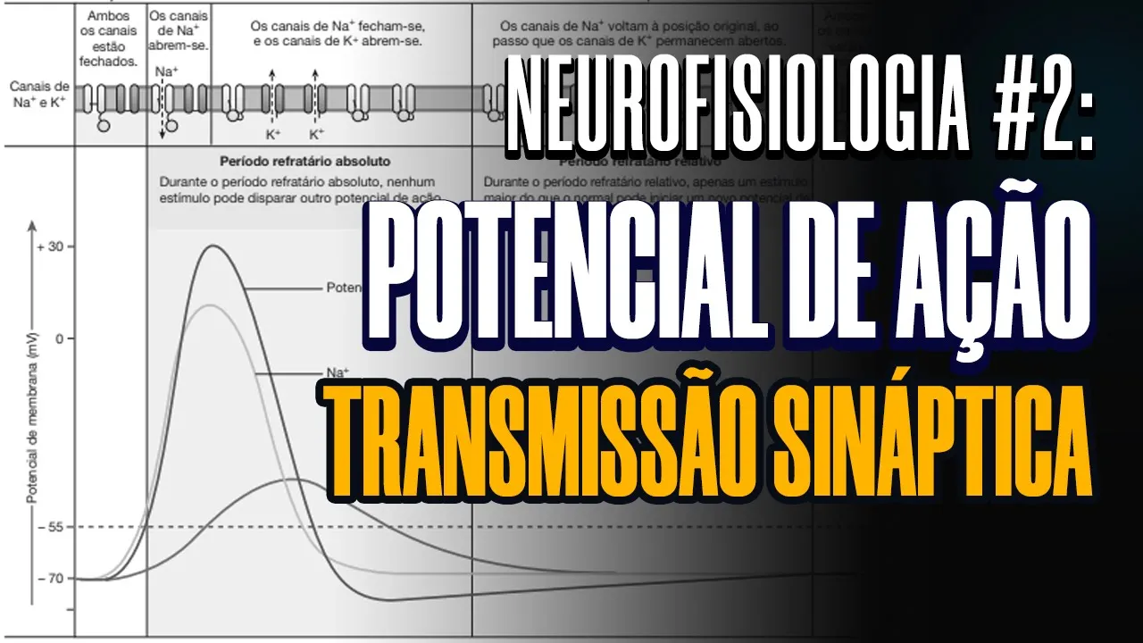
POTENCIAL DE AÇÃO E TRANSMISSÃO SINÁPTICA / AULA NEUROFISIOLOGIA #2
5.6K
Action potentials are rapid, all-or-nothing electrical signals that propagate along neurons through sequential opening of voltage-gated sodium channels (causing depolarization), followed by potassium channels (causing repolarization and hyperpolarization), with refractory periods preventing immediate re-firing; synaptic transmission involves chemical signals (neurotransmitters) released from presynaptic neurons that bind to postsynaptic receptors, generating excitatory or inhibitory potentials that integrate through temporal and spatial summation to determine whether an action potential will be triggered.
Click to watch →

Prof. NANCY BONINI, Ph.D.- BIG QUESTIONS IN NEUROSCIENCE
1.1K
Neurons communicate electrically through action potentials - all-or-none electrical events triggered when sufficient depolarizing signals summate at the axon hillock. These signals travel down the axon to synaptic terminals where neurotransmitter-containing vesicles release their contents across the synaptic cleft. Neurotransmitters bind to receptors on the postsynaptic neuron's dendrites, continuing the signal transmission. This electrochemical communication forms the basis of all neural signaling.
Click to watch →

O que é o Potencial de Ação e Sinapse? | Introdução à Neurociências | 004 - Parte 2
135
Neurons communicate through electrical signals called action potentials that travel along axons, and chemical signals called neurotransmitters that cross synapses between neurons. Action potentials are generated when ion channels open, allowing sodium and potassium ions to flow across the cell membrane, creating a rapid change in electrical charge that follows an 'all-or-nothing' principle. The process involves depolarization (cell becomes positive), repolarization (cell returns to negative state), and hyperpolarization (cell becomes more negative than resting state). At synapses, neurotransmitters are released from vesicles in the presynaptic neuron, cross the synaptic gap, and bind to receptors on the postsynaptic neuron, transmitting information. This communication is regulated through reuptake, enzymatic degradation, and autoreceptors.
Click to watch →
Deep Dive
The concept of chemoreception, specifically how chemical molecules (ligands) interact with cellular receptors.

Cамый быстрый нос, язык для обоняния и нос вместо рук. Как твари чувствуют запахи?
9.5K
Chemical reception (chemoreception) is the first sense that evolution developed. Living organisms have specialized receptors on their cell surfaces that detect chemical molecules. These receptors function like locks that specific molecules (keys) fit into, causing the receptor protein to change shape and send a signal to the organism. This allows bacteria and other simple organisms to detect chemical information in their environment and move toward favorable conditions or away from harmful ones.
Click to watch →
Aula 2: Neuroquímica - Parte 1 - LANeC OPNeuro
776
Cells perceive their environment through a system more refined than human senses, consisting of interactions between ligands and receptors. A ligand is a molecule with a specific chemical structure that interacts with a receptor protein located in the plasma membrane. When a ligand binds to its receptor, it activates the receptor and induces an intracellular response. This mechanism allows cells to detect environmental changes and respond appropriately, serving as the fundamental basis for cellular communication and survival.
Click to watch →

Fizyoloji 4. Ders, Bölüm 1:Hücreler arası haberleşme
681
Chemical communication typically involves ligand-receptor interactions. Ligands are extracellular chemical molecules that act as signaling molecules, while receptors are proteins located on the target cell membrane that specifically bind to ligands.
Click to watch →

TODO sobre SEÑALIZACIÓN CELULAR 📞🦠 mensajeros, receptores, etc
33.4K
Receptors are specialized proteins that recognize and respond to specific ligands (messenger molecules). Each receptor has a unique shape that matches its ligand both geometrically and chemically, ensuring highly specific signaling. Not all cells possess all types of receptors, which explains why signaling is so specific—ligands only bind to cells expressing their corresponding receptors. Contrary to older beliefs that ligands themselves produce effects, modern understanding shows that receptors undergo conformational changes upon ligand binding, and it is this modification that initiates the signaling cascade. The same ligand can produce different responses in different cell types depending on which receptors are present.
Click to watch →

Chemoreceptor
2.3K
The gustatory system employs G protein-coupled receptors (GPCRs) and ion channels to detect five basic tastes: salty/sour through ion channels, sweet/bitter through GPCRs, and savory (umami) through glutamate activation. Contact chemoreceptors are uniporous structures with single pores at their tips, found primarily in mouthparts but also on antennae and legs. Recent research reveals primary cilia function as cellular antennae coordinating multiple signaling pathways related to cell division and differentiation. Physiologically, chemoreception enables detection of food, habitat, mates, and predators through chemical emissions. Receptors on head cells change upon contact with environmental emissions, transmitting signals electrochemically to the central nervous system, which generates appropriate survival responses.
Click to watch →
Deep Dive
Anatomy of the human nasal cavity and the general location of the olfactory epithelium.
![ANATOMÍA DE LA CAVIDAD NASAL 👃[explicación fácil y rápida]](https://i.ytimg.com/vi/DirR-MygB-s/maxresdefault.jpg)
ANATOMÍA DE LA CAVIDAD NASAL 👃[explicación fácil y rápida]
99.3K
The nasal cavity is divided into three regions: the vestibule (covered by skin with sweat and sebaceous glands and vibrissae), the olfactory area (with olfactory epithelium containing smell receptors), and the respiratory area (lined with pseudostratified ciliated columnar epithelium); its walls include the roof (frontal, ethmoidal, and sphenoidal portions), the floor (hard palate), the medial wall (nasal septum formed by osseous and cartilaginous components), and the lateral wall (containing superior, middle, and inferior conchae that create meatuses for sinus drainage).
Click to watch →

Olfaction Anatomy and Function | MCAT Biology | Khan Academy
419.2K
The olfactory epithelium is a specialized region in the nasal passage that contains olfactory receptor cells. It is located in the upper part of the nasal cavity and is responsible for detecting odor molecules.
Click to watch →
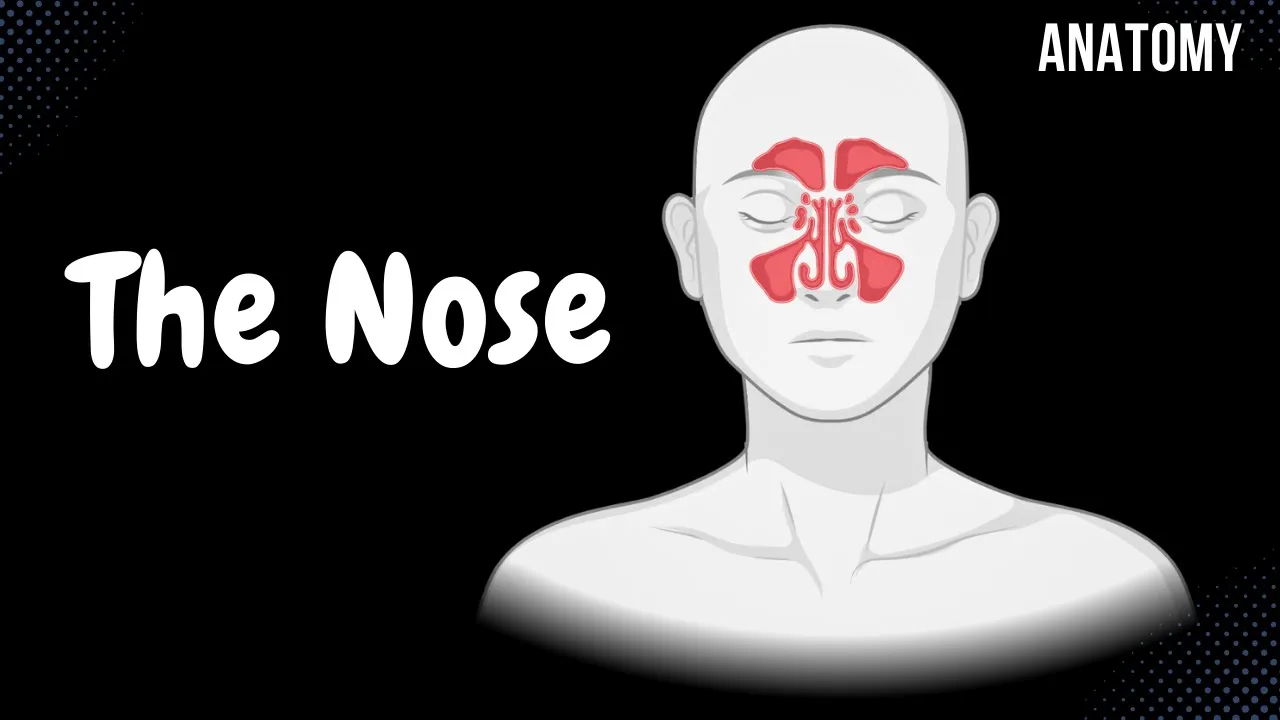
Nasal Anatomy (Cartilage, Nasal Cavity, Sinuses, Meatuses, Nasal Mucosa)
584.5K
The nasal cavity contains specialized structures serving distinct functions: the olfactory part (upper region) detects smell through olfactory epithelium with receptor cells, sustentacular cells, and Bowman's glands, while the respiratory part (lower region) filters and conditions air using pseudostratified ciliated epithelium with goblet cells and mixed mucus glands; the paranasal sinuses (frontal, maxillary, ethmoid, and sphenoid) open into specific meatuses (middle, superior, and inferior) and serve to reduce skull weight, enhance voice resonance, warm and humidify inhaled air, and produce protective mucus, with their drainage pathways being clinically significant for conditions like sinusitis caused by infections, allergies, nasal polyps, or deviated septum.
Click to watch →
ДЫХАТЕЛЬНАЯ СИСТЕМА (ЛЕКЦИЯ ПО ГИСТОЛОГИИ)
3.3K
The nasal cavity is divided into the vestibule (anterior, cartilaginous, lined with skin containing vibrissae and vestibular glands) and the proper nasal cavity (posterior, lined with respiratory epithelium). The mucosa is functionally divided into olfactory region (upper parts, olfactory epithelium, Bowman's glands) and respiratory region (main part, respiratory epithelium). Olfactory epithelium is pseudostratified columnar, 60 μm thick, containing olfactory receptor cells (bipolar neurons with cilia), supporting cells (predominant, provide structural support and secrete mucus), and basal cells (stem cells for continuous renewal). Brush cells have chemoreceptors for bitter taste. The olfactory epithelium is supported by a wide lamina propria containing numerous Bowman's glands (seromucous glands) that secrete mucus rich in odorant-binding proteins and antimicrobial substances.
Click to watch →

Shotgun Histology Nasal Cavity
64.8K
The upper respiratory tract follows a fundamental principle: epithelial type depends on what substance contacts it. Air-exposed surfaces develop pseudostratified ciliated columnar epithelium, while areas exposed to harder substances like food develop stratified squamous epithelium. The nasal cavity contains three conchi/turbinates separated by meatus (superior, middle, inferior). The superior region hosts specialized olfactory epithelium where smell sensation occurs, featuring exceptional cilia and nerve cells. Bowman's glands secrete mucus to dissolve odor molecules. The underlying tissue is rich in veins that warm incoming air. Non-olfactory regions contain standard pseudostratified ciliated columnar epithelium with goblet cells producing protective mucus.
Click to watch →
Deep Dive
The distinction between primary sensory organs and the central nervous system processing centers.

HISTOLOGIE UE5 : Les organes de sens.
111
Sensory organs are specialized structures that receive environmental information through five senses: vision, audition, gustation, olfaction, and tactile sensation. These organs contain specialized nerve elements enabling interaction with the external environment. A sensory organ is defined as a specialized receptor containing sophisticated sensory receptors that perceive external stimuli, refine reception, and transmit information to the central nervous system. Sensory organs consist of three cell types: primary sensory cells (true neurons that detect stimuli and transmit information to the brain), accessory or pseudo-sensory cells (specialized epithelial cells that detect stimuli but cannot transmit information independently), and support cells (epithelial cells that provide structural support, nourishment, and protection). Primary sensory cells are classified into three categories based on embryological origin: placodial neurosensory cells (originating from the epiblast and remaining in the nasal wall, such as olfactory neurons), ganglionic sensory cells (located in ganglia along cranial and spinal nerve roots serving as relay stations), and central sensory cells (integral parts of the central nervous system from the beginning, such as retinal cells). All three types share the common ability to generate nerve impulses themselves, differing only in their developmental origin and anatomical location. The sensory system is divided into conscious and unconscious processing based on brain destination. Conscious sensory processing (exteroception) involves information reaching the cerebral cortex for perception of the external world through the five senses. Unconscious sensory processing includes interoception (monitoring internal organs and blood vessels for parameters like blood sugar and pressure) and proprioception (awareness of body position detected by proprioceptors in muscles, joints, and tendons). Sensory receptors are classified by stimulus type: statoreceptors for equilibrium, phonoreceptors for hearing, photoreceptors for vision, chemoreceptors for taste and smell, mechanoreceptors for touch, thermoreceptors for temperature, and nociceptors for pain detection. Sensory organs are classified physiologically by the complexity of the relay between stimulus and brain: primary sensory organs have the simplest system where the stimulus-receiving cell is directly a sensory neuron, with only one cell involved between the outside and the brain. The olfactory organ is the only primary sensory organ, where olfactory cells both detect odors and transmit information to the central nervous system. Secondary sensory organs require two neurons: a pseudo-sensory cell at the periphery that receives the stimulus and transmits excitation to a primary sensory neuron in ganglia. Tertiary sensory organs involve three neurons and no accessory cells. The olfactory organ is the only primary sensory organ, consisting of the olfactory mucosa in the upper nasal cavity. It appears yellow-brown due to pigment granules in support cells containing carotenoids and the richness of mucus in carotene. The olfactory organ has an epiblastic origin and develops through three stages: nasal placodes (week 4), olfactory pits or cupules (week 5), and olfactory grooves as pits deepen and elongate.
Click to watch →

SISTEMA SENSORIAL: Receptores sensoriais, Vias aferentes e Centros de processamento | MK Fisiologia
207K
The sensory system consists of three main components: sensory receptors that detect and encode physical or chemical stimuli into electrical signals called receptor potentials through sensory transduction; afferent pathways formed by primary, secondary, and tertiary neurons that transmit encoded information to the central nervous system; and processing centers in the cerebral cortex (such as the somatosensory, visual, auditory, gustatory, and olfactory cortices) that process information and make it conscious. Sensory stimuli are classified as physical (pressure, temperature, light, sound) or chemical (odor, taste), and only stimuli processed in the cerebral cortex become conscious awareness.
Click to watch →

NERVOUS SYSTEM PART 2
188
The central nervous system (CNS) processes information from sensory organs and coordinates responses. Sensory organs detect environmental stimuli and convert them into electrical signals that travel to the CNS for processing. The CNS then generates appropriate responses, which may involve motor neurons sending signals to muscles or glands.
Click to watch →

SISTEMA SENSORIAL: ORGÃOS DOS SENTIDOS - biologia
62.5K
The brain is the central processor of sensory information, while sensory organs (eyes, ears, nose, tongue, skin) capture and transmit information. The brain is divided into two hemispheres: the left hemisphere handles logical thinking, while the right hemisphere handles creativity. Sensory cells are specialized cells that transmit information to the brain, including neurosensory cells (where neurons transmit information) and epithelial sensory cells (modified epithelial cells). Different receptors are specialized for specific information: thermoreceptors detect temperature, photoreceptors detect light, mechanoreceptors detect pressure, and chemoreceptors detect chemical substances like food flavors.
Click to watch →
Neuroanatomy: Nervous System Basics | CNS & PNS
3.8M
The nervous system is organized into central (brain and spinal cord) and peripheral components. The CNS receives sensory input, processes information by comparing new data with stored memories, integrates experiences, and generates motor responses. Sensory information is classified into special senses (vision, hearing, olfaction, taste, equilibrium from specialized body regions) and general senses (touch, temperature, pain from multiple body parts). This foundational organization enables perception, cognition, and coordinated response to environmental stimuli.
Click to watch →
Prerequisite Knowledge
- Concept 01Basic concepts of neurobiology, including how neurons transmit signals via action potentials and synapses.
- Concept 02The concept of chemoreception, specifically how chemical molecules (ligands) interact with cellular receptors.
- Concept 03Anatomy of the human nasal cavity and the general location of the olfactory epithelium.
- Concept 04The distinction between primary sensory organs and the central nervous system processing centers.
Subsequent Learning
- Step 01The detailed biochemical cascade of olfactory transduction, including GPCR activation, cAMP production, and ion channel opening.
- Step 02The integration of olfaction with the limbic system (amygdala and hippocampus) to explain smell-induced memories and emotions.
- Step 03The multisensory integration of taste (gustation) and smell (olfaction) in creating the perception of flavor.
- Step 04Clinical conditions associated with olfactory dysfunction, such as anosmia, hyposmia, and their diagnostic significance in neurodegenerative diseases.
Olfactory start
Receptor cells
Odor detection
Cranial nerve I
Bulb convergence
Cortical relay
Cortex areas
Olfactory start
0:04Playing Section
- 1
Discusses the sense of smell and its beginning in the olfactory epithelium.
- 2
Explains the location of olfactory epithelium lining human nasal cavities.
The Vibration Theory of Olfaction (Quantum Olfaction)
While the standard scientific model explains olfaction through the "shape theory"—where odorant molecules fit into receptors like a lock and key—a significant alternative perspective is the Vibration Theory of Olfaction. Popularized by biophysicist Luca Turin, this theory proposes that olfactory receptors detect the molecular vibrations of odorants rather than just their physical shape, utilizing a quantum mechanics phenomenon known as inelastic electron tunneling. Proponents of the vibration theory argue that shape-based models fail to explain why molecules with identical shapes but different isotopes (such as deuterated compounds) can smell distinctly different, or why molecules with completely different structures but similar vibrational frequencies can smell identical. Although highly controversial and actively debated within neuroscience, the quantum vibration theory serves as a major critical counterpoint to the established, purely structural explanation of how the brain perceives smell.
Deep Dive
The detailed biochemical cascade of olfactory transduction, including GPCR activation, cAMP production, and ion channel opening.
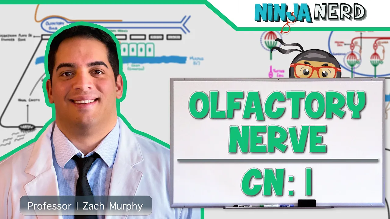
Neurology | Olfactory Nerve: Cranial Nerve I
543.4K
Olfactory transduction involves a complex signaling cascade beginning when odorants bind to G-protein coupled receptors on ciliary membranes. This binding activates G-olfactory proteins that stimulate adenylate cyclase, converting ATP to cyclic AMP. Rising cAMP opens ion channels allowing sodium and calcium influx while chloride exits, depolarizing the membrane to generate action potentials. Each olfactory neuron expresses only one receptor type, yet single receptors respond to multiple odorants, and single odorants activate multiple receptors—a combinatorial system enabling detection of thousands of odors. Calcium ions mediate adaptation, reducing neuron responsiveness during prolonged odor exposure.
Click to watch →
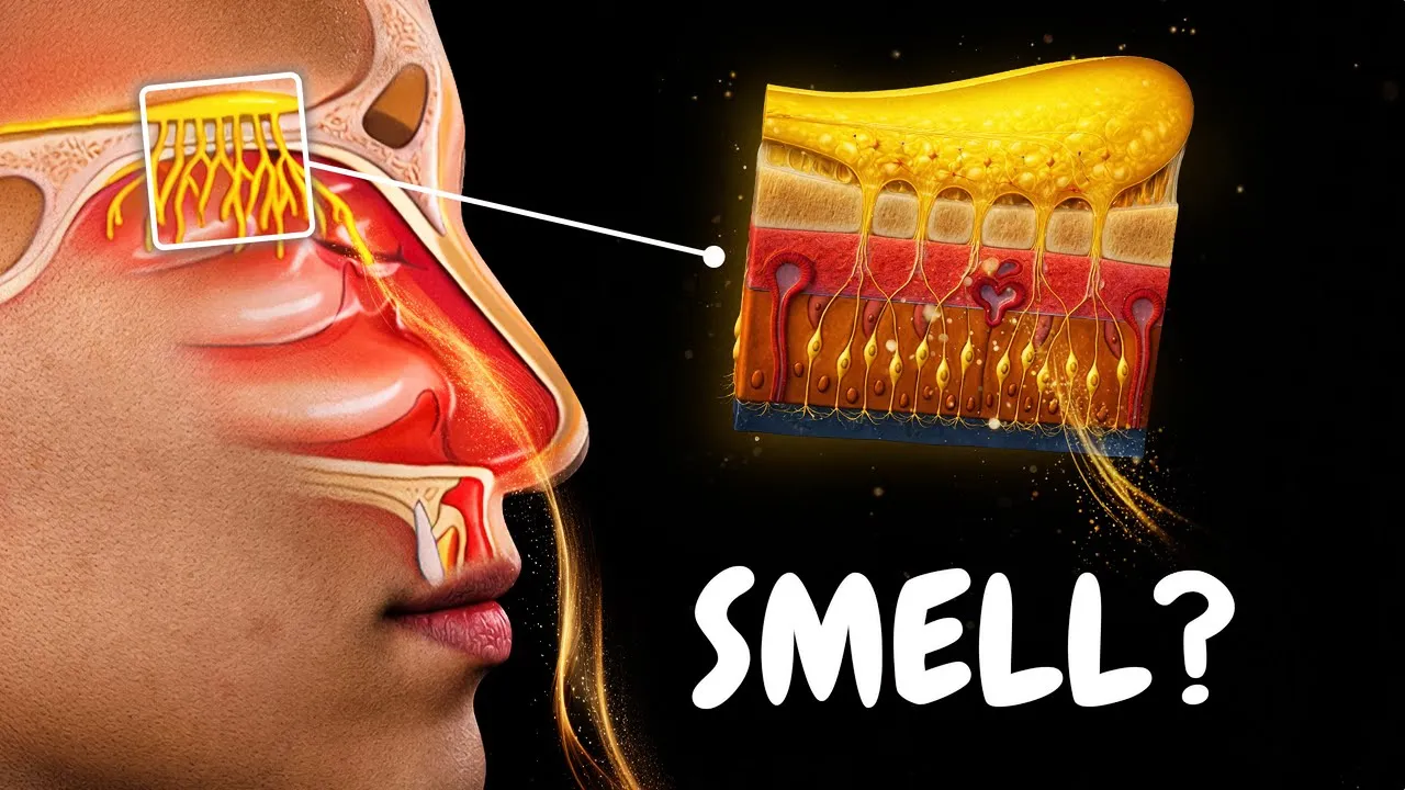
Olfactory System | How Smell Works (Special Senses)
1K
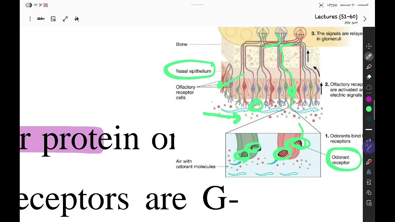
Odorant binding to receptors triggers a seven-step signal transduction cascade: odorant binding activates Golf G-protein, which activates adenylyl cyclase type III converting ATP to cyclic AMP; rising cAMP opens cyclic nucleotide-gated ion channels allowing Na+ and Ca2+ influx; Ca2+ activates calcium-activated Cl- channels, and since intracellular Cl- concentration is unusually high, Cl- efflux further depolarizes the membrane; if threshold is reached, an action potential is generated. Olfactory receptor neuron axons bundle into filaments passing through the cribriform plate to the olfactory bulb, where they synapse onto second-order neurons in glomeruli. All neurons expressing the same receptor converge onto the same glomerulus, which then projects to mitral and tufted cells.
Click to watch →
شرح لمحاضرة رقم ٥٤ فسيولوجىChemoreceptor functionsمع حل الكويز
553
The olfactory transduction process involves: (1) Odorant molecules dissolve in mucus and bind to olfactory receptor proteins on cilia; (2) Receptor activation triggers G-protein (Golf) activation; (3) Activated G-protein stimulates adenylyl cyclase to produce cAMP from ATP; (4) Increased cAMP opens cyclic nucleotide-gated (CNG) channels, allowing calcium and sodium influx; (5) Calcium influx activates calcium-activated chloride channels, causing chloride efflux; (6) The net effect is membrane depolarization and action potential generation. This mechanism converts chemical signals into electrical signals that the brain can interpret.
Click to watch →

NOSE | SENSE ORGAN
17.7K
Olfactory signal transduction converts chemical energy from odor molecules into electrical signals. When odor molecules dissolve in nasal mucus and bind to olfactory receptor proteins on cilia, they activate G-protein coupled receptors. This activates adenylyl cyclase, which converts ATP to cyclic AMP (cAMP). cAMP opens ion channels, allowing sodium and calcium ions to enter the cell, depolarizing it and generating an action potential. The action potential travels along the axon through the cribriform plate to the olfactory bulb. The strength of the action potential corresponds to odorant concentration, allowing the brain to encode both identity and intensity of odors.
Click to watch →
CN 1: Olfactory Nerve (Scheme, Pathway, Clinical Relevance) | Neuroanatomy
104.3K
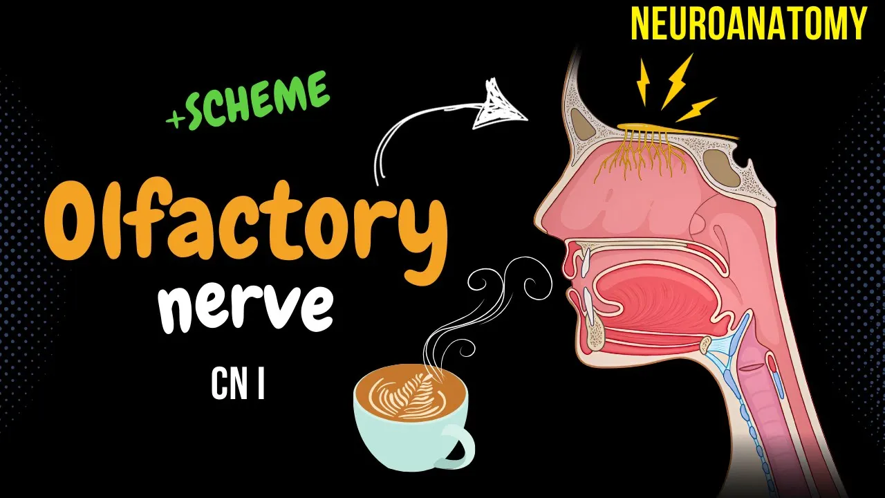
The olfactory epithelium on the nasal cavity roof contains four cell types: basal cells (stem cells), sustentacular cells (support), Bowman's glands (produce mucin), and bipolar olfactory receptor neurons with dendrites bearing sensory cilia and axons projecting to the olfactory bulb. Odorants enter through two pathways: nasal opening and posterior nasopharyngeal transfer. Mucus in the nasal cavity traps odor molecules, which bind to proteins and are transported to cilia receptors. Each odorant binds multiple receptors, and each receptor binds multiple odorants, creating combinatorial diversity enabling discrimination of approximately one trillion odor mixtures. The signal transduction cascade involves G-protein-coupled receptors (7-pass transmembrane proteins) activating adenylyl cyclase to produce cyclic AMP, which opens ion channels causing depolarization and action potential generation. Axons form fila olfactoria bundles creating the olfactory nerve, the shortest cranial nerve capable of partial regeneration.
Click to watch →
Deep Dive
The integration of olfaction with the limbic system (amygdala and hippocampus) to explain smell-induced memories and emotions.
Homunculus: Crash Course Psychology #6
3.3M
Smell perception is closely tied to emotions and memories because the olfactory system connects directly to the limbic system, including the amygdala (emotional registry) and hippocampus (memory keeper). Even if we cannot immediately name an odor, our brains store and recognize scents by their associations. This explains why certain smells can instantly transport us to specific memories, like walking into a bakery and feeling happy while thinking of a beloved grandmother.
Click to watch →

Sensation vs. Perception: What's the Difference?
143.5K
Unlike other senses, smell directly connects to the limbic system—the brain's center for emotions, memories, and survival instincts. The olfactory cortex communicates with the hippocampus (for recognizing familiar smells and explicit memories) and the amygdala (for triggering fear and strong emotional responses to potentially dangerous odors like smoke or rotting food).
Click to watch →

Los sentidos y las propiedades sensoriales (parte 1)
557
Olfactory information is processed in the olfactory bulb and directly connected to the limbic system (amygdala and hippocampus). This direct connection explains why smells can trigger strong emotional responses and memories. The olfactory system is unique among senses in its immediate connection to brain regions involved in emotion and memory. Olfactory detection thresholds vary by compound, ranging from parts per billion to parts per million, depending on volatility and molecular structure.
Click to watch →

100% Realtalk 181 | Michael Nehls | Impfungen | Orwell & Huxley | Maßnahmen & Weltregierung | A.I.
124.9K
The olfactory system (smell) has a direct connection to the hippocampus, which is why smells can trigger vivid memories. This connection is evolutionarily ancient, as even fish use smell to navigate back to their birthplace. The hippocampus and olfactory system are both part of the limbic system, which processes emotions and memories.
Click to watch →
Rhinencephalon | Olfactory Brain, Limbic Connections & Clinical Correlations 🧠✨ | USMLE Step 1👩⚕️📚
348
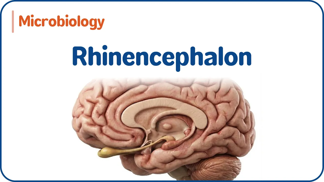
The limbic system integrates olfactory, emotional, and memory functions through interconnected structures. The amygdaloid body (amygdala) processes emotional responses to odors and is located near the caudate nucleus tail. The parahippocampal gyrus connects to the hippocampal formation, which is essential for memory consolidation. The fornix connects the hippocampus to mammillary bodies and hypothalamus. The paraterminal gyrus and Habenular nuclei modulate autonomic and emotional responses. Together, these structures form the olfactory-limbic circuit, explaining why smell is deeply connected to emotion and memory.
Click to watch →
Deep Dive
The multisensory integration of taste (gustation) and smell (olfaction) in creating the perception of flavor.

The enchanting smell (and revolting flavor) of rain
469.4K
Flavor is created through multisensory integration where the brain combines signals from taste (tongue) and smell (nose). Research using tubes delivering molecules directly to the back of the throat demonstrates that the same compound produces different perceptions depending on whether it reaches the nose alone or combines with taste signals. This explains why geosmin smells pleasant outdoors but tastes muddy in food, showing that the brain integrates sensory information differently based on context and input pathway.
Click to watch →
Everything I Thought I Knew About Flavor Was Wrong
219.3K
Flavor is a complex multisensory experience involving taste (gustation), smell (olfaction), sound, touch (somatosensory), and vision, all processed through the brain's amygdala and hippocampus which connect flavor to emotions and memories; the brain integrates these diverse sensory inputs to create our perception of flavor, which explains why identical foods can taste completely different to different people based on their individual sensory sensitivities and life experiences.
Click to watch →

Psychology for Beginners 14. Sensation and Perception (V)
114
Smell and taste are distinct but integrated senses that work together to create flavor; smell receptors in the olfactory epithelium bind with odor molecules and send signals to the olfactory bulb and limbic system, while taste buds detect five basic tastes (sweet, sour, bitter, salty, umami), and together these senses create our complete experience of flavor, as demonstrated by the simple experiment of holding one's nose while eating to isolate taste versus releasing it to experience full flavor.
Click to watch →

AP Psychology Unit 1: Biological Basis of Behavior Review
350K
Taste and smell interact closely to create the full sensation of flavor. Taste buds detect basic tastes while olfactory receptors identify aromas released from food. Together these inputs are processed by the brain to produce different flavors. Without smell, taste sensations become muted or not experienced—when you try Skittles without being able to smell them, they all start to taste the same.
Click to watch →
![Understanding Sensation [AP Psychology Unit 1Topic 6]](https://i.ytimg.com/vi/5ZM2JetHI3s/hqdefault.jpg)
Understanding Sensation [AP Psychology Unit 1Topic 6]
20.5K
Chemical senses include olfaction (smell) and gustation (taste). Olfactory receptor cells in the nasal cavity transduce odor molecules into electrical signals sent to the olfactory bulb, then to emotional/limbic brain regions explaining smell's power to trigger memories and emotions. Pheromones are chemical signals affecting same-species behavior. Gustation detects six basic tastes: sweet (energy-rich foods), sour (spoiled foods), salty (electrolyte regulation), bitter (toxin warning), umami (protein-rich foods), and fatty (fats/oils). Taste buds contain receptors transducing chemical signals. Individual variation creates supertasters (intense taste experience), medium tasters, and non-tasters (reduced sensitivity, especially to bitter). Taste and smell interact closely to create complete flavor perception.
Click to watch →
Deep Dive
Clinical conditions associated with olfactory dysfunction, such as anosmia, hyposmia, and their diagnostic significance in neurodegenerative diseases.

Basics of Olfaction, Taste and Swallowing | Prof. S R Chandra | The White Army
1.1K
Olfactory dysfunction presents with distinct clinical features: anosmia (absence), hyposmia (decreased), hyperosmia (increased, seen in pregnancy/migraine), dysosmia (distortion), phantosmia (hallucinations), and olfactory agnosia (inability to verbally identify odors). Peripheral dysfunction causes simple loss, while central dysfunction produces altered perception. Psychogenic loss cannot occur in isolation—patients retain flavor perception and cannot identify irritants (trigeminal nerve-mediated). Olfactory dysfunction is an early sign of Alzheimer's disease, frontotemporal dementia, and esthesioneuroblastoma, making it clinically significant for early diagnosis.
Click to watch →
Neuroanatomía y fisiología del olfato
831
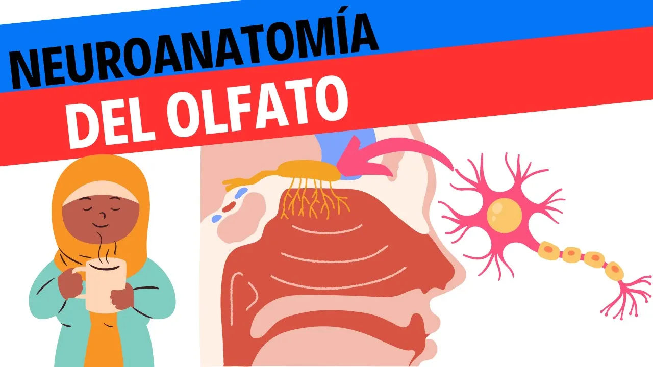
Olfactory information travels via two parallel pathways: to the olfactory cortex for conscious perception, and directly to limbic structures (amygdala, entorhinal cortex) for emotional responses. This explains why odors trigger intense emotions. Olfactory dysfunction includes anosmia, hyposmia, and dysosmia. Causes include neurodegenerative diseases (Parkinson's, Alzheimer's), rhinitis, infections, tumors, medications, intubation, and aging. Approximately 70% of taste perception derives from olfaction, explaining why blocked noses make food taste bland.
Click to watch →
Neurology | Olfactory Nerve: Cranial Nerve I
543.4K
The olfactory bulb processes sensory information through glomerular structures where olfactory nerve axons synapse onto mitral cell dendrites. Granule cells provide inhibitory GABAergic lateral inhibition, creating a competitive network where only the most strongly activated mitral cells transmit signals. The olfactory tract projects to multiple targets: the piriform cortex (primary olfactory cortex), hippocampus, amygdala, and entorhinal cortex for conscious perception and emotional processing; and the subcallosal gyrus and orbital frontal cortex for odor evaluation and reward assessment. Most fibers project ipsilaterally, though some cross contralaterally. Anosmia results from nasal infections, tumors, trauma, or CSF rhinorrhea. Critically, anosmia can indicate early-stage neurodegenerative diseases including Alzheimer's, Parkinson's, and Lewy body dementia, making it clinically significant beyond mere sensory loss.
Click to watch →

Olfaction in neurodegenerative disease
367
Comprehensive diagnostic evaluation includes detailed patient history, ENT examination with nasal endoscopy to exclude structural pathology, psychophysical testing using validated tools like Sniffin' Sticks (TDI test) for threshold, discrimination, and identification, and MRI assessment evaluating olfactory bulb volume (normal 40-60 mm³) and olfactory sulcus depth. Classification uses Sniffin' Sticks: identification scores of 6 or less indicate functional anosmia; 7-10 indicate hyposmia; 11-12 indicate normosmia. Post-infectious olfactory loss typically presents with parosmia and has the best prognosis, with 40-60% recovering normal function through olfactory training (daily practice for 12+ weeks using four essential oils). Currently, no treatments improve olfactory function in neurodegenerative diseases, though olfactory training shows promise. Patients with olfactory dysfunction face real dangers including inability to detect spoiled food, gas leaks, or smoke, requiring patient and family education about safety concerns.
Click to watch →
Sensation Of Smell |Olfactory Pathway - Physiology
150.3K
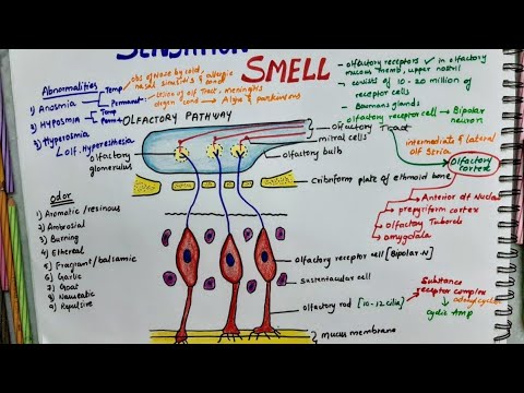
Olfactory transduction converts chemical energy to electrical signals: odorant molecules dissolve in mucus, bind receptor proteins, activate adenylyl cyclase to produce cyclic AMP, open sodium channels generating receptor potentials that trigger action potentials. Odors are classified into nine categories including aromatic, ambrosial, burning, ethereal, fragrant, garlic, goat, nauseating, and repulsive types. Clinical disorders include anosmia (total loss, temporary from nasal obstruction or permanent from tract damage/meningitis/Alzheimer's/Parkinsonism), hyposmia (reduced detection, most common disorder), and hyperosmia (increased sensation in brain injury/epilepsy/neurotic conditions).
Click to watch →
Olfactory start
Receptor cells
Odor detection
Cranial nerve I
Bulb convergence
Cortical relay
Cortex areas
Olfactory start
0:04- 1
Discusses the sense of smell and its beginning in the olfactory epithelium.
- 2
Explains the location of olfactory epithelium lining human nasal cavities.
The Vibration Theory of Olfaction (Quantum Olfaction)
While the standard scientific model explains olfaction through the "shape theory"—where odorant molecules fit into receptors like a lock and key—a significant alternative perspective is the Vibration Theory of Olfaction. Popularized by biophysicist Luca Turin, this theory proposes that olfactory receptors detect the molecular vibrations of odorants rather than just their physical shape, utilizing a quantum mechanics phenomenon known as inelastic electron tunneling. Proponents of the vibration theory argue that shape-based models fail to explain why molecules with identical shapes but different isotopes (such as deuterated compounds) can smell distinctly different, or why molecules with completely different structures but similar vibrational frequencies can smell identical. Although highly controversial and actively debated within neuroscience, the quantum vibration theory serves as a major critical counterpoint to the established, purely structural explanation of how the brain perceives smell.
Basic concepts of neurobiology, including how neurons transmit signals via action potentials and synapses.
The concept of chemoreception, specifically how chemical molecules (ligands) interact with cellular receptors.
Anatomy of the human nasal cavity and the general location of the olfactory epithelium.
The distinction between primary sensory organs and the central nervous system processing centers.
The detailed biochemical cascade of olfactory transduction, including GPCR activation, cAMP production, and ion channel opening.
The integration of olfaction with the limbic system (amygdala and hippocampus) to explain smell-induced memories and emotions.
The multisensory integration of taste (gustation) and smell (olfaction) in creating the perception of flavor.
Clinical conditions associated with olfactory dysfunction, such as anosmia, hyposmia, and their diagnostic significance in neurodegenerative diseases.
303.7K views3.7Klikes1:59@neurochallengedOriginal Release: 2018-01-31
Welcome to 2 minute neuroscience, where I explain neuroscience topics in 2 minutes or less.
In this installment I will discuss olfaction.
Olfaction refers to the sense of smell, which begins with a specialized collection of cells called the olfactory epithelium.
In humans, the olfactory epithelium lines the nasal cavities.
The olfactory epithelium contains millions of olfactory receptor cells.
These cells have a single dendrite that extends to the outermost layer of the epithelium, where cilia emerge from the end of the dendrite and spread over the surface of the olfactory epithelium.
When odorants enter the nasal cavity due to inhalation or by rising from the mouth during the chewing of food, they stimulate receptors on the cilia, depolarizing the olfactory receptor cells and initiating action potentials that travel down the axon of the receptor cell into an adjacent structure called the olfactory bulb.
These axons that travel from the olfactory epithelium to the olfactory bulb together make up the first cranial nerve.
In the olfactory bulb the axons of the olfactory receptor cells converge on the dendrites of olfactory bulb neurons in small clusters called glomeruli.
In these glomeruli, the receptor cells form synaptic connections with several types of olfactory bulb neurons, including cells called mitral cells and tufted relay neurons.
Both of these cells project into the olfactory tract, a bundle of fibers that carries olfactory information to the olfactory cortex, where most olfactory processing occurs.
The olfactory cortex consists of a collection of cortical areas that receive information from the olfactory bulb, including the piriform cortex, an area of cortex surrounding the amygdala known as the periamygdaloid cortex, entorhinal cortex, and two regions known as the olfactory tubercle and anterior olfactory nucleus.
Up Next

5:38
Olfactory Neuroanatomy: Brain Dissection Lab
@EHSLDigitalPublishing
55.8K views•2015-08-13

21:55
Deciphering Neural Circuits Controlling Anorexia in Mice | Richard Palmiter
@AllenInstitute
2.3K views•2011-10-10
2:00
Hippocampus Anatomy & Memory Function: 2-Minute Neuroscience
@neurochallenged
425.2K views•2015-06-02

13:03
How Exercise Benefits Your Brain: Science Explained
@TED
11.4M views•2018-03-21
Related Study Plans & Knowledge Roadmaps
Structured learning paths in Neuroscience
neuroscience
Appetite Regulation: Brain Circuits & Hormones
01Foundations of Endocrinology and Brain Anatomy
02Introduction to Energy Homeostasis
03Peripheral Signals: Leptin, Ghrelin, and Insulin
+ 2 more learning modules
14h5 modules
Explore Roadmap
neuroscience
Neuroeconomics: Value, Risk & Brain Mechanisms
01Foundations of Neuroscience and Brain Anatomy
02Introduction to Neuroeconomics and Subjective Value
03Dopaminergic Prediction Errors & Reinforcement Learning
+ 3 more learning modules
18h6 modules
Explore Roadmap
neuroscience
Spatial Navigation: Cells, Grids & Brain Maps
01Foundations of Brain Anatomy and Memory
02Place Cells and Head-Direction Cells
03Grid Cells: The Brain's Coordinate System
+ 2 more learning modules
12h5 modules
Explore Roadmap
neuroscience
BCIs: Neural Decoding & Prosthetic Control
01Neuroscience Foundations: How the Brain Signals Intent
02Signal Acquisition: Recording the Brain's Activity
03Neural Decoding: Translating Brain Waves into Data
+ 2 more learning modules
25h5 modules
Explore Roadmap