














![ASMR Cranial Nerve Exam [YOU are the DOCTOR]](https://i.ytimg.com/vi/TL8DwqXstJ4/hqdefault.jpg)

![[본방 종합] 향기가 사라졌다... 파킨슨·알츠하이머의 첫 번째 신호는 후각장애! 냄새와 함께 기억도 잃는 이유! I KBS 20251105 방송](https://i.ytimg.com/vi_webp/274IbgrG1Gw/maxresdefault.webp)



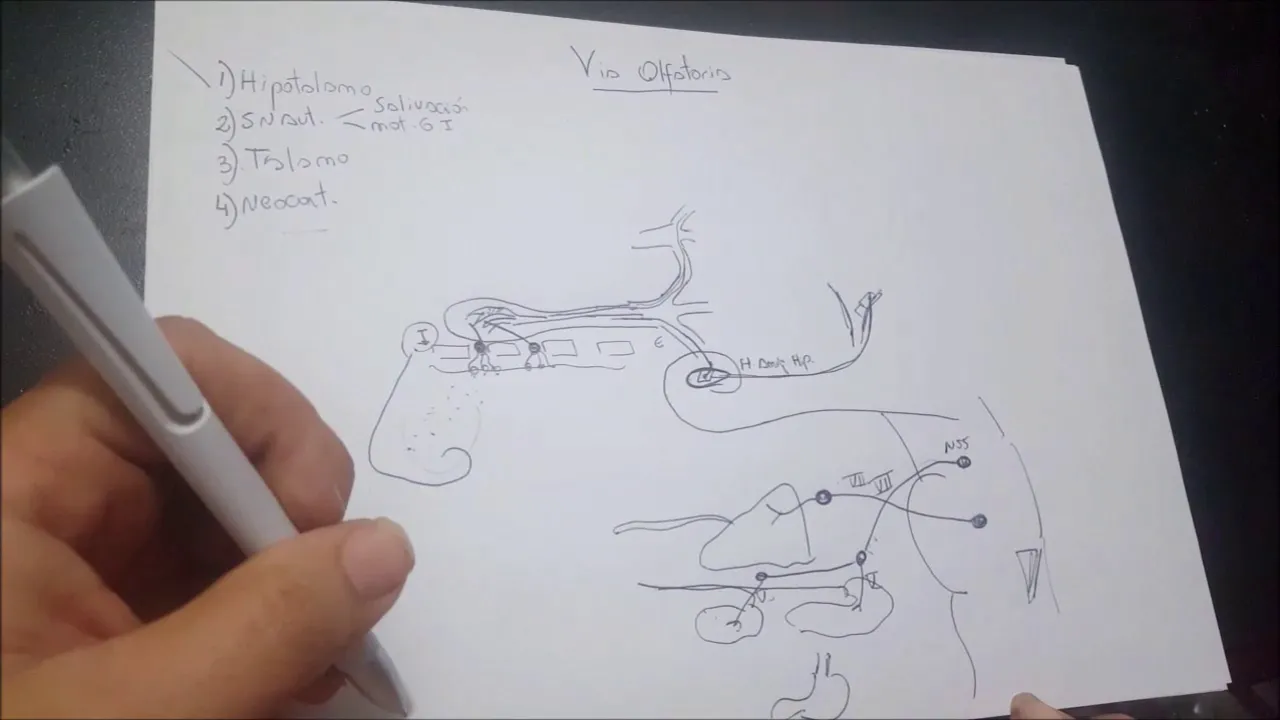
The olfactory system consists of bipolar neurons in the olfactory epithelium (turning over every 30 days with 350 receptor types) whose axons pass through the cribriform plate to synapse in the olfactory bulb, then project via the olfactory tract to the primary olfactory cortex on the temporal lobe and amygdala, bypassing the thalamus; this pathway can be disrupted by trauma causing irreversible anosmia or by meningiomas growing at the crista galli, and degenerative diseases like Alzheimer's accelerate olfactory neuron loss, demonstrating the critical integration of olfaction with taste in flavor perception.
Olfactory Neuroanatomy: Brain Dissection Lab
Added:Basic neuroanatomy of the human brain, including the locations of the temporal and frontal lobes.
The structure and function of the cranial nerves, specifically Cranial Nerve I (the olfactory nerve).
The skeletal anatomy of the anterior skull base, particularly the cribriform plate of the ethmoid bone.
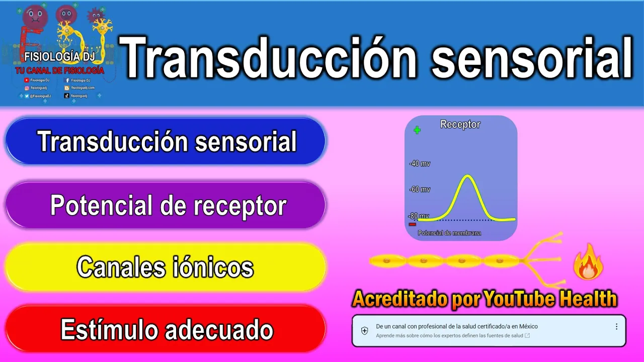
Fundamental concepts of sensory transduction, particularly how chemical stimuli are converted into electrical neural signals.
The functional connection between the olfactory pathway and the limbic system, governing memory, emotion, and behavior.
Clinical neurological examination techniques for assessing olfactory function and diagnosing localized cranial nerve deficits.
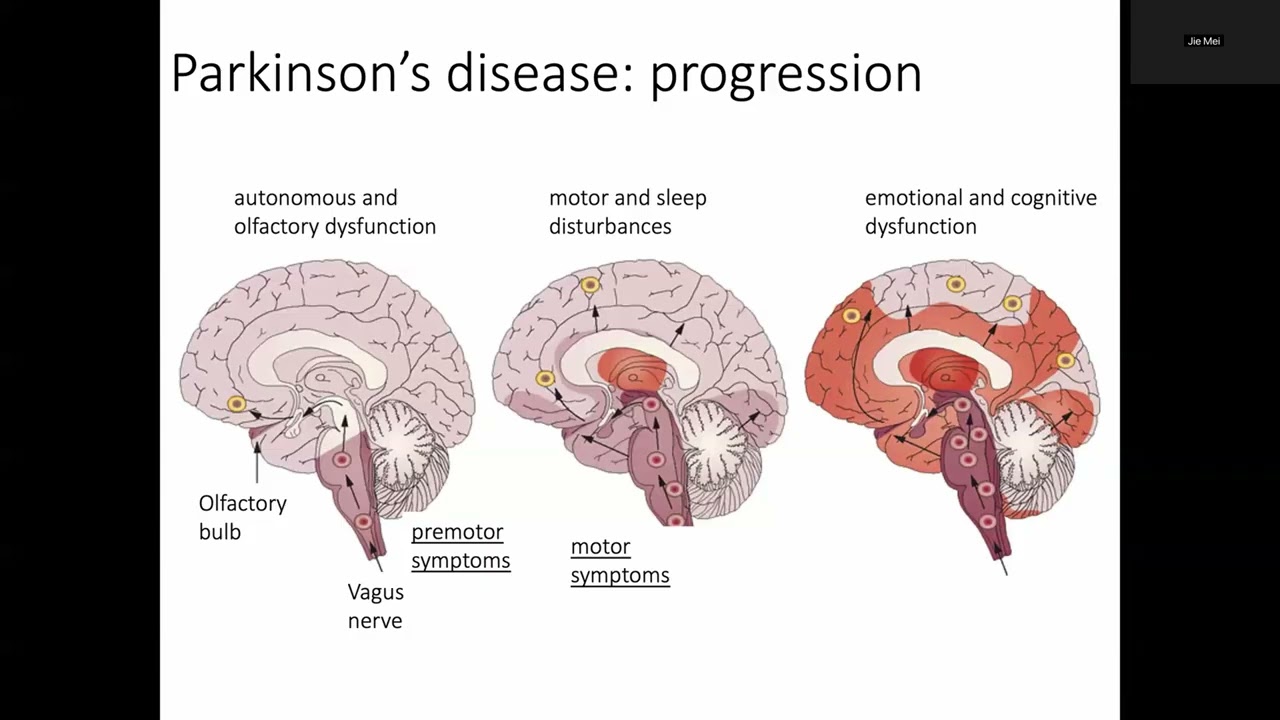
The pathophysiology linking early olfactory dysfunction (anosmia/hyposmia) to neurodegenerative disorders like Parkinson's and Alzheimer's disease.
Neurosurgical approaches to the anterior cranial fossa, such as resecting olfactory groove meningiomas or repairing CSF rhinorrhea.
55.8K views857likes5:38Original Release: 2015-08-13
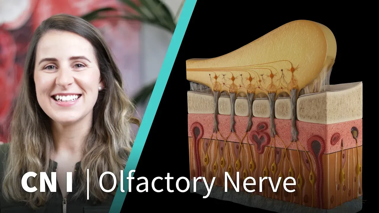
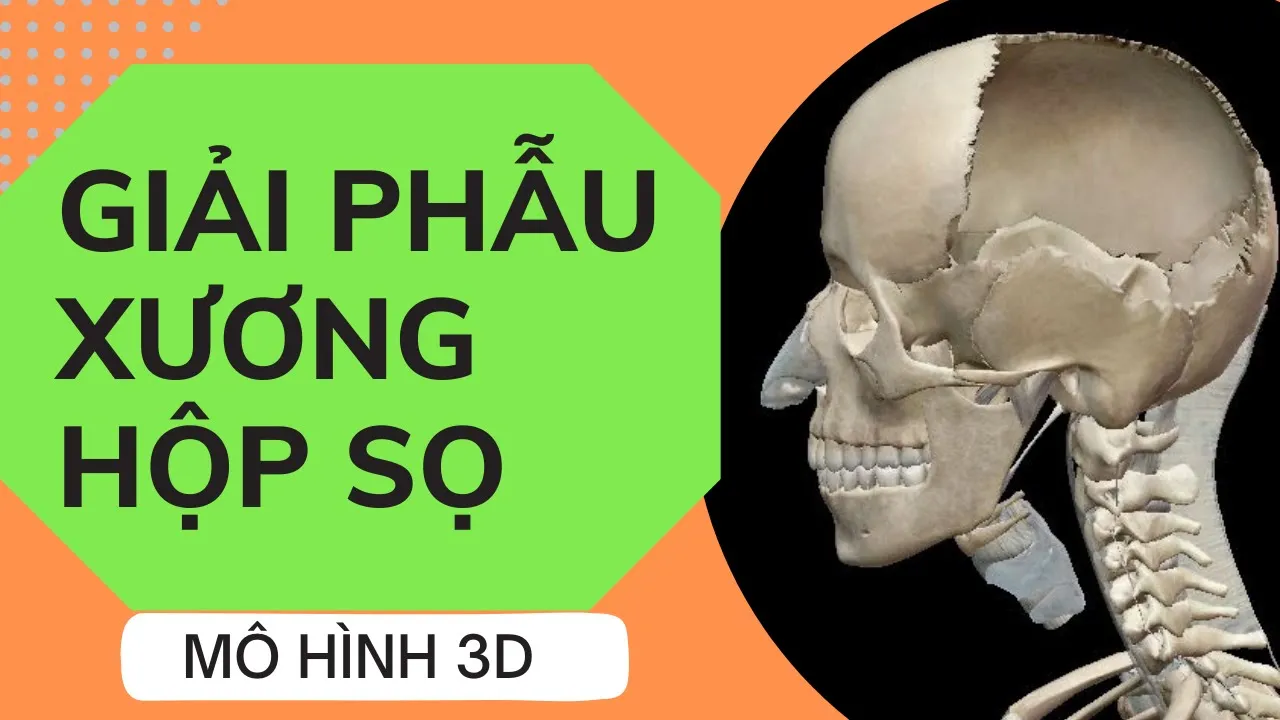
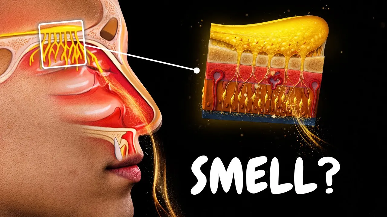
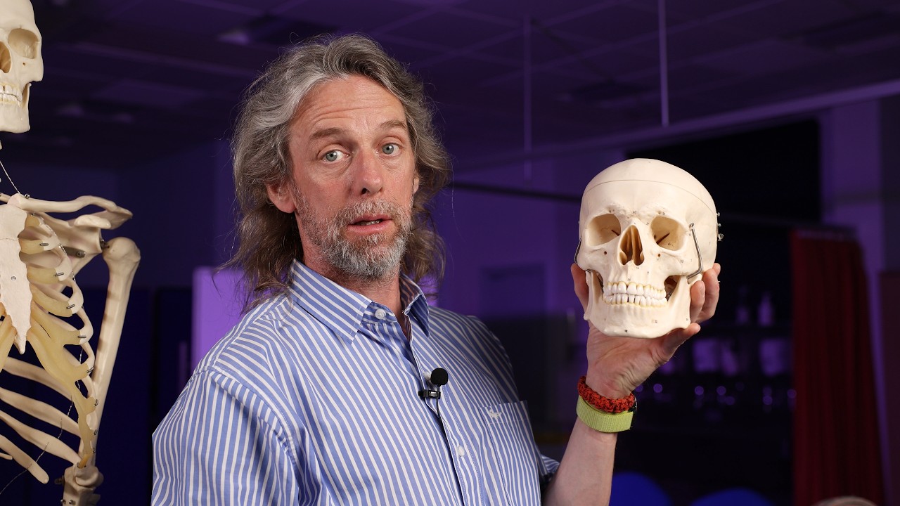
>> The olfactory system is often neglected because it's not as clinically important as some of the other sensory modalities like audition, vision, balance and sensation from the face and body but we're going to briefly discuss it today and we're going to start with the skull here and looking into the nasal cavity we can see the superior concha here which is covered with an epithelium and the epithelium contains neurons, bipolar neurons.
These neurons are not long lived.
They turn over about every 30 days, which is quite unique.
Another unique factor is that they each have a different receptor.
Three hundred and fifty types of receptors means you have 350 different kinds of receptor cells that are important in binding the odors that lead to transmission and information into the central nervous system.
So these axons now we're going to look at and see where they come through into the main cranial cavity and make connection with the brain.
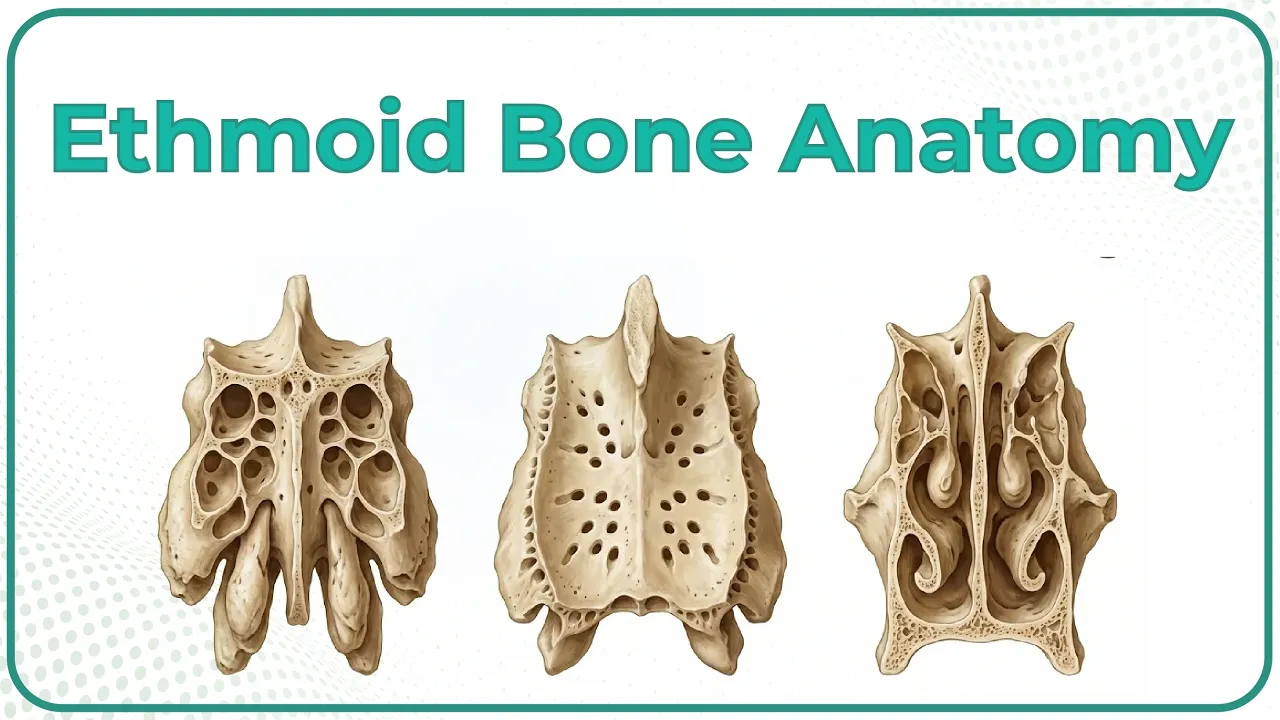
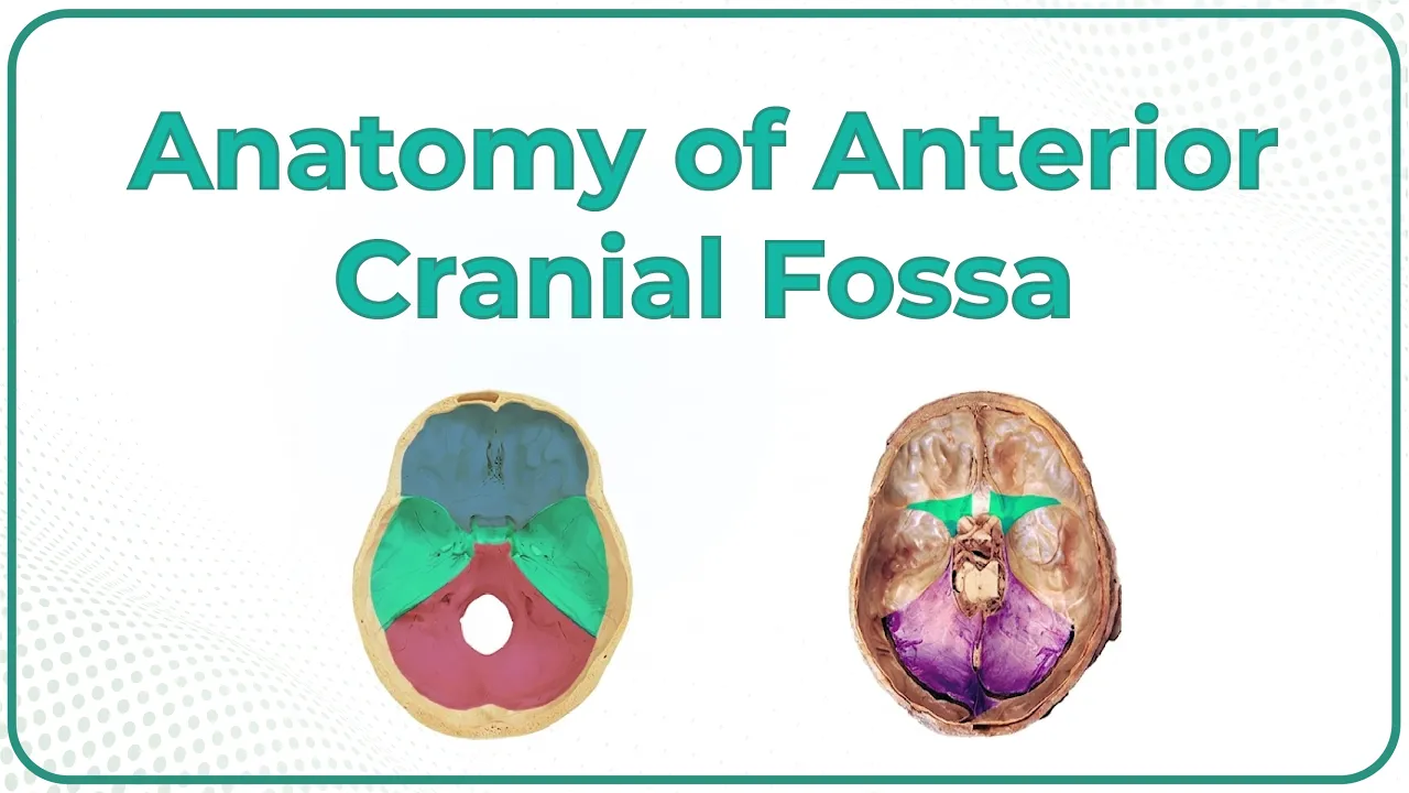
Okay, let's look now at the inside of the skull with a bit of orientation to the posterior fossa, the middle fossa where the temporal lobe sits, and the anterior fossa where the orbital cortex of the frontal lobe sits, given its name because it is the roof of the orbital cavity and in the midline is a little sieve-like plate which is part of the ethmoid bone and through those little holes pass the axons of these primary olfactory neurons coming from our olfactory epithelium and they're going to synapse in the olfactory bulb which lies right above this plate and which we should look at next on a gross specimen.
Here on the ventral surface of the brain we can see the olfactory bulb, and then coming from it is the olfactory tract and these axons then are carrying processed information from the connectivity of the neurons in the olfactory bulb and are traveling down here to this ventral forebrain area right near the internal carotid artery underneath that on the surface and over onto this bulging area of the temporal lobe called the uncus.
So the next synapse or primary olfactory cortex is on the surface here of the temporal lobe and a little bit of it is on the surface of the amygdala, right contiguous with it.
So that is primary olfactory cortex.
Notice that there was no synapse in the thalamus like we saw in other sensory systems.
Then information goes from here to association olfactory cortex some of which is on this inferior surface of the orbital frontal cortex.
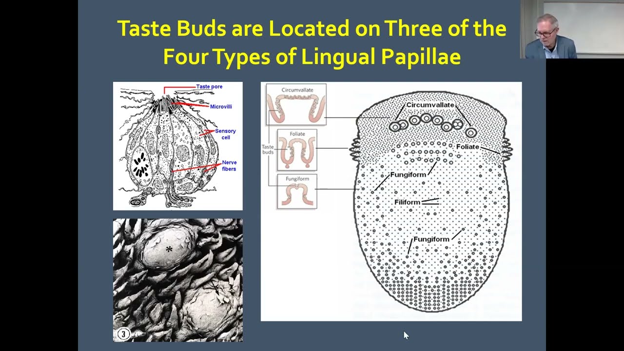
An interesting fact about this orbital frontal cortex is that it's thought that information coming from the taste buds is combined in this area with information from the olfactory system and together they make up what we call flavor and when we have damage or loss of olfaction food does not taste nearly as good and similarly in older people over the age of 80 it's estimated that about 75% of neurons in the olfactory system either in the epithelium or the bulb have degenerated and so food does not taste nearly as good without that olfaction and as a matter of fact people with Alzheimer's disease, Parkinson's disease, other degenerative neuron diseases, seem to have an accelerated loss of neurons in the olfactory system.
So in conclusion let's come back to our skull and look at this cribriform plate and recall that these delicate axons that are passing through here can easily be sheared if the brain is traumatized, let's say you hit your head against the windshield of a car and shear those axons going to the olfactory bulb, damage the olfactory bulb, and lose some of those connections.
That results in a condition called anosmia, which is irreversible.
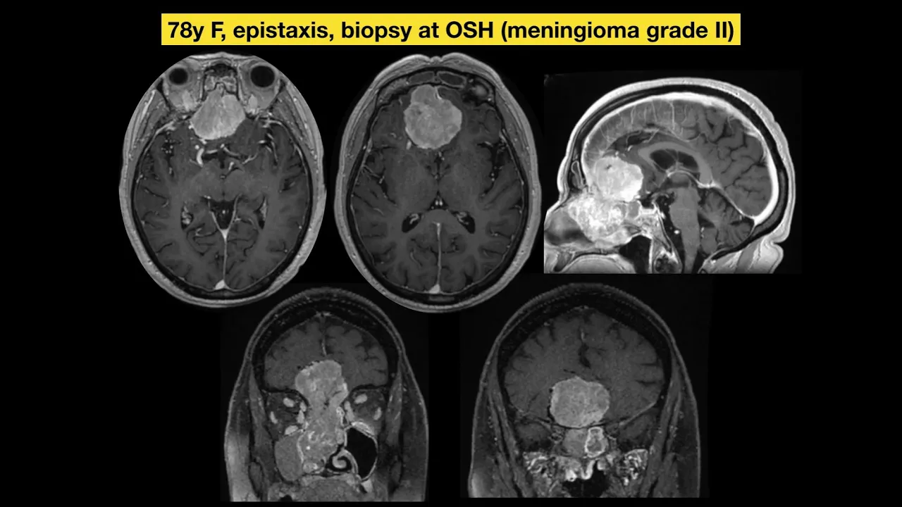
So trauma can be a cause of loss of olfaction as well as meningiomas which tend to grow here because the dura attaches right here on the crista galli of the bone here and the dura is an area in the midline where meningiomas like to grow, so olfactory meningiomas would also occur in this area and cause partial or complete anosmia.
Up Next

82:31
Facial Nerve Anatomy: Cranial Nerve VII Explained (Neurology)
@DoctorNajeeb
193.3K views•2022-07-20

21:55
Deciphering Neural Circuits Controlling Anorexia in Mice | Richard Palmiter
@AllenInstitute
2.3K views•2011-10-10

4:10
Vagus Nerve (CN X): Anatomy, Nuclei & Functions Explained
@Alilamedicalmedia
305.2K views•2022-10-31

13:03
How Exercise Benefits Your Brain: Science Explained
@TED
11.4M views•2018-03-21
Related Study Plans & Knowledge Roadmaps
Structured learning paths in Neuroscience
neuroscience
Appetite Regulation: Brain Circuits & Hormones
01Foundations of Endocrinology and Brain Anatomy
02Introduction to Energy Homeostasis
03Peripheral Signals: Leptin, Ghrelin, and Insulin
+ 2 more learning modules
14h5 modules
Explore Roadmap
neuroscience
Neuroeconomics: Value, Risk & Brain Mechanisms
01Foundations of Neuroscience and Brain Anatomy
02Introduction to Neuroeconomics and Subjective Value
03Dopaminergic Prediction Errors & Reinforcement Learning
+ 3 more learning modules
18h6 modules
Explore Roadmap
neuroscience
Spatial Navigation: Cells, Grids & Brain Maps
01Foundations of Brain Anatomy and Memory
02Place Cells and Head-Direction Cells
03Grid Cells: The Brain's Coordinate System
+ 2 more learning modules
12h5 modules
Explore Roadmap
neuroscience
BCIs: Neural Decoding & Prosthetic Control
01Neuroscience Foundations: How the Brain Signals Intent
02Signal Acquisition: Recording the Brain's Activity
03Neural Decoding: Translating Brain Waves into Data
+ 2 more learning modules
25h5 modules
Explore Roadmap