


![Neuroanatomie 1 : Moelle spinale et tronc cérébral [Anatomy of the spinal cord and brainstem]](https://i.ytimg.com/vi/KnaQzcuH6O4/maxresdefault.jpg)




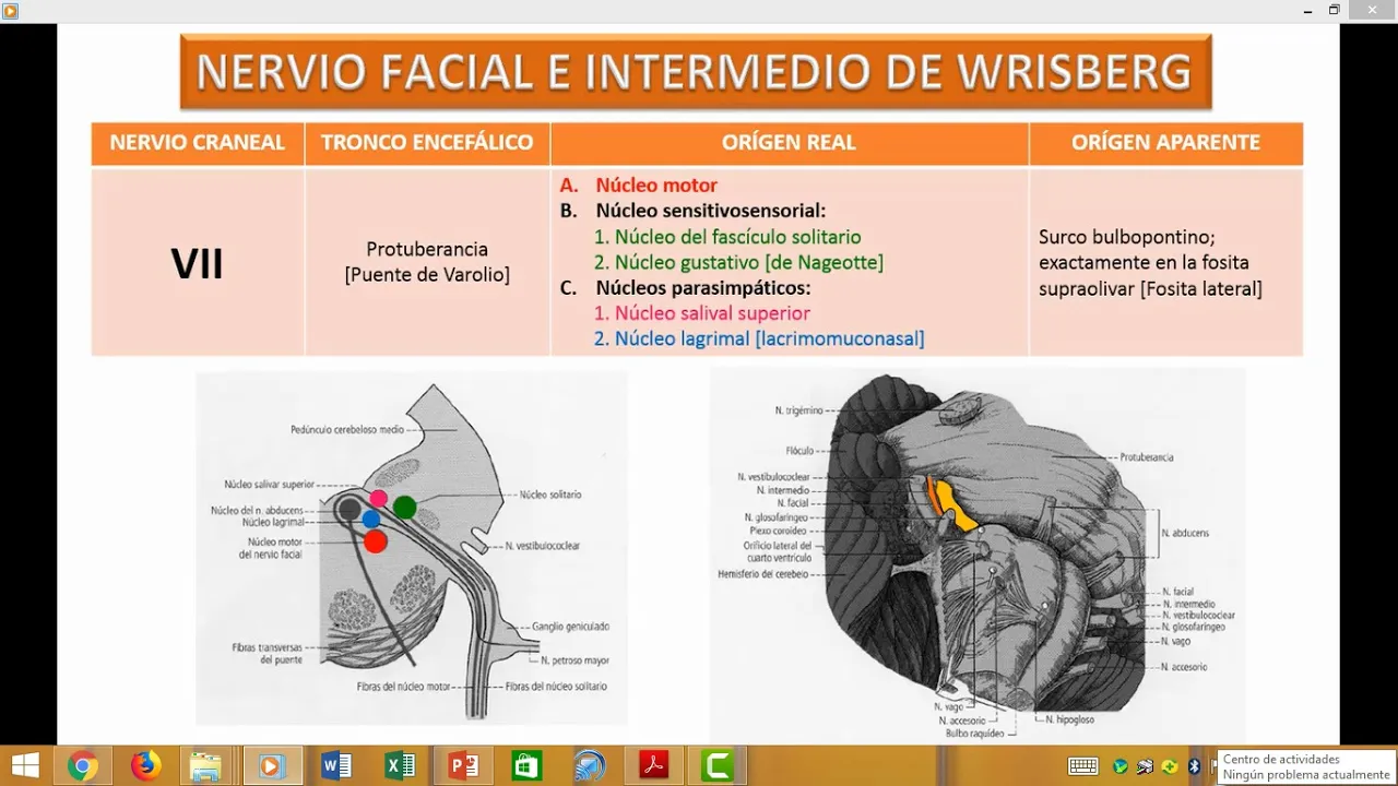


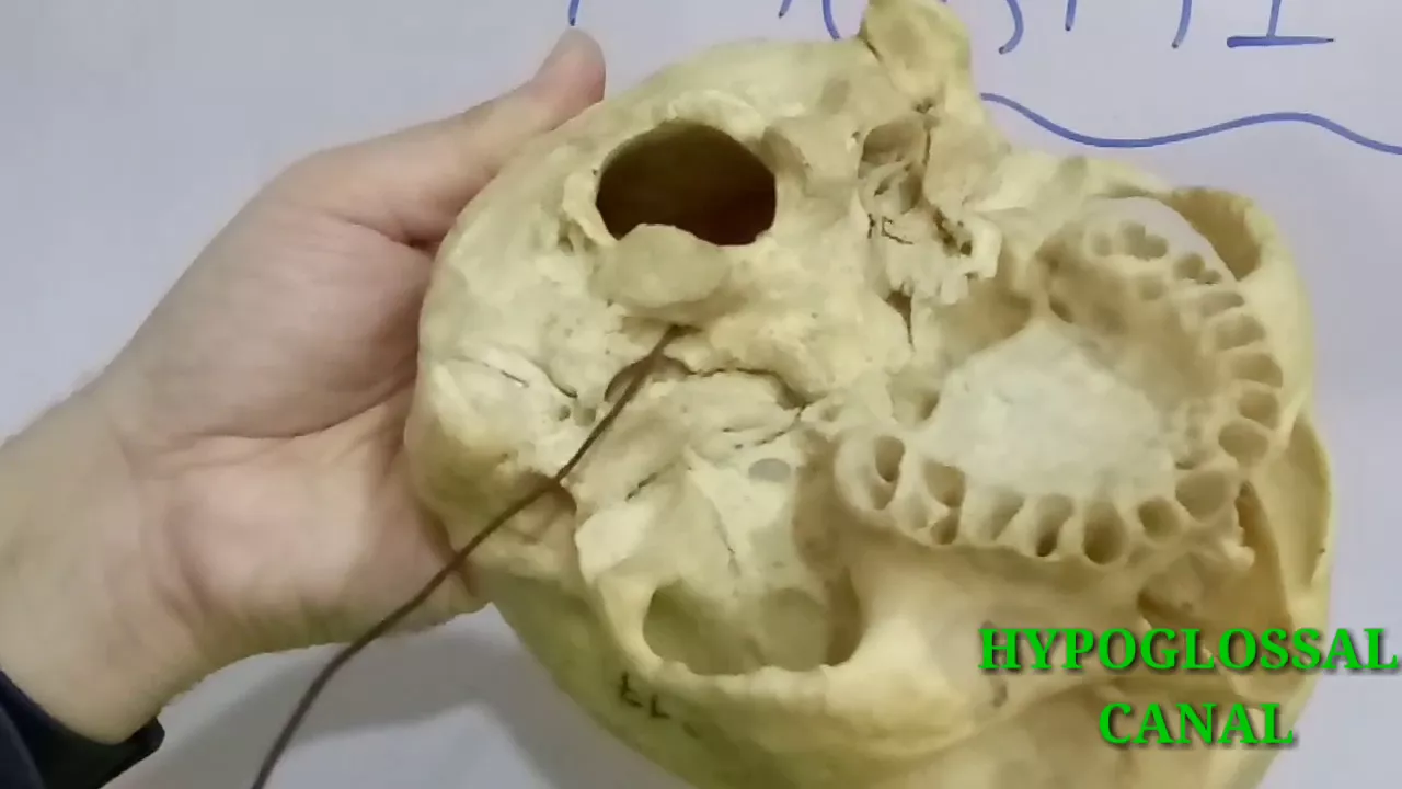
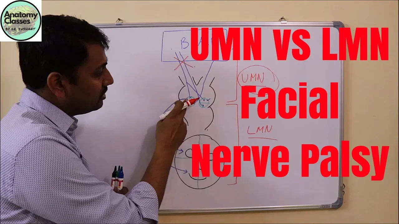
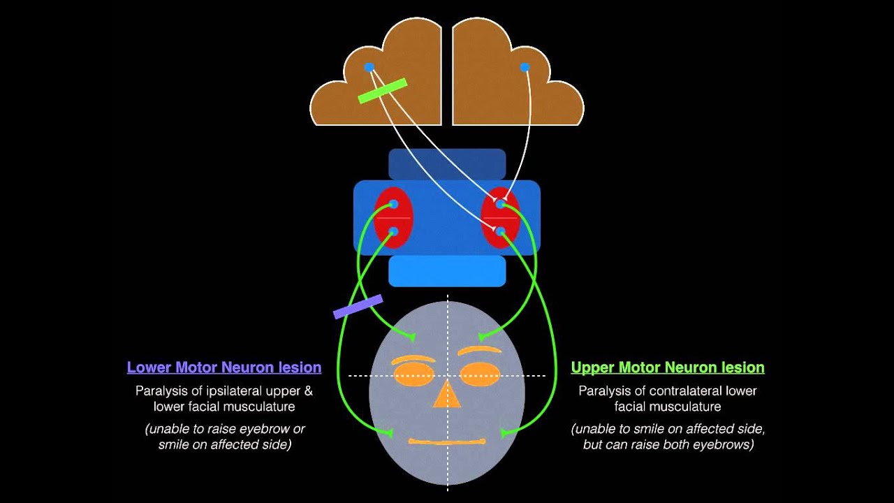


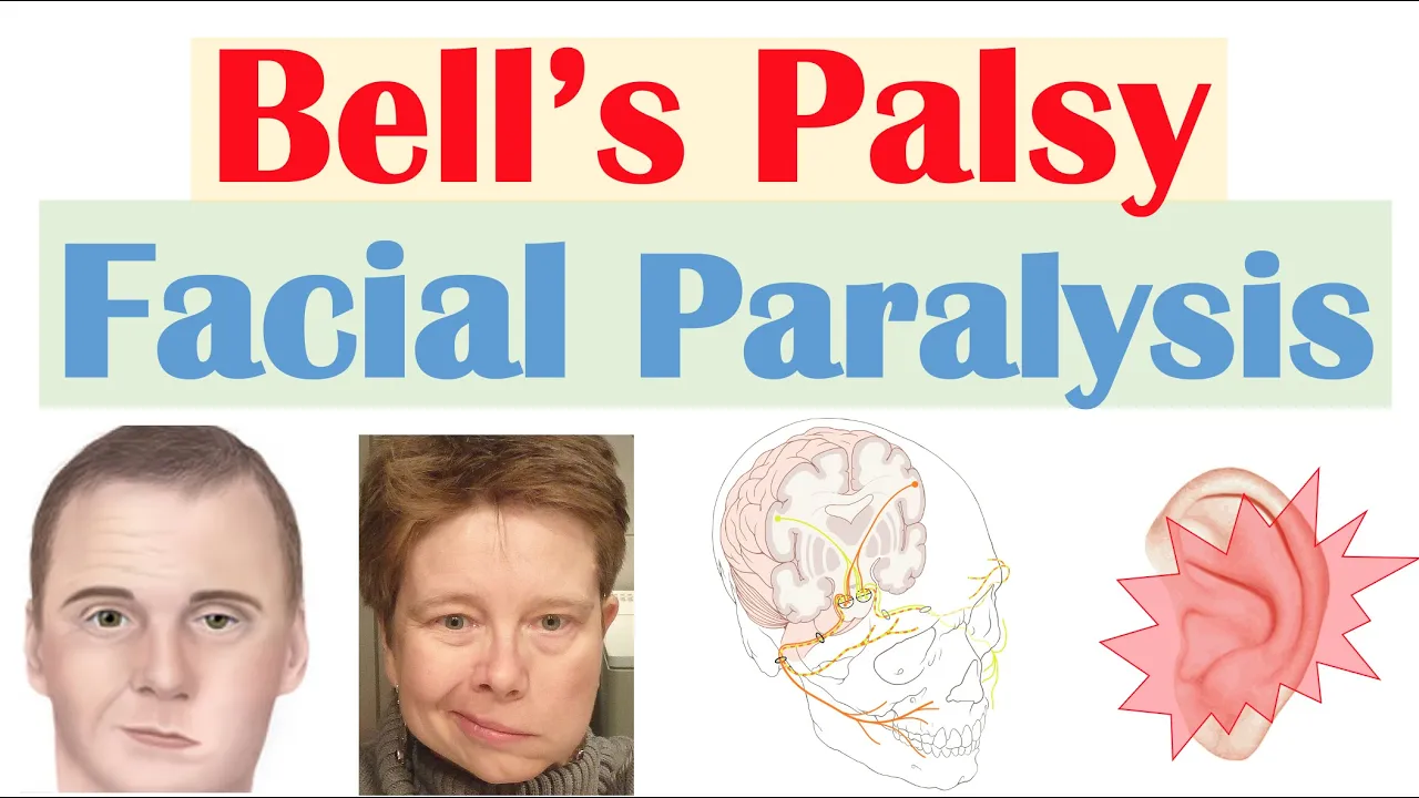
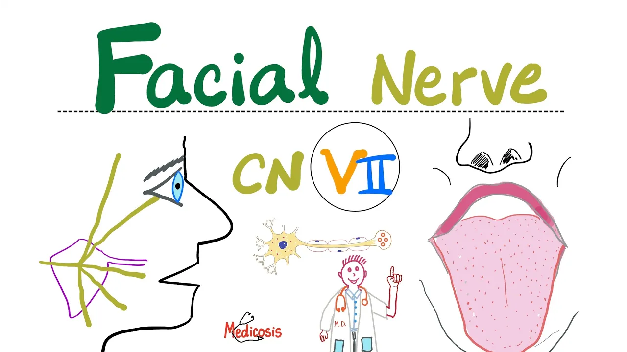
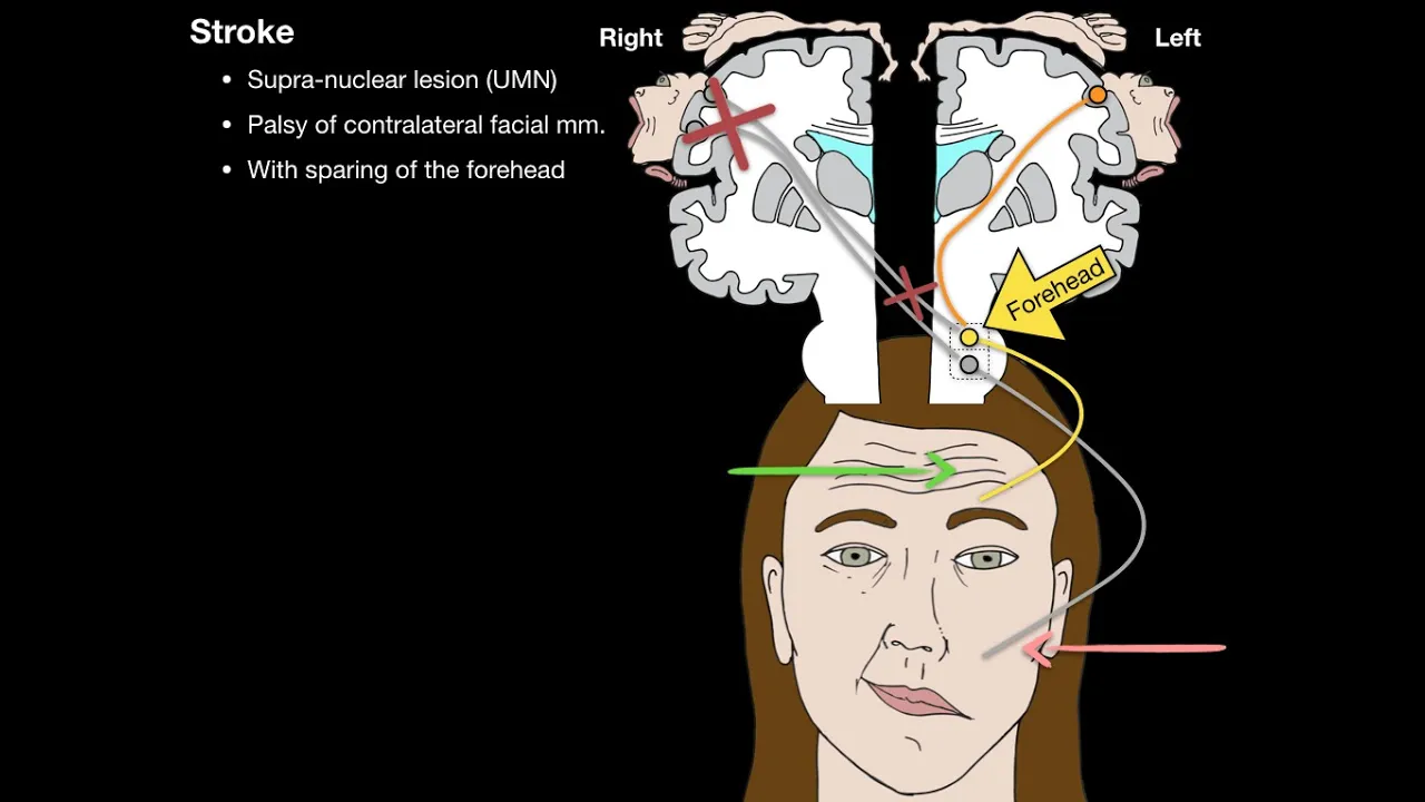






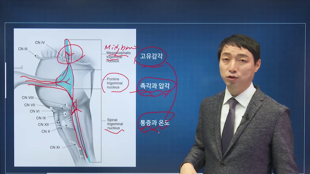
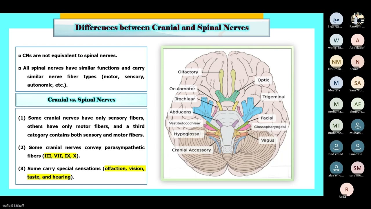
The facial nerve (CN VII) is a mixed cranial nerve containing four major fiber types: branchiomotor fibers (controlling facial expression muscles from the principal nucleus in the pons), parasympathetic fibers (controlling lacrimation and salivation from the superior salivatory nucleus), taste fibers (from the anterior two-thirds of the tongue terminating in the nucleus of tractus solitarius), and general somatic afferent fibers (for external ear sensation connected to the trigeminal system). The nerve exits the brainstem at the ponto-cerebellar junction, travels through the internal acoustic meatus to the geniculate ganglion, then proceeds through the facial canal in the middle ear medial wall before exiting at the stylomastoid foramen. From there, it gives branches to the stapedius muscle, posterior auricular region, and enters the parotid gland where it divides into five terminal branches: temporal, zygomatic, buccal, marginal mandibular, and cervical, supplying all facial expression muscles. Parasympathetic fibers reach their targets via the greater petrosal nerve and chorda tympani pathways, while taste fibers travel with parasympathetic fibers to the nucleus of tractus solitarius.
Facial Nerve Anatomy: Cranial Nerve VII Explained (Neurology)
Added:okay today we are going to talk about facial nerve core concepts right facial nerve core concepts right now you must be knowing that facial nerve is a master of facial expressions right it regulates control the muscle the facial expression everyone knows and it is so much master on facial expression and emotions that if you are very sad facial nerve helps you to bring tears and when you are angry it helps you to produce lot of saliva you can throw it on others yeah right so let's start how it really works right first we will see that what kind of fibers are present in facial nerve what type of fibers are present in facial nerve it means we have to go back in the central nervous system that in the central nervous system right facial nerve is taking what kind of fibers and it is connected to what type of different nuclei right so let's draw brainstem here is your suppose midbrain and here is palms and here is medulla right and your beloved spinal cord cerebellum now number one facial nerve have fibers motor fibers first i will talk about the motor fibers and then i will talk about sensory fibers fission nerve motor fibers which mainly control the muscle the facial expression right they are called brainchild motor fibers you must be knowing that muscle of fish muscle the facial expression are derived from the second brinkle arch right and those fibers of facial nerve which are connected with the muscles of the facial expression they are called brain cure motor fibers right now these fibers have their cell bodies in the lower part of the pons right in the lower part of the pons there are group of cell bodies here which make a nucleus we call it facial nerve proper nucleus right this group of cell bodies here in the lower part of pawns rostral pawns right and this group of cell bodies make a small nucleus and this nucleus is called principal nucleus of facial nerve or facial nerve proper nucleus or brain key or motor nucleus of the facial nerve right and usually motor fibers usually motor fibers from central nervous system they exit forward but there is something funny about these fibers you know what happen rather than they directly move forward and outward first they go backward anyone can tell me why they are going backward actually they heard something that there's a tent here on the back you know what is this tent can you tell me what is the stent fourth ventricle and i think these fibers heard somewhere that there's some sex going on here so they start moving backward but by the time they reach here they were very sad you know why they were very sad they came to know that was not sex that was six six of nucleus is here they misheard it so when they miss realize their mistake they just move around this what is this nucleus and go to their normal route have you seen men the nutty men when they are supposed to go forward straight sometimes they hear there's something good there and they go back but rather than sex they found the six and when they found the six they just turn back and go to the real destination is that right now these facial nerve fibers when they move backward and loop around the sixth of nucleus they produce a little elevation yes a little elevation in the floor of fourth ventricle this is called facial coliculus what is it called facial colliculus and then they turn forward and downward and they exit here what is this place where they they are exerting right it is ponto middlery junction but on the side there is cerebellum because cerebellum is on this side you are getting it so we also call it pontoseri barrel junction you are understanding so that is the point ponto cerebral junction where facial nerve is exerting outward let's see a frontal view right let me draw mid brain from the front and brain stem this is the brain stem from the front right bones medulla and what is on the side yes sorry bellum thank you for knowing and six nerve come out here right seventh nerve is exerting at this point seventh and seventh right and of course just lateral to the seventh which now is coming out eighth vegetable layer you're getting it at ponto medellin junction as you move from medial to lateral the nerves which are coming out there or they are attached at this junction that is sixth seventh and eighth abduction facial and vestibular cochlear is that right but especially because 7th and 8th are much laterally placed so we can also say that they are exiting at punto cereberal junction right so these facial nerve fiber which are going to be connected eventually with them mainly with the muscle the facial expression right they are coming out from here and they make the facial nerve proper this is one group of fiber then there is another type of motor fibers which come out of the brain stem and become part of the facial nerve right this everyone knows the facial nerve has fibers for the muscle the facial expression but facial nerve has more fibers right now second group of motor fibers which are coming for the facial nerve right those motor fibers are basically parasympathetic fibers and those parasympathetic fibers help you to lacrimate and salivate is that right now those parasympathetic fibers which are part of the facial nerve and eventually they supply the lacrimal gland and which salivary gland submandibular and sublingual salivary gland is that right they are connected to which nucleus in the brain stem anyone what my question is listen my question is facial nerve has parasympathetic fibers right facial nerve is parasympathetic fiber my question is in the central nervous system in the brain stem these fibers are coming from which parasympathetic nucleus yes anyone what happened you don't want to tell me anything okay you like to hide your knowledge as you are reading the book can you tell me still you couldn't find it okay let me tell you there's a nucleus here look this is also lower part of the pons you can see right this nucleus is parasympathetic nucleus right here it has oh this is vp nucleus and drooling nucleus this nucleus is called superior salivatory nucleus what is it called superior salivatory nucleus but actually it should be called lacrimetry celebratory nucleus right but i don't know why so in some books they write just lacrimetry nucleus and in some books right just superior salivatory nucleus but you must remember this nucleus has the neurons cell bodies of the neurons which are sending the fibers right which actually eventually are going to influence the lacrimation and salivation by which glands sublingual and submandibular right so this is superior salivatory nucleus and rostral part of pawns in the lower part of palms you can say and this is having cell bodies of preganglionic parasympathetic fibers preganglionic parasympathetic fibers and these fibers they come out of these what is this this parasympathetic fiber come out right and they also go with the facial nerve superior salivatory nucleus right remember inferior salivation nucleus is just below it and inferior salivation nucleus gives us fiber to which now ninth nerve glossopharyngeal right but superior salivatory nucleus gives fiber to seventh nerve and superior salivator is not only salivatory but it is also lacrimatory is that right now these are the two motor type of fibers which are component of facial nerve now we come to the sensory fibers with the facial nerve right what kind of sensory fibers are present in facial now yes my friend oh my god he really is a sexy boy he's saying that facial nerve has some sensation from the tip of the tongue what kind of sensation it's really not from the tip of the tongue it is interior two-third of the tongue right i know you at your age you are very bothered about the tip but i understand your preoccupation uh but facial nerve is little more practical right you know here is your tongue what is the color of your tongue blacker red okay some people do have black tongue i think anyway so this is the tongue interior to third and posterior one third is it right facial nerve have sensation collect the sensation from anterior two third of the tongue which sensations taste sensation don't try to be sour sweet just say taste sensation right facial nerve through lingual nerve which will go into detail that the taste sensation from interior two-third of the tongue is the taste sensation from interior to third of the tongue remember touch pain and temperature sensation from anterior two-third of the tongue are part of the trigeminal system through the lingual nerve right but interior two-thirds of the tongue taste sensations right they pass through the facial nerve right and posterior one third of the tongue taste sensation go by which nerve glossopharyngeal and this is facial and posterior most and from the you can say belly area around the epiglottis taste sensations go by tenth nerve is that right so my friend these sensations are not only at the tip fine so interior two-third of the tongue has taste sensations which are taken by facial nerve so we can say i will teach you later how these fibers really reach there right but just right now we say these are fibers for the taste okay what are these fibers taste fiber right gastric fiber sensory fibers right they are connected of course they are bringing the taste sensation from the periphery to the center right ah in the center of a system these fibers are connected where which nucleus in the central nervous system is related with the taste what happened someone put your mute button any nucleus in central nervous system where all the taste sensation come you heard of that nucleus right i'm going to make that nucleus here right it is dancing a lot because it can taste taste sensation come here what is this nucleus nucleus of practice solitarius nucleus of tractor solitarius has many connections some of the very important connections are for the test right and of course because seventh nerve facial nerve collect that taste sensation from interior to third of the tongue is that right so samantha should be connected with which nucleus nucleus of practice solitarius right so these are samantha fiber which will be connected with the yes oh thank you for knowing it right so this is connected with practice solutions let me tell you little thing about practice solutions practice solitaire says you know nucleus for the taste it means all the nerves which bring the taste fibers they should have connection with which nucleus tractor solutaries all the nerves which bring the taste fiber from the tongue right right all the nerves which bring the taste fiber from the tongue eventually should be connected to which nucleus it's all logical so gastrectomy fiber taste fiber of seventh gestative fiber from posterior one third of the tongue and the strategy fiber from the 10th nucleal s all of them centrally end up on on nucleus of tractor solid areas is that right so anyway i have shown here this is the connection of which nerve seventh nerve tetrasolitarias nucleus takes some other sensations also i will not right now talk about that so this is how many two motor fibers part of facial nerve and one sensory fiber there's one more sensation which are part of the facial nerve who will tell me one more sensation anyone facial nerve has touch pain and temperature fibers which are called general somatic offerings just to put a little academic situation uh touch pain and temperature fibers are also in facial nerve few fiber which are coming from the ear right posterior part of what is this external autoimmutators and external air right so those are which fiber touch pain and temperature what are these fibers touch pain and temperature they are also part of facial nerve and these stretch pain and temperature fibers when they go into central nervous system of course they cannot be connected to motor nuclei and they are not connected with the nucleus for the test they are connected with the major nuclear system in the brain stem which is connected with all those nerves which are concerned with touch pain and temperature on face which is a major nuclear system for touch pain and temperature trigeminal system have you heard of trigeminal system right trigeminal system so you can say here is mesencephalic nucleus of trigeminal and here is pontine nucleus or trigeminal have you heard of it and then there is another trigeminal nucleus what is this oh my god this is not a he's saying corticospinal it is not extending from cortex to spinal cord oh my god you are adding to my knowledge it is called only spinal nucleus of trigeminal right actually listen mesencephalic nucleus of trigeminal pontine nucleus of trigeminal and spinal nucleus or trigeminal all three nuclei connect collect the touch pain temperature from phase right because most of the touch pain temperature sensation go through the trigeminal nerve and trigeminal nerve is heavily connected with these nuclei so this nuclear system is called trigeminal nuclei but now we know that even some little touch pain temperature sensation or somatic sensation some of the somatic sensation may go through seventh nerve ninth nerve tension nerve but because the attach pain temperature sensation they have to go to the nuclear system which is related with touch pain and temperature and what is the nuclear system for touch pain and temperature trigeminal nuclear system so if i say 7th nerve has fibers for touch pain and temperature or somatic offerings from the external air when they enter into central nervous system they should be eventually connected to yes what is this business valid nucleus sorry spinal nucleus of triangle so these are the four main fiber then facial nerve right so first of all what we have learned up to now the facial nerve has importantly clinically there are four types of fibers which are present in facial nerve number one brain cure motor fiber which are going for muscle the facial expression mainly right then there are parasympathetic fibers which are mainly concerned with lacrimation and salivation then there are taste fibers with anterior two-thirds of the tongue and then there are touch pen temperature or somatic afferent which are with external air right but as you should know these fibers are connected and periphery where you should also know how they are connected in centrally right so brain tumor motor fibers in the periphery are connected with muscle the facial expression and centrally they are connected with principal nucleus of facial nerve brain to motor nucleus parasympathetic fiber peripherally they are connected with the lacrimal gland and also with the submandibular and sublingual gland eventually they are controlling their secretion centrally superior salivatory nucleus test fiber in the seventh nerve peripherally they are connected with the anterior two-third of the tongue for the taste centrally they are connected with practice solid areas that paint temperature somatic affront of the facial nerve peripherally they are innervating external layer centrally they are connected with the trigeminal nuclear system spinal component am i clear now once all these fiber exert these fibers make two bundles they are arranged into two bundle all these three fibers they are part of one bundle they are part of one bundle right they become component of they make one tight bundle right in this way facial nerve is divided into two parts one part has mainly brain tumor motor fibers and other part has parasympathetic fibers as well as test fiber as well as somatic offerings right this component of the facial nerve is called nervous intermedius what is this component called nervous intermedius now this nervous intermedius in most of the book is it is written it is the sensory component of facial nerve and this is called facial nerve proper which is also called motor component right this brachial motor fibers right they make a thick bundle and this is called facial nerve proper this is facial nerve proper right and this is nervous intermediate even though usually we say conventionally the nervous intermedius is sensory component of course it's having uh taste sensation and also that's been temperature sensation but we should not forget that even though we call it sensory but it also has parasympathetic motor fiber is that right clear now we know these are the fibers which are component of facial nerve now next work is that we have to see how these fibers reach to their final destination because right now these fibers when they exit out of the brainstem at pontosary barrel junction they are inside the cranial cavity right we have to see from this point on right how they reach to the final destination for that purpose i i will draw first some landmark of head and neck some landmark features of head and neck then we'll see how facial nerve and branches move about inside that system right so let's make it some landmark of head and neck okay so let's suppose it is interior cranial fossa right of course this must be orbit this is anterior cranial fossa here it is middle cranial fosa and here it is yes this is posterior cranial fossa is that right here it is what interior cranial fossa and of course there's no fun in telling that under the orbit under the orbit what is this maxilla if you forget you can touch on your own face and know it under the orbit there is maxilla right so i will just draw some important structure so we can see and under the maxilla of course if you open your mouth you will see your tongue i think someone else will see your tongue right here is your tongue ah yes so anterior canal fossa middle cranial fossa posterior cranial fossa here it is brainstem right and this is orbit of course there is there is no fun in telling there is something like this here you know and yes anyone tell me what is this area if you move on the orbit backward under the orbit and on the back of the maxilla upward what is this area called i'm about to be impressed by someone what is this area called you have heard it so many times you move on the orbit backward and maxilla backward and upward and deep inside right there is a fosa here all of you know that fossa you have heard of it but you don't want to tell me i know you are nasty guys there you go palatine fosa why don't you tell me have you heard of tirigo paladin my friend if you don't know where it is how oh very sad what is this at least now you tell me what is this there you go palatine fosa right anyway this says something very important related with the facial nerve so we will talk about that right then there is another very important landmark related with the facial nerve here and that is what is this this is middle ear i want to show you relationship of facial now with the middle ear now let me make the middle ear exactly more clearly i am going to draw the middle layer here and related with the external ear also let's suppose this is external uh you know middle ear is something like a cube this is middle ear cavity i'm drawing the structure on the right side and if you move on this side of course then what is that here and this is maybe of a lady this is external here from external layer if you move external auditory mediators is that right from external autoimmunities what is this wall yes very good tympanic membrane you get it this is the lateral wall of middle ear this is lateral wall and what is this wall interior wall what is this roof of middle earth this is the roof of middle ear this is interior wall of middle air directed forward this is the roof and this is lateral wall of middle ear am i right you are clear about this this is the right ear okay now it's very important to understand the very intimate relationship of facial nerve with the ear right because many clinical problems occur when facial nerves in intimate relationship with the ear passing through that area so let me show where the facial nerve is related actually if i remove the lateral wall let's suppose i remove the lateral wall what is left there this is medial wall this is the posterior wall this is the floor this is floor this is medial wall this is posterior wall and there were lateral wall here which i have removed okay this is interior wall now listen this is interior wall of what is this middle ear cavity this is what is this posterior this is floor and what is this medial wall middle ear medial waller yeah am i clear everyone is clear about this diagram from bringing this diagram here right to show you the relationship of facial nerve okay now again i will repeat this is the middle ear here was tympanic cavity i have removed the temperature what i have done i have removed the tympanic cavity i have removed the roof now you can look at the middle ear cavity this is the what is this this is the interior voila middle ear i don't need to make something here to show this is the posterior end of posterior wall right and this is the floor and that is which wall what is this wall on the medial side it's clear to everyone now facial nerve has very very special relationship with the medial wall and the posterior wall how let me tell you actually facial now when it is entering into this area right from here it enters into yes it enter into internal auditory mediators right what is this external order determinators right in the same way there is internal auditory mediators which connect the posterior kinema fossa and bring it connection up to where medial wall of the middle ear so what is this internal acoustic meatus facial nerve will enter into that right in fashion i will enter into that now i will make this area more clear that this is the medial wall posterior yes wall floor and what is this this is interior this is interior wall right okay now actually if this is the from here there is a hole from where the facial knob will enter in the petrous part of temporal bone right and this is having this is ending up at this point now this canal this canal which is within the bone of the skull this is called internal auditory meatus or internal auditory canal facial now enter into this and if you keep on moving inside eventually it will reach where on which wall of the medial wall of middle ear medial wall in upper corner or lower corner upper corner interior corner posterior corner so now you know that actually facial nerve through the auditory canal reaches to intro superior corner of what medial wall of middle ear any question here now up to this all fiber the facial nerve come together all of these fibers come together here is that right they are very loyal you know at least for a while now but there is a special friend with them because this canal is used by two nerve number one facial nerve of course facial nerve has two component facial no proper and nervous intermedius facial nerve goes through internal auditory meters what is the other nerve which also goes with the facial nerve vestibulo cochlear nerve eighth now right so basically seventh nerve is entering here and with that in this canal what other nerve is entering vestibular cochlear why i'm telling you this that if there is leon in this area in internal auditory meatus it will damage the seventh nerve as well as damada its nerve vestibular cochlear component anyway now i will show you fibers one by one right now how what is the plan of the lecture we will understand one by one first of all i will talk about how fibers what are these fibers visceral motor without motor fiber bring sorry brain tube motor fiber reach their destination then i will talk about how parachute fiber reach their destination then i will talk about how the taste fiber are connected with the periphery and in the last i will discuss how the general somatic afferent are connected with the periphery first of all we talk about these fibers facial now proper fiber they enter into where they have entered now you will tell me what is this internal auditory meat is very good and the fiber rich there right now here there is a ganglion there is a ganglion right and from this ganglion facial now take a very sharp turn backward right this fiber reach up to this ganglion but remember these fibers don't don't relay in the ganglion these are branching motor fibers they just pass by the ganglion or within the ganglion but they don't stop there these fibers pass through the ganglion and then they move what is this now they are moving forward or backward backward very good they are moving backward now these fibers are going backward but where they are going through a bunny canal there's a long canal here and this canal is boni canal right so you can say facial now proper fiber which are coming from this proper facial nucleus up to now they have two bend two important band one band is they move backward and then around the nucleus suddenly move forward right then they enter into internal acoustic meat as internal artery canal they reach up to that geniculate ganglion i will tell you later what fiber the what cell bodies are present in genuclear ganglion but from here facial nerve fiber right have another beautiful bend right and this bend now they move backward you know like beautiful woman session of proper or facial now has two bands one band is here and other band is there right now because bend is also called genu so that is why this uh what is this ganglion is called genuclear ganglia it's a genus you know in turn anyway the fashion of proper fibers they are going backward into this bony canal and this bonil bony canal is running horizontally within the medial wall of middle ear i mean this was the medial wall of middle air right and this is the canal here this is a boni canal you are getting it this this is a long tube in the bone and this goes first backward and then from here it turn yes where this can all turn downward now my friend look at it it's downward nothing can go back forever somewhere it has to go down also now what i'm saying listen it came from internal auditory meatus internal auditory canal then facial canal right facial canal which facial canal is the canal in the bony system of the medial wall right now these fibers are going backward but when it reaches to the posterior wall of middle ear from here this can alter where downward this can all turns yes downward right and it moves down down down down down and now you have seen there's a canal in the bone right in the medial upper medial upper part of medial wall of the middle ear and then it is moving downward through which wall posterior wall and then this bony canal open at a foramen this foramen is on one side there is mastered process you know mastered process this is mastered process right other side there is styloid process right so this foramen where the all open between the spoiler process and mastered process this is called which foramen stellar mastered foramen what is it called stellar mastered foramen right so it will reach here this is the master process and deep to that there is styloid process and it will come over here now these fibers okay i will make a little window here so you can see how these fibers are moving so these fibers freshen up proper fiber branchial motor fiber are efferent fibers for muscle the facial expression from the geniculate ganglion they enter into facial canal they move backward right within the middle ear medial wall and then they turn downward again continuing within the bony canal now this bony canal is a part of yes posterior wall from there they go downward downward and downward downward and eventually these fibers exit facial nerve proper fibers exit at what is this point stellar mastered for a man am i clear now on the way these fibers bring your motor fibers right they give a branch which is very important branch here they give a small branch here which comes out here small branch right and which will control a special bone here maybe you know if your ear anatomy is good that there is a bone here this bone is called stapes what is this bone called stapes is that right right and this bone is controlled by a muscle and that muscle is called stapedius what is this muscle called very good stapedius muscle some one of you know about it that's great so now where is exactly the stapedius muscle you remember what is this wall please what is this wall anterior posterior say it loudly my friend everyone has a big tongue so what is this posterior in the posterior wall there is a muscle which is coming forward like this and what is the name of this muscle stapedius and stapedious muscle come out of posterior wall and catch a bone what is that bone stapes and the stapedius muscle control the stapedius stapedious muscle control the step is born and does not allow it to excessively vibrate when we hear the sound for example if you talk to me very loudly tympanic membrane will vibrate and if there is excessive vibration the panic membrane will vibrate melee as incarcerates which will produce vibration in inner ear but if you talk very loudly there's a chance stapedius stapes move too much to control the excessive movement of trapeze there's a muscle called what is this stapedius and this muscle is given a branch from facial nerve proper so this is what is this muscle stapedius what is this bone strap is and what is this branch coming this is the first branch of brinjal motor what is this branch this branch is for step is clear and this branch if it is damaged then step is move less for more more and if this branch is not functioning stapes stapedious muscle become paralyzed paralyzed or weak and step is with when someone talk when there is a loud sound step is moved too much and that becomes annoying right so anyway so this is one branch after that this has gone down and come out at which point shallow mastered foramen when it come at out at the style of mustard for men from this foramen it gives different branches first of all it gives a branch which goes backward right and this goes backward and upward and this is on the this branch comes from here and goes up backward and upward so what is it called posterior auricular branch because it is on the going on the back of the ear what is this branch called posterior auricular and this posterior articular branch right it will give some muscle control here but these are not very strong muscles you know some articularis muscles or frontal occipital occipital part right but it's not very important right plus later on i will tell you with this motor branch some sensory fiber also come that i will discuss later but anyway the first branch which came out from here was stapedius branch for strapedious second is posterior auricular and then another branch which come out and give to two muscles muscle stylohide and posterior belly of digestive right after that this facial now proper jump into jump into periodic gland it moves forward there is a protein gland here is that right these days it is still here right so fission nerve what it has done when it was going down it gave a branch to the stapedius then it went further down it came out of stellar mustard foramen it gave posterior articular branch right and then it gave motor branches to stellar height muscle and posterior valley of digastry and then it jump into the rotted gland and when they jump into protein gland i will make a this is a protein gland here and facial nerve jump into protein gland within the substance of protein gland this these fibers a facial enough proper they divide into five branches five terminal branches right and these five terminal branches they will come out of this let me make a little face here okay now these fibers are going to the muscle the facial expression right so some fiber will go upward look fiber come from here and goes upward what is this area temple so what is this branch called temporal then some move forward and little upward what is this bone zygomatic what is this branch zygomatic right and some fibers move like this and they are going to supply the muscles around the mouth what is this area buckle so what is this called buckle and then and some fibers come down and they are moving with the mandible what is this branch called marginal mandibular because this is the margin of mandible and some really go down and supply some muscles in the neck like platysma so what is that called cervical is that right so how many branches look here from protein gland terminal branches of facial nerve are coming out what is this branch going say loudly temporal what is this zygomatic what is this buckle don't say mouth branch buckle and what is this marginal mandibular and what is this cervical so there's no need to remember this you just remember what is this temple zygomatic buccal mandible you can never say mandible is here i hope right mandibular and of course cervical is here right next so just we put it here what is this branch going upward templar and what is this zygomatic what is this buckle not vocal buckle and what is this marginal mandibular and what is this cervical these are the terminal branches of facial nerve which are going to supply the muscle the facial expression am i clear any question up to this so now you know that what is this facial now proper brain tumor motor fiber from where they originate and how they eventually pass through the skull and where they terminate any question up to this any question no now we come to parasympathetic fibers they are really very naughty fiber you know they are going to control for the lacrimation and salivation now the question is that if these parasympathetic fibers are going to control for lacrimation they have to go to lacrimal gland lacrimal gland is here and if these parasympathetic fibers are also supposed to control salivation by submandibular gland and sublingual gland they have to come here now we will see how these parasympathetic fibers eventually pass through different areas of head and neck and eventually end up in controlling lacrimation and salivation right now let's suppose i talk about that two fibers are starting from here you can say mainly lacrimetry pathway fibers and what is the other salivatory right so here i am showing it as one group so what they have entered into which area now please can you tell me what is this internal auditory meatus from here these fiber are going through that and reach up to which ganglion geniculate ganglion and these parasympathetic fibers what they do in the geniculate ganglia do the synapse there or not these parasympathetic fibers do they synapse in geniculate ganglion or not who said yes you should know that he's wrong yeah the reason being at least you should know as good student that head and neck has basically only four parasympathetic ganglion ciliary ganglion silvery ganglion perigo palatine ganglion artic ganglion and some mandibular ganglion head and neck has four parasympathetic uh ganglion cellular ganglion herigo palatine ganglion submandibular ganglion or ganglion and geniculate is none of these channiculate ganglion is now it is not a parasympathetic anglion later i will tell you which fibers are there really which cell bodies are there but not for parasympathetic fibers don't synapse here they don't do any activity here like these fiber they just float here and pass by right these parasitic fibers but there's one thing there were two types of parasympathetic fiber coming up to this point what were those two fibers lacrimatry and salivatory after reaching up to the level of geniculate ganglion they separate lacroix battery fibers go to different route and what is this salivatory fibers go to different route these salivatory fibers go along with the facial nerve proper where they go i will talk later right what are these fibers coming luckily material or salivatory salivatory right i will talk later how they reach to the submandibular ganglion but first we talk about these fibers what are these which are going to the lacrimal gland am i right these are parasympathetic preganglionic fibers which are centrally connected with superior salivatory nucleus and along with the facial nerve as a part of nervous intermedius they have reached up to the geniculate ganglion from here these parasympathetic fibers separate and these fibers go to the what is this what is this middle cranial fosa these fibers go to the middle kind of fossa and when they are moving there they find there is some hole here they find a foramen here and i don't know like many people they love to jump into some hole so these fibers will jump into this hole right these are parasympathetic fibers from geniculate ganglion they are eventually going to be lacrimal gland connection so they jump from geniculate ganglion and they go to the middle cranial fossa from there they jump into which foramen for an oval no my friend foramen is foreign don't try to give me a little twist this is like a lesser rated foramen foramen lesseram okay i know you don't know these lacerated things anyway so this nerve jump into foramen lacing in the front of the foramen laserum right it finds the hole the front of the wall of the foramen laserum it find a hole right and these fiber love to enter into another hole right but there is one thing one front friend will join them you must be knowing there is something called internal carotid artery and around the internal carotid artery there is a plexus of sympathetic fibers you know that some of the sympathetic fibers from here also come and join these parasympathetic fibers are you understanding what has happened that parasympathetic fibers are coming from geniculate ganglion right from there they jump into middle class fossa they jump into formal electron and enter into this special canal but before entering they are joined by these parasympathetic fibers are joined by sympathetic fiber which are coming from plexus around internal carotid is that right then these two fibers together move through this canal they move through this canal and come into which foramen which area my friend i told you in the beginning of the lecture there you go palatine fosa and in this tiricopalatin fosa there is a ganglion here right and this ganglion is which ganglion yeah what is this ganglion there you go paladin ganglion so these fibers this parathyroid fibers end up into this ganglion and from here post kinglionic fiber will start and sympathetic fibers sympathetic fiber just pass through this ganglion right now let me tell you what are these nerves now please be attentive for a while what are these important connection parasympathetic fibers lacrimetry fiber from superior salivatory nucleus or electromatic nucleus from lower part of the pons as a part of nervous intermedius which is a part of facial nerve they enter into internal auditory motors and eventually reach up to geniculate ganglion their lacrimetry fibers depart separate from the celebratory fiber lacrimatic fiber move from geniculate ganglion making a very special nerve which bothers the medical stones a lot because all of you have heard of this nerve but i know you will not tell me the name of this nerve from this nerve they will jump and reach up to here but someone may tell me what is this nerve called this branch of facial nerve from geniculate ganglion having parasympathetic fiber mainly fuse sensory fiber and going to middle kingdom fossa jumping into formal acid what is this nerve you have heard of it it is called greater petrosal nerve have you heard of greater petrol oh my god you have not heard so many things so this is greater petroleum is that right and from sympathetic plexus around the internal carotid artery sympathetic fiber jump and they also become fused with the greater part of the nerve now these sympathetic fibers these sympathetic fibers which are jumping from the internal crotched artery towards the greater petroleum nerve this branch what is this called yes yes you are right deep petrosal now what is that deep petroville nerve so greater of the nerve and deeper through the nerve they come together and they move through this canal this canal which is moving from the interior wall of from a lesser up to which fosa digital this boni canal this boni canal this boni canal is called tiri guide canal what is it called teri guide canal and this nerve which is now made by the greater petroleum nerve and deep petroleum nerve and both of them together move and reach connected to the ganglion this combined nerve is called nerve to trigger canal what is it called nerve to terry guide canal the another name for this nerve from here up to here this another name for this now is vidian nerve what is this median nerve median nerve or nerve to trigger kerala so in this way sympathetic fibers from the facial nerve right they have reached therigo paladin ganglion now you know that your friend what is this here lacrimal gland you know lacrimal gland is here so preganglionic fiber end here from here this poor skin any fibers have to somehow reach to what is this lacrimal gland and how they reach there right before i really move forward i want to tell you now this was interior cranial fossa what is this middle cranial fossa right from middle cranial fossa let's come in another area there's a very big ganglion here what is this ganglion called trigeminal ganglia heard of trigeminal ganglia which has uh one of thalamic dominion other maxillary tibian and another mandibular deviant you must be knowing of thalamic divine fibers enter through superior orbital fissure and they divide into nasal cellular branch and they also divide into frontal branch they also divide into okay what is this nerve ophthalmic division of thalamic dominant divide into three branches one is neither celery there's no celery other is frontal and another is lacrimal and this lacrimal branch comes here right to the lacrimal gland okay i remove this you know that it's there now if these parasympathetic fiber have to reach to the electrical gland somehow they should reach to lacrimal communicate to the lacrimal love how they do actually here is another foramen it is very much round for a man what we call it foramen rotundum and from here what is coming down yes and forward what is this this was thalamic dominion what is this maxillary dominion what is this mandibular right now this maxillary nerve it is passing from here and you know now actually these parasympathetic fibers from tyrio pellet and ganglion jump to maxillary nerve right maxillino give here through the inferior orbital fissure missile enough give a branch which is called zygomatic branch right of maxillina these naughty fibers they jump from the ganglion to the missile nerve and then move with which branch zygomatic branch is that right this is zygomatic branch of maxillary nerve from here they make a communication to what lacrimal love and with that the reach to wear lacrimal gland so this is likely military pathway of parasympathetic fibers let me recap right start from the beginning when a girl is weeping right what is the connection started i'm talking about central nervous system yes or a baby is weeping right which nucleus is firing superior salivating nucleus which is also called lacrimatic nucleus will rapidly now you will tell me what are the fibers superior salivatory nucleus these are sympathetic parasympathetic fibers parasympathetic these are preganglionic postganglionic preganglionics because ganglion is there these three ganglionic fibers are part of facial proper or nervous intermedius nervous intermediate they are passing through which can all know internal alternators the risk to which ganglion geniculate ganglion they don't synapse their their lacrimatic fibers separate from salivatory fibers these lacrimetric fiber from the geniculate ganglion this fiber exit and move towards the middle canal fossa as greater greater yes say it loudly i don't hear which now greater part of the love greater patrol now jump into foreign and that reaches to the rigor paladin ganglion from there freezingly any fiber end post can any fibers from here mainly they jump to the mixing of the zygomatic branch and then communication of zygomatic branch to lacrimal branch and with that this fiber reach to lacrimal gland this is the minimum you should know about this pathway lacrimatry but of course all these parasympathetic fibers which are coming through the up to this point of course many of them are going to lacrimal gland but some of them go to nose also so nasal gland some of them come down and palatine glands some of them fibers from here go to paranasal sinuses like feminine sinus or frontal sinus or maxillary sinus right so a good doctor will remember that from trigopalatine ganglion postganglionic parasitic fiber go for lacrimation but excellent doctor will remember these parasympathetic fiber not only go to the not only go to the lacrimal gland but from here through other branches of the palatine ganglion they go there they go to the nasal glands they go to paranasal sinuses glands they also go to the palatine and palatine glands am i clear any question no thank god so these parasympathetic fibers are done with lacrimatory pathway next let's deal with the what are these fibers salivatory pathway right now if we talk about the salivatory pathway these parasympathetic fibers are coming with the facial nerve proper they're moving through the facial canal backward then these fibers go downward they are moving with this but when you look here they don't come out from here where they have gone listen carefully these parasympathetic fiber which are going for salivation and stimulate what sublingual and some mandibular gland from the geniculate ganglion these celebrity fibers you you get it they move with the facial nerve proper into facial canal first they move in the horizontal component of facial canal they move backward and then the posterior wall of the middle ear in the facial can also move downward but if you look at stellar mastered foramen they never exist from here where they have gone yes ma'am where they have gone the magic has been done there anyone where these fibers have gone parasympathetic they don't come they do enter into facial canal but they don't come out at the silo mastered forum and somewhere on the way they disappear they jump where they jump they jump into middle air right the problem is that they were very happily going with that but suddenly they realize if they keep on going they will end up on the face and grades are not on the face glad they're inside so as soon as they realize they are in the with the wrong partner and going for the wrong destination they have to jump off the track and take a new road now where they have to go they have to go sublingual gland what is this gland here sublingual gland and what is this gland here submandibular gland now these parasympathetic fibers need to go to sublingual gland and some mandibular gland now they have to somehow jump from the track and reach to some nerve which is going near to the tongue are you understanding the nerve which go to the tongue one of the very important nerve is lingual nerve is it difficult to remember that lingual nerve goes to the tongue it's easy it means these fiber parasympathetic fiber somehow should jump from the seventh nerve and go to which now say loudly my friends they have to jump to which now lingual now now we see fruit where is the lingual nerve a little bit whereabouts of lingual now you must be knowing there is something called foramen ovale here one of you was trying to push some other nerves there you know from an oval here through foramen where this is the mandibular divian which comes you you understand it mandibular divine one of the branch of mandibular v and divided into rdpn and posterior deviant i will not go into detail one of the main branch of manipulation is called lingual nerve and this lingual nerve goes to the tongue and give touch pain and temperature from the interior to third of the tongue lingual nerve collect which sensation from interior to third of the tongue touch if someone touch your where is the tip boy yes if someone touch the tongue into your two third of the tongue right so which nerve is going to take fibers of the touch yes lingual nerve it takes touch and if someone bites there again it will take pain touch pain and if put a hot potato in your mouth again this so lingual nerve take the fiber of touch pain and temperature but if someone has in his mouth hello and mouth chocolate also so then interior two-thirds of the tongue has to taste the chocolate right taste is not done by this component of lingual nerve because this is coming from trigeminal system trigeminal system is concerned with somatic sensation which are touch pain and temperature now you remember that this celebratory okay so this these fibers were passing through internal artery meters january ganglion facial canal suddenly when they were going down in the facial canal they realized oh my god i'm going to go out of the cranial on the surface of face so they decide to jump off from here from here they should jump off i think here their brain worked here the fibers suddenly realize that they are going for wrong destination is to follow the facial nerve proper so now where they have to jump yes my friends to lengual now now how they have to jump they are on the posterior wall of they are inside a bunny canal which is present in the posterior wall of middle ear so first they have to make a hole and jump out of this canal so actually this canal has a hole here this canal has a hole here this is called posterior canaliculus right from this canaliculus these parasympathetic fibers move and jump into here jump into which cavity yes my friends middle layer from here now they have to move backward or forward for lingual now forward they jump through that like this right while they're jumping like this then from here there is a special fissure we call this fissure fisher what we call it there you go tympanic fisher from there they jump out and as they jump out they see their friends going what is this now going live one now this is an infra temporal fossa so in the infrared and porofossa these parasympathetic fibers will jump to what is this lingual nerve they will go with the lingual nerve they will go with the lingual nerve but again they don't want to go to the tongue they just find here what is this ganglion sub mandibular ganglion they separate from the lingual nerve and preaching any fibers end up into which ganglion sub mandibular ganglion from here post kingdom fibers go to what is this sublingual gland and also go to submandibular gland so salivation am i clear so we have dealt with the the final destination of this visceral mode or sorry branch motor fibers we have also dealt with the fibers in the facial nerve coming from superior salivating nucleus which are lacrimetry fiber then celebratory fiber now we are connected with what what is this dancing nucleus nucleus of practice solitarius is that right now nucleus of tractor solitary as i told you special now bring the taste fiber from interior to third of the tongue and they are connected with nucleus rectus solutaries so what really happened now touch pain and temperature from interior to third of the tongue is taken by lingual nerve but this is the taste fiber from anterior two-third of the tongue right they are also going with the lingual nerve these a taste fiber and these are that pain temperature hot potato here but chocolate here is that right taste fibers go from here they are going as a part of what what lingual nerve here it was lingual nerve now these are which fibers just please taste taste fibers are part of the lingual nerve right now and they go they leave the lingual nerve because they don't want to end up into trigeminal system they have to end up into tractor solutions nucleus so they actually they move with the parasympathetic fibers from here they jump into what is this cavity middle ear cavity right from here they enter where posterior canal and from here they go upward taste fibers what are these fibers taste fibers and yes they are connected to the practical solute areas right now so it means their salivatory pathway fibers parasympathetic fiber for salivatory pathway and what is this taste fibers they have little common thing that taste fibers actually their sensory fiber they should be explained from periphery to the center but i will do it from center to periphery just to make it easy to your mind so fibers are which fiber connected gastric taste sensory fiber these fibers move with the parasympathetic fiber into which fashion of proper nervous intermediaries nervous intermedius taste fibers go through again with the facial nerve tract into internal automata taste fibers are passing through geniculate ganglion now this is the point to remember actually generate ganglion has the cell bodies of this test fiber geniculate ganglion has cell bodies of these taste fibers and from these cell bodies peripheral process go to the tongue and central process go to the nucleus cactus solid areas in the geniculate ganglion there are cell bodies of taste fiber and later on i will tell you some cell bodies are touch point temperature fiber from here touch pain and temperature fibers from here right now so geniculate ganglion is not a parasympathetic ganglion geniculate ganglion is a ganglion which has cell bodies of sensory neuron related with the taste taste from interior to third of the tongue and also cell bodies for some sensory fibers coming from the external air right now so from here the taste fibers they are going along with the facial proper and parasympathetic fibers but when parasympathetic fiber decide to jump out of facial canal taste fiber also jump out of the facial canal so parasympathetic and taste fiber both jump through what tympanic cavity right and they exit in front of the tympanic cavity and then they become part of which now lingual nerve the parasympathetic fiber on the way leave to the submandibular ganglion but test fiber continue with the lingual nerve until the they are able to inner weight and tear two third of the tongue for the fungi form papillae or taste you can say this sensation system clear now thank god most of the things are done we are left with one fiber what are these that's been temperature from the external ear is that right these fibers also have their cell bodies in where genuine ganglion these their central processes go in the center and get connected with what is this spinal nucleus or trigeminal system and the peripheral process move with what facial nerve proper into facial canal backward and then facial can all downward but these stretch pain temperature fiber keep on going down that's pain and temperature fibers keep on going down down down and they come out of what is this funny thing stellar muscled foramen and they're going to posterior articular branch and posterior articular branch gave sensation touch pain and temperature or somatic general somatic sensation to the posterior part of external ear right and thank god that's complete the basic orientation or clear orientation or core concept related with the facial nerve i have not talked about the clinical points that we'll discuss in the next lecture right but before really a close i will test you right now you will tell me what what is there right say it loudly first of all we are going to start the fiber from here and connect to the periphery what are this fiber in facial nerve break your motor right seventh nerve fission now proper then they move forward they are moving through what's this part moving through facial nerve proper through internal artery test and then through the geniculate ganglion they move backward into what facial canal in the medial wall then they move downward in facial canal and posterior wall then first branch is given by what is this this bring your motor fiber to which muscle stapedious and then these fiber come out at stellar mastered for a man and they give posterior branches posterior a regular branch goes to muscle with it some sensory fiber also supply that what is this external air sensation touch pain and temperature then these fibers give a little branches to the posterior valley of digestive and steroid and then main facial proper enter into which gland protein gland and from the protein glands they divide into which branches temporal then zygomatic buccal then marginal medullary and cervical for muscle the facial expression and platysma claro clear now after this we come to the parasympathetic fibers parasympathetic fiber are two types of fiber mainly lacrimetry and salivatory they start from which nucleus say it loudly which nuclear superior celebratory nucleus they move as a part of which the facial proper and nervous intermedius nervous intermediates up to the geniculation their lacrimetry pathway and celebratory pathway goes through a nerve which is called greater petroleum love which meat into ferment lesser with deep petroleum together these fibers go as enough to parasympathetic fiber end then from here lacrimatic fiber jump to maxillary zygomatic branch of masonry and from there they jump to the lacrimal branch of ophthalmic and reach to electrical gland and some other branches go to nasal gland and paralysal gland and palatine glands done then salivatory fibers salivatory fibers separated from electromagnetic pathway at genuine ganglion they moved with the facial proper interfacial canal they are moving backward then downward but from there through posterior canaliculus they jump into middle layer and they are having which other fiber with them taste fiber now this nerve the jumping nerve from this posterior part of facial canal this jumping nerve is passing through the tympanic cavity when you throw light in the on the tympanic membrane when you throw a strong light on tympanic membrane you will look at the shade of this nerve jumping across the cavity you know what is the name of this nerve this part called our timpani have you heard of corda timpani have you heard of carter tympani in past this is chordae temporary what is corda tympani this is a chord in the tympanic membrane tympanic cavity what is this chord it's a neurons a nerve is jumping across a temporary cavity this nerve has rich fibers it has parasympathetic preganglionic fibers and also the test fiber from anterior two-third of the tongue and parasitic fiber going through the submandibular ganglion right so what is this nerve yes chorda tympani so we can say basically chord or temporary fuse with the lingual nerve right so what i was saying that submerged a parasympathetic fibers from here they jump as called a temporary along with the taste fibers and they exit at the irico tympanic fissure and they in the infra temporal fossa they meet with lingual nerve as a part of neural lingual nerve they move a little and eventually depart and bringing any fibers end into which ganglion submandibular ganglion and from the submandibular ganglion was posting alien fibers go to uh some metabolic land and sublingual plant done third taste fiber taste fiber truly that we should explain them from the periphery that is fiber from anterior two thirds of the tongue they move centrally yes as a part of lingual nerve but from the lingual nerve so matt touch pain and temperature fiber go to the mandibular divine and taste fiber become part of carda tympani and through the quarter template they jump through the middle ear cavity and they reach to the facial nerve in the facial canal here the tails fibers move upward right then they move forward into facial what is this canal and at january ganglion they have their cell body from this cell body the central processes move with nervous intermedius through the internal acoustic meters and eventually go to the brainstem and get connected to nucleus of tractor solution done then touch pain and temperature somatic often from ear they start from the ear they enter into posterior articular branch of facial nerve and from there they enter into facial canal they move upward right and then they move forward through facial canal into middle middle ear medial wall the reach to jennifer ganglion these fibers also have the cell body here and the central processes move as a part of which which now nervous intermediate is part of facial nerve and eventually they enter into pontocity barrel junction right they enter into brain stem and they get connected inside the brainstem with the spinal part of trigeminal nuclei any question up to this so this completes our core concept basic concept about the facial nerve in the next lecture we will talk about wealth policy and other facial nerve reliance classlessness
Up Next
How Food Affects Your Brain: Diet & Mental Health
@SciShowPsych
212K views•2021-07-01

Deciphering Neural Circuits Controlling Anorexia in Mice | Richard Palmiter
@AllenInstitute
2.3K views•2011-10-10
Hypothalamus Structure & Function | Neuroanatomy Lecture
@DoctorNajeeb
335K views•2017-09-27

How Exercise Benefits Your Brain: Science Explained
@TED
11.4M views•2018-03-21
Related Study Plans & Knowledge Roadmaps
Structured learning paths in Neuroscience