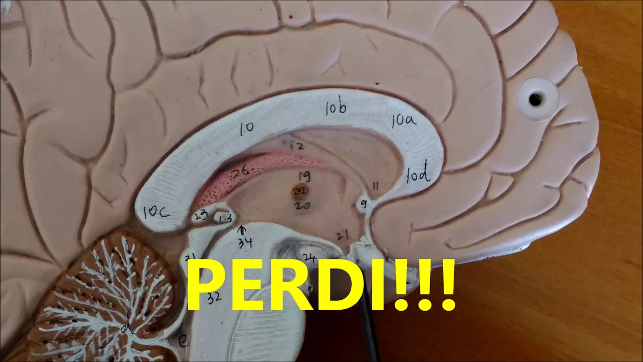


















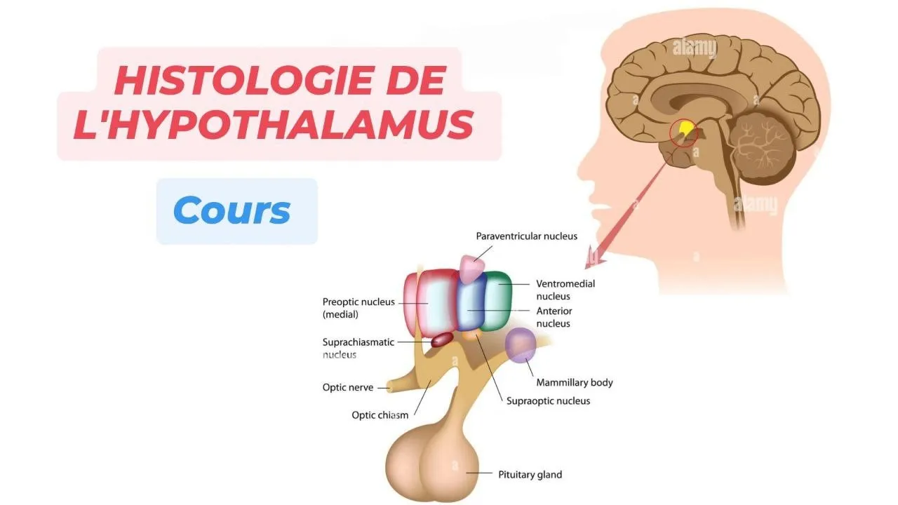

The hypothalamus is a small sheet of gray matter (approximately 4 grams) located in the diencephalon, extending from the optic chiasma to the upper midbrain and forming the lateral walls and floor of the third ventricle; it serves as a critical integration center linking the nervous system to the endocrine system via the pituitary gland, with three primary functions: participating in the limbic system for emotions and memory, controlling the autonomic nervous system, and regulating hormone release through releasing and inhibiting factors; structurally, it is divided into right and left halves, each further divided into medial and lateral regions by the fornix pathway, which connects to the mammillary bodies and thalamus as part of the Papez circuit for memory processing.
Hypothalamus Structure & Function | Neuroanatomy Lecture
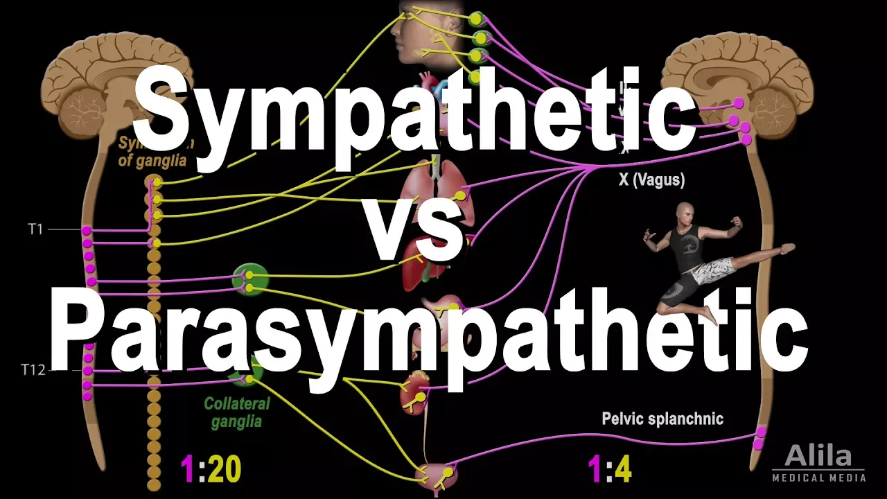
Added:today we are going to talk about hypothalamus right hi would all of you know their hypothalamus is a very important area of Diane Cephalon right and major functions of the hypothalamus basically it has three basic functions number one that it is concerned with the limbic system it's part of the limbic system you know limbic system is that part of the central nervous system which is concerned with your emotions behavior and recent memory secondly hypothalamus is intimately related with the function of autonomic nervous system and thirdly hypothalamus is also very important Center in the central nervous system which is having a very powerful influence on the endocrine system so it is also having endocrine functions right so it has functions related with the part of the limbic system so it is concerned with the emotions behavior and recent memory through the autonomic nervous system it influences the sympathetic and parasympathetic output in the body and through producing different releasing and release inhibiting factors for different hormones from the interior pituitary and also releasing hormone through posterior pituitary it hypothalamus observed a very powerful control on the endocrine system in the body right so before we really go into detail first we will see exactly where the hypothalamus is situated in the central nervous system is that right first of all now we will discuss where exactly in central of a system hypothalamus is present after that we will go into detail of hypothalamus now if I draw a central nervous system from the side right you know that here is your what is that midbrain pons medulla right spinal cord of course and now exactly where is hypothalamus let me tell you just above the midbrain there is a structure called thalamus a structure which is called Kalama s-- right now actually anthro inferior to the thalamus there's a sheet of gray matter there's a sheet of gray matter which is going anteriorly and inferiorly from the from the thalamus is that right and this structure this sheet of gray matter it is called hypothalamus and it extends from midbrain right upper part of the midbrain up to the optic chiasma it extend up to the optic chiasma right or we can say from the optic chiasma hypothalamus extend up to the yes what is this up to the midbrain or we can say up to the interpret and color sources and here the important structures related there is a swelling underserved on the undersurface of the hypothalamus and from here the stark and interior pituitary end post a officially this component is also the part of hypothalamus this is a right and there are bilateral swellings from posterior part and this well in is called the memory bodies and mammillary bodies are also part of hypothalamus but pituitary stock is not the part of hypothalamus so actually hypothalamus is this structure from here right so what is this hypothalamus is mainly a sheet of gray matter extending from the optic chiasma backward up to the upper part of the midbrain right and under surface of the hypothalamus has one median swelling one central swelling called to Bursa nerium and to swelling on the posterior part of the hypothalamus which can be visualized from inferior area right that is mammillary body this is the right this is one way to look at hypothalamus now let me draw the structure of hypothalamus with little more detail let me draw here your third ventricle you remember your third ventricle this is a roof of that third ventricle posterior wall of the third ventricle right here it is floor of third ventricle is that right and you must be knowing that there's one structure on this this side of that third ventricle what is it calamus very good there's another structure on this side of the third ventricle this is the right Columbus is there right so third ventricle is in between the thalamus but it extends forward beyond the thalamus is there right and you must be knowing what is the structure under here what is that it is midbrain right pons medulla right and already you know there's a which nucleus here have another nucleus have another nucleus and here the swelling backward what is that final body this is heavy NOLA Kamisha is that right now in this diagram now I will draw hypothalamus is that right hypothalamus expands from here it's a sheet of gray matter which is extending forward from the thalamus now from here what are these two mammillary bodies right and i actually third ventricle extends even much farther much interiorly now ROC hypothalamus is basically a sheet of gray matter around this part of 3rd ventricle so here this is the third ventricle posteriorly third ventricle is surrounded by tell mi and D nearly and inferiorly third ventricle is surrounded by hypothalamus so we can say hypothalamus is a sheet of gray matter which is present on the lateral walls of the third ventricle and in the floor of third ventricle so another way to look at hypothalamus hypothalamus is present in the lateral wall of 3rd ventricle then inferior or the floor of the third ventricle is that clear there's no problem here now once you know that position of the hypothalamus you already know it was extending from here what was the structure here yes optic chiasma and here it was upper part of midbrain or interpeduncular fossa is that right now any question in this orientation now you can see another thing the third ventricle is in between some part of the hypothalamus is on the right side of the third ventricle other part of the hypothalamus is on the left side of the third ventricle it means hypothalamus has right area and left area if someone can come here yes who want to come please come here let me give you an example exactly how is the hypothalamus he is not hypothalamus right no look there look there okay just now look make a structure like this right now what we can imagine it should be like this okay like this okay now actually this is my this box is what is this hand is Maya's hand is thalamus what is this part of my hand thalamus so these are - my hands are - hell am i right these are to tell am I in between what is this structure third ventricle and third ventricle having this lateral walls and the floor what are these structures hypothalamus so hypothalamus has right half and the left half what is the hypothalamus is a sheet of gray matter here and sheet of gray matter here right surrounding interior part of the third ventricle and what is the structure here optic chiasma and now you come from here from the optic chiasma suppose this was your eyeballs here come the optic chiasma right optic chiasma moves backward as object right now from here this is the undersurface off what is this under surface of hypothalamus and this is ventricular surface of hypothalamus this is the inferior surface of hypothalamus now if you move in inferior surface backward it become it make a central swelling like this the central swelling is called doober sine areum the central swelling come down and make a longitudinal stark we call it in front of Homer put your posterior in front of Elam all stock of which ovary which convert into anterior pituitary and posterior pituitary then here was the to Bertha Nerium and then we move a little more posteriorly you will find that there is a one millinery swelling on the right side and other mammillary swelling is on the left side so what are the mammillary bodies is a message of gray matter which can be seen on the posterior part on the posterior part of hypothalamus from inferior aspect and there are two swelling with one right one will be body and the left mamela way body so one central swelling what is that do percent helium and two para medium swellings which are normally body is that right now thank you now you will tell me what are the structure I am going to tell you now if I say the mile this is my right hand and this is my left of course you are intelligent to understand it right now these two surfaces this is making hypothalamus now what are the structure here and here hello my is it right these are tell'em I what is the structure crossing here optic chiasma what is the central swelling here 2% idiom going down as in frontof lump and then they charge a posterior tree and on the back there is a swelling here and there's a swelling here what are these normal everybody just from the back what is here midbrain any question about it so you let me know where is the hypothalamus present and of course you can understand hypothalamus has left sheet and right sheet left side and the right side any question up to this there is no question okay you know hypothalamus is very small area the weight of hypothalamus is just 4 grams 4 grams meaty it is equal to the nail of your little finger but it influences every tissue from head to toe through whatever make nervous system or and through endocrine system now you will recognize these structures what is this this is midbrain and what is it under it palms superior colliculi inferior is there right now where is exactly hypothalamus in this relation look here hypothalamus will start from here and from here it will expand of course I've made it out of proportion can you understand what is the exact location of hypothalamus that when you are going on the back it is fusing with the midbrain is that right now this part of the midbrain this part of the midbrain is called interpret and cooler fossa so it is coming up to that it is coming extending up to interpret and cooler fossa this is left side of the hypothalamus this is the right side of hypothalamus any question here there is no now I will draw the structures and you will name them one putting so much time in orientation of where in hypothalamus because in my career I came across many doctors who knew a lot about hypothalamus but if you ask them where in the central nervous system hypothalamus is the start looking at your nose yes they don't know what is it my male bodies of course this is from the top and this is from the side and what is this structure yes tuber sigh nerium very good and what is this structure in front of elam and this is posterior which rotary here is interior picture tree and what is this fear third ventricle is there right and this is which part of the hypothalamus left side and this is a right side any question up to this and of course you are so intelligent you must be knowing now what are the structure here yes clear no problem okay from here if I raise this area okay we'll do that later what is here thalamus and what is this this is of course this is the lateral wall of hypothalamus or we can say actually this is hypothalamus from the lateral left lateral side of the third ventricle and if I make here this structure do you understand what is it and then yes what is this right half of the hypothalamus left half of the hypothalamus under surface of the hypothalamus any question there's no question okay right now we will if someone asked you that identify the under structures what is this structure optic chiasma what is this structure you were say Nerium in that this all is to Bursa Nerium in the two percent helium this swelling is called median swelling what is that normal every body there's a right no problem okay after this now we go into detail structure of hypothalamus now I will draw on the board only hypothalamus and we will see now what are the details of hypothalamus now of course you know what is it right there's no need to ask you again I hope am i clear and what was this third going to do is that right now you have to tell me what it is let's hope who can identify it what is it it is four necks now what we really see that for this is originating from parahippocampal formation in the Perico kemple formation right form a fornix derives most of its fibers from a special area which is called that special area is called sue B Coulomb right from subiculum is special area and parahippocampal formation most of the fibers originate and they become the part of yes Forness is their right and form is as it moves backward then upward and then forward and then eventually downward and it embeds into substance of substance of calamus it embedded it enters into substance of telomers this is right and this in the same way there is this was the right for nickel system there is also yes left for nickel system and it embeds in which hypothalamus left side of the hypothalamus is this structure clear to your mind what is happening that parapro kemple formation gives rise to the fornix which is a bundle of what white of course a bundle of white matter right and these neuronal fibers move from the parahippocampal formation backward upward forward and eventually downward and again backward and it embeds into the embeds into the hypothalamus of course there's one right for instance this left forints both of them enter into right and left part of hypothalamus now within the hypothalamus now listen very very carefully yes this is one of mammillary body and this is other memory bodies is there right actually once the right form is Center to right side of the hypothalamus then within this within the substance it moves up to mammillary body and now it has arrived into my memory nuclei this is the right side the same in the left side also it passes through the substance of the hypothalamus and eventually it reaches yes we're left monitor body am i clear and some of you must be knowing that there is a very important structure here I will give it a different color and this structure is okay I'll make it ground colored and of course you will immediately recognize that structure yes Tala my right and you must be knowing that found the mobility body a very important relationship yes what is this from the mammillary body what is this pathway miller thalamic fibers and from here also there will be which fiber below tell omit fibers any question up to this now you may be wondering why I'm specially brining this specially for Miguel's system that how Perico can pool system goes into the Miller body and then from from Miller body there is no mellow telomere track what's the importance of this drawing actually I want to tell you something very important and that is that if you take that this is the middle after this is the right half of the hypothalamus and here it is what is this this is the right half of hypothalamus and this is the left half of the hypothalamus actually where the fornix enters it hypothetically divide the right hypothalamus into lateral and medial area in the same will left fornix divide the left side of the hypothalamus into medial and lateral area right so what we can see here now that this part of hypothalamus this is this part of the hypothalamus is called yes right medial hypothalamus in the same way there must be what is that left medial hypo calamus this is the right so there's a right hypothalamus and then left hypothalamus right and both have not the hypothalamus substances are receiving the fornix and releasing what mama Lothal emmett pride is the right fornix and mommy locally McBride divided the one side of hypothalamus into lateral area medial area is there right so here is your what is it this is right lateral hypothalamus and what is this this is left lateral hypothalamus so what did we learn up to now that hypothalamus is a sheet of gray matter on the left interior part of lateral wall of third ventricles and also floor of the third ventricle and hypothalamus is a right half and the left half is the right and both half of the hypothalamus are divided into medial and lateral side by an imaginary plane which is demarcated by entrance of the fornix and as er Tov kamila helmet tract any question up to this yes there is no question what do you think in hypothalamus their happiness area and sadness area or not yeah it has some areas related with the emotion reward centers and punishment centers we'll talk about that later once we have learned up to now now go detailed structure of the lateral hypothalamus and medial hypothalamus
Up Next

Deciphering Neural Circuits Controlling Anorexia in Mice | Richard Palmiter
@AllenInstitute
2.3K views•2011-10-10

Dynamic Functional Connectivity in fMRI: Methods & Challenges
@MGHMartinosCenter
326 views•2025-04-04

Facial Nerve Anatomy: Cranial Nerve VII Explained (Neurology)
@DoctorNajeeb
193.3K views•2022-07-20

How Exercise Benefits Your Brain: Science Explained
@TED
11.4M views•2018-03-21
Related Study Plans & Knowledge Roadmaps
Structured learning paths in Neuroscience