













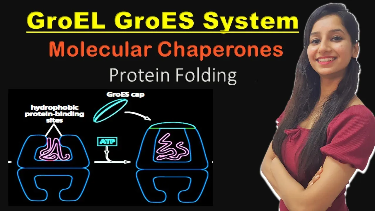








Molecular chaperones, particularly Hsp60 chaperonins and Hsp70 proteins, assist protein folding by recognizing exposed hydrophobic surfaces on non-native polypeptides that would otherwise aggregate; Hsp60 chaperonins use ATP-driven conformational changes to encapsulate unfolded proteins in a hydrophilic chamber where they can fold without aggregating, while Hsp70 proteins bind to short hydrophobic stretches of extended polypeptide chains to prevent premature aggregation during protein synthesis and translocation.
Arthur Horwich (Yale/HHMI) Part 1A: Chaperone-Assisted Protein Folding
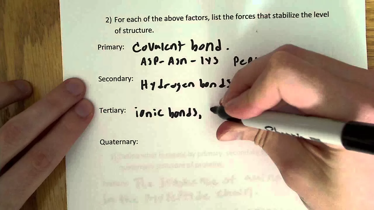
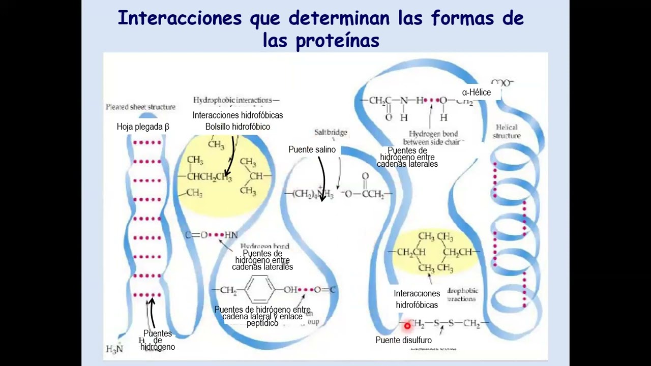
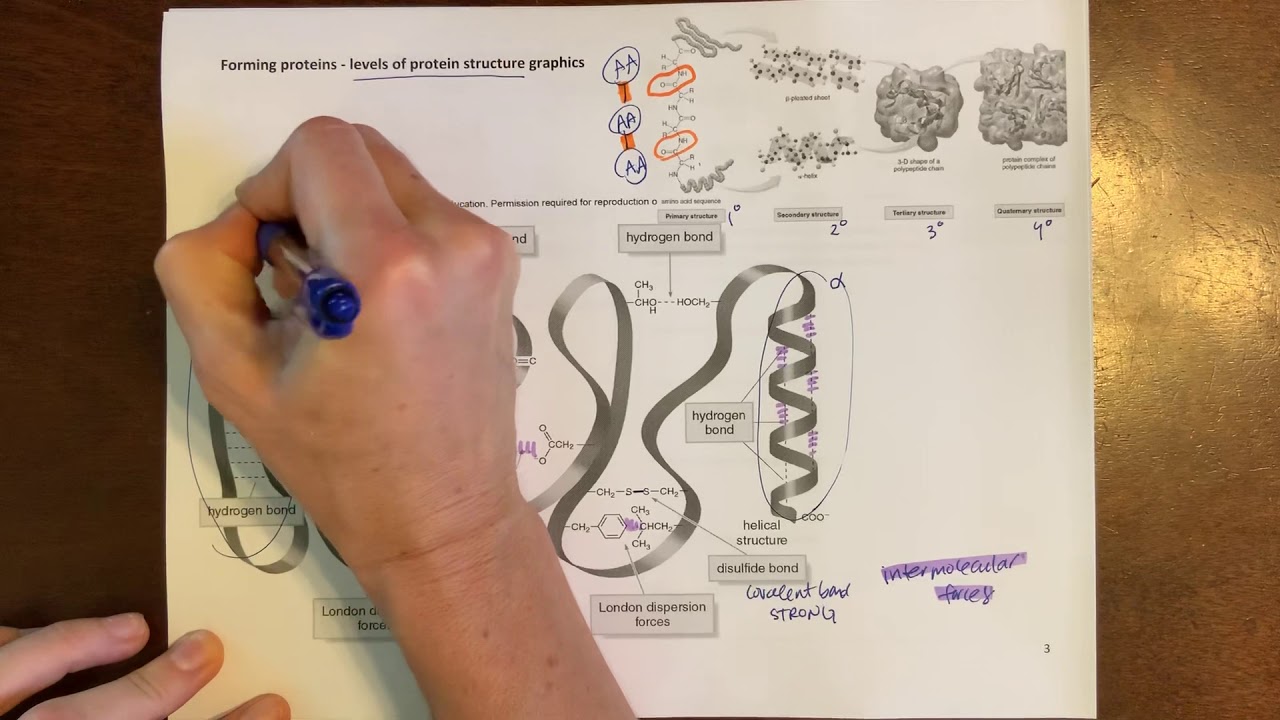
Added:The levels of protein structure (primary, secondary, tertiary, and quaternary) and the chemical forces driving protein stability.
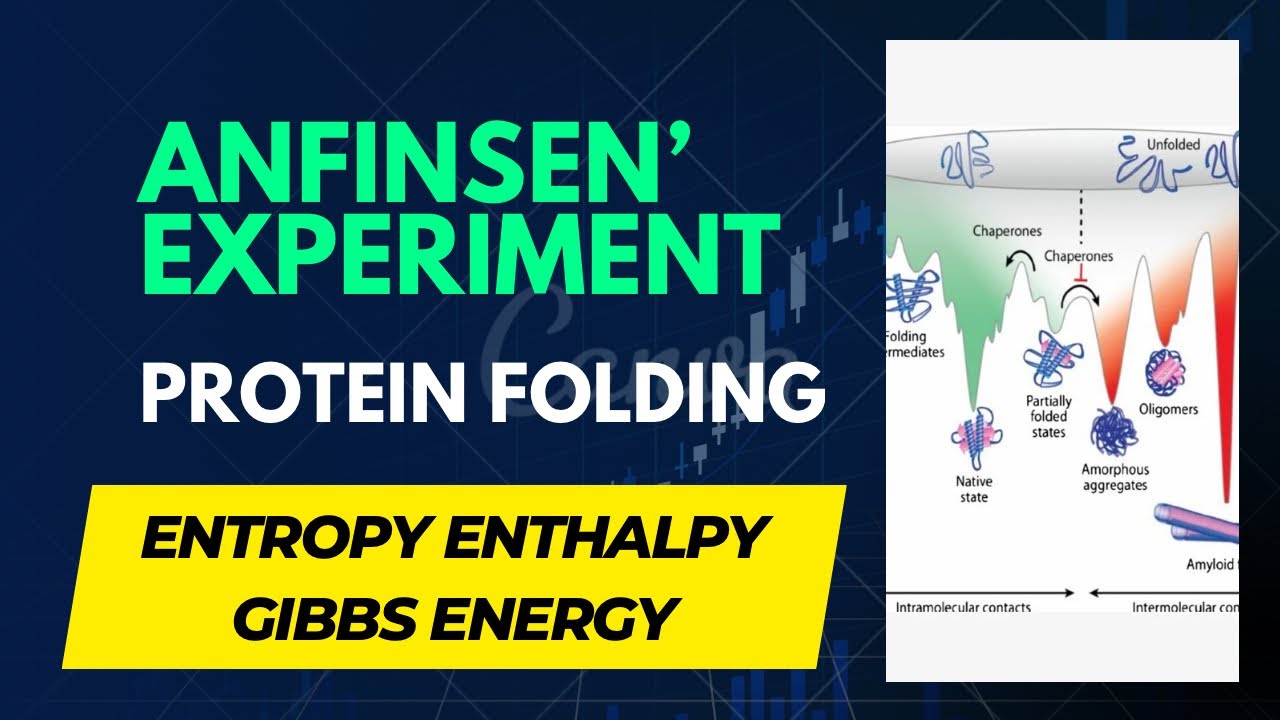
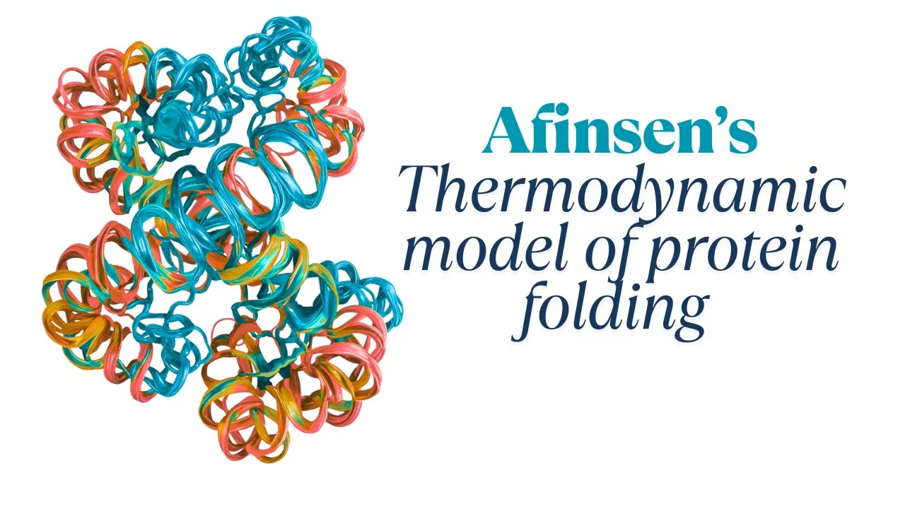
Anfinsen's dogma and the thermodynamic principles governing spontaneous, sequence-determined protein folding.
The basic mechanics of translation, including how nascent polypeptide chains emerge from the ribosome.
The concept of macromolecular crowding within the cytoplasm and its potential to cause non-specific protein aggregation.
The detailed, ATP-driven conformational cycle and mechanism of the GroEL-GroES (Hsp60/Hsp10) chaperonin chamber.
The cellular clearance mechanisms for irreversibly misfolded proteins, specifically the Ubiquitin-Proteasome System (UPS) and autophagy.
The pathophysiology of proteopathies (protein misfolding diseases) such as Alzheimer's, Parkinson's, and Huntington's disease.
Therapeutic interventions targeting protein folding, including the development of pharmacological chaperones and proteostasis network regulators.
56.7K views691likes38:09Original Release: 2014-01-21
hi I'm art which I want to talk to you about Ronan and chaperone protein folding a cellular mechanism that we've been working on for many years and I want to give you a sort of history of how this field came about I want to talk about the two main chaperone families that have been fairly well studied the HSP 60 and hsp70 families and then I want to spend a little bit of time surveying several other chaperone families that have great importance in the cell as well I want to talk briefly about a stress detection system because chaperones are sort of effectors of protein folding in the cell and they in particular help the cell under stress conditions and so how is the stress transmitted such that one makes more of these effector proteins to help the cell and Dyer when it's in dire straits and finally I just want to comment on the relation or increasing relation of molecular chaperones and protein misfolding in disease particularly neurodegenerative disease so um by the way of history let start by indicating that we're talking about the final step of information transfer in the cell so there have been machines that have been identified decades ago and characterized more recently in great detail that are involved with producing RNA from encoding DNA with translating RNA at ribosomes to produce polypeptide chains but until recently less was known about what happened to newly made polypeptide chains leaving the ribosome and I want to give you some of the background to that so for those unfamiliar polypeptide chains are composed of strings of 20 different amino acids and the particular sequences of amino acids dictate ultimately a final folded structure that is a unique and relatively stable structure that is involved in carrying out cellular function so for example enzymes cytoskeletal components channels hormones receptors and a large variety of the effectors in our cells are uniquely folded polypeptide chains that carry out biological activities so the real granddaddy experiment in this related to this last step of information transfer protein folding was carried out in the late 1950s by anfinsen and his co-workers a remarkable experiment in which they started with an enzyme ribonuclease a native folded structure of a hundred odd amino acids with a bunch of disulfide bonds in it completely unfolded the protein in urea and reductant and then asked whether the essentially random coil chain could find its way back to the native state when they removed the denature and reductant and remarkably the protein fairly efficiently refolded and resumed its enzymatic activity so this suggested that all of the information to properly fold a polypeptide chain is contained in its primary amino acid sequence in addition to which when it reaches the native state it's at some sort of an energetic minimum a sort of stable minimum so this was an astonishing experiment I remember being an undergrad at Brown University at that point and hearing in 1972 that the Nobel Prize was being conferred for this experiment and thought it was really one of the most indelibly beautiful experiments I'd ever heard of I never thought I would have anything more to do with protein folding it's really a bit serendipitous that I should ever wind up having something to do with this so one can think of an FinCEN's experiment in terms of protein folding on what is called a smooth energy landscape so at the top of this energy landscape are a host of random coil confirmations represented by D 1 and D 2 here there's a large number of weekly structured conformers with a lot of entropy and very little in the way of stabilizing contacts but as the polypeptide chain folds it gains stabilizing contacts there are fewer confirmations and it's going down the energy landscape effectively sort of like a sea slope and the polypeptide ultimately winds up at the bottom of this slope in the native state a unique sort of energetic minimum and so this is sort of an energetic explanation of the anfinsen experiment but after anfinsen is great experiment what was noticed was that a lot of polypeptide chains could not fold refold spontaneously in a test tube following dilution from denature and they tended to wind up in miss folded aggregates in white material that could be sedimented to the bottom of the tube furthermore in the biotech industry when people went to try to express their favorite mammalian protein in e.coli they often wound up with the same kind of result miss folded aggregated inclusions inside of the bacterial cell usually at its terminal invasive ecoli and so this raised questions as to whether proteins could be having kinetic difficulties in cells that is an Vinson's principles were certainly operative that the primary amino acid sequence was directing a fold to an energetic minimum but things could go wrong under cellular conditions of relatively high temperature and very high local solute concentration and the energetic interpretation of that kind of situation is that in the cell one has a rugged energy landscape where the folding polypeptide chain can get stuck it can get kinetically trapped behind one of these energy barriers and on the timescale of what's needed in the cell the protein effectively never reaches the native state and has to be degraded or fails to or winds up in an aggregate and thus there was it seemed that there was a need for kinetic assistance inside cells well there was a class of proteins identified independently of all the things I've been telling you about in the mid to late 70s called heat shock proteins and these are proteins that under cellular stress conditions particularly thermal stress were highly transcribed and translated and became abundant proteins in the cell and they were classified according to their molecular mass the hsp90 proteins of roughly 90 KD size the hsp70 proteins and for example small heat shock proteins of roughly 20 KD size and however the function of heat shock proteins remained really quite unclear there were postulates that they had something to do with glucose metabolism that they had something to do with stabilizing nucleic acids and there were really a myriad of models for what they might be doing the answer as to what they might be doing really came from Hugh Pollan working at the LMB he noticed that the abundant heat shock protein called heat shock protein 70 or 70 KD molecular mass when supplied in more significant amounts by transfection to cells that were undergoing heat stress could accelerate the recovery of nuclear morphology after heat shock and this started out as sort of a vague idea that maybe other proteins were being helped or maybe other nucleic acids were being helped to maintain their active forms under stress conditions but Pelin very quickly reduced this to the level of protein protein interactions with a variety of in-vitro experiments carried out with isolated nucleoli this is taken from a cell new commentary in 1986 he generated a model that suggested that in the presence of ATP hsp70 could bind to incipient lee aggregating proteins and through the action of hydrolysis pull them effectively apart from each other and now release itself in the presence of a DP from these dissociated proteins you could then go through another cycle in which it acquired ATP again and multiple rounds would would lead to disaggregation that's not quite exactly how hsp70 s worked but he came awfully close to a correct model even at that early point in time really a remarkable synthesis and so this defined now that heat-shock proteins had something to do with protein conformation and with protecting the cell from protein aggregation the question remained open however as to whether such components as these so-called chaperones could have any role in de novo protein folding under normal conditions it seems and it's been quoted that nature leaves nothing to chance and that seems to be the case with de novo protein folding as well so I have to spend just a minute to tell you about the system that we were working on because it's a little bit off the beaten track but we were busy studying how proteins enter mitochondria so it turns out that most of the proteins of mitochondria are nuclear encoded and are synthesized on cytosolic poly ribosomes and then following synthesis on the ribosome an n-terminal ticketing peptide a targeting peptide directs the protein to specifically two receptors on the mitochondrial surface and the recognized protein is then translocated through the mitochondrial membranes and there was a very important experiment published by Gottfried Schatz and his co-workers in 1986 that showed for that for newly made mitochondrial proteins to transit the mitochondrial membranes they had to be unfolded this was demonstrated using essentially a dihydrofolate reductase protein that had never seen mitochondria it had a signal peptide attached to it and it could go into mitochondria as long as you didn't force the dhfr to be folded so for example if you supply to methotrexate ligand the protein couldn't get into my Kandra if you took that ligand away now the protein wanted to mitochondria a very elegant set of experiments but the question that we were in a position to address was what happens on the other side of the mitochondrial membranes do proteins fold refold spontaneously to reach their native active form or could there be some form of machinery necessary to assist the refolding of an imported protein and so we carried out a set of experiments on a yeast library that was a conditional lethal yeast library that we had generated in which a bank of temperature sensitive lethal yeast mutants was used and each mutant was screened individually to ask whether an imported mitochondrial protein could reach its biologically active form and the reporter used for our screen was this mitochondrial matrix protein of liver origin ornithine transcarbamylase a homo trimeric protein of the urea cycle it turns out that yeast do not have an OTC in mitochondria and we had ablated the OTC that isn't a cytosol a localized version in yeast the arch of deleting the arch 3 gene and so we could measure essentially the presence of OTC activity as reflecting intactness in our temperature-sensitive mutants of the entire pathway presumably of import through the membranes cleavage of a signal peptide and then folding in homo try memorization of OTC to its native state and so initial temperature sensitive mutants that we analyzed that were defective in production of OTC and somatic activity turned out to affect the cleavage of its signal peptide and so these were multiple subunits of a matrix processing peptidase that's an essential protein that Cleaves the leader' peptides off of important mitochondrial proteins and one night it occurred to us that maybe there could be such a mutant as would affect in fact the folding of newly imported OTC subunits imported into the matrix space and no sooner did we look for such a mutant than we actually found one we didn't quite quite know what to make of it it seemed heretic 'old that there would be a machinery that assists the folding of newly made proteins or newly translated proteins in this case so we started to look at endogenous yeast proteins the f1 beta subunit of the ATPase here that sits in the the stalk of the ATPase and is involved with energy transduction was found imported into the mitochondrial matrix but non assembled and rather aggregated in the matrix compartment and other proteins were affected as well we couldn't really make heads or tails of this until we received a phone call from aurash hardl and walter nobert who said we understand you're studying some mitochondrial mutants and what happened and the long the short of it is that they helped us further characterize this particular mutant and indeed in their hands proteins were imported completely into the mitochondrial matrix compartment where they failed to reach their native active form and one protein particular was of interest this iron-sulfur protein excuse me right here is a monomeric protein that is imported into the mitochondrial matrix and undergoes several cleavage events before it winds up in the inner mitochondrial membrane and in this particular mutant none of the cleavage events occurred the protein was found in a miss folded aggregated state so this suggested that monomeric proteins not just all these all-america assemble proteins were affected and that suggested that it was polypeptide chain folding that was affected not so much at the subsequent event of all chimeric protein assembly and irish carried out an elegant in vitro experiment with yokomo Sterman showing that if you deliver dihydrofolate reductase to the mitochondrial matrix it became associated with a large assembly and in fact in our mutant as well dhfr could not the native state and so what was this assembly what did any of this have to do this work so we rescued our mutant with a bank of yeast clones and the clone that rescued and coded a mildly heat inducible protein that had been recognized a year beforehand by Richard Hallberg out in Iowa he was busy sequencing the yeast version he'd originally identified at tetrahymena version we called him out up and asked him does a sequence of this gene that rescues our mutant matched the sequence of HS this heat shock protein that you're studying and he said yes indeed we could match this their sequences base where base and so collectively we dubbed this protein heat shock protein 60 but it's a bit of a misnomer because this component is essential under all conditions is crucial for folding under all conditions and so that suggests that indeed protein folding requires kinetic assistance not just under heat shock conditions but under normal conditions of growth so the reaction carried out by this assembly was reconstituted in a test tube within a year of the time of our original observation this was initially carried out by George Lorimer and his co-workers at DuPont and then by aurash and George Martin with us sort of as bystanders in Munich and the reaction could really be broken down into two steps in a first step a chaperone and ring assembly here the bacterial homolog of HSP 60 called Gro L becomes complexed with a non-native protein that for example is diluted from denature it and this forms a binary complex in which the non-native polypeptide as we could show by electron microscopy both Hartl and Baumeister in our own group with joe wall the non-native polypeptide is bound in a whole in a central hole in the ring assembly then and in in that context the non-native polypeptide has no enzymatic activity but is stabilized against aggregation in a second step one could add Co chaperonin another ring assembly also a seven membered ring of 10 KD subunits called gros es along with hydrolyzable ATP and something would happen a remarkable thing would happen in fact the non-native polypeptide would be released in its native form from the assembly over a period of a few minutes and so understanding this reaction really took us a good 15 or so years to work out in a series of experiments carried out by our group or Isha's group georges Laura's group and many other groups worldwide I'm one of the things that we approached was to try to get a decent x-ray structure of grow Yale as a sort of guide to structure of studies and so after four years work we were able to finally generate from a variant version of grow L with two benign mutations in it decent crystals of the grow L chaperonin it's so here you see the double ring assembly that had been seen in the electron microscope each of the subunits is divided into three domains an equatorial domain and these collectively make the waistline of the cylinder and our sort of a stable base to the cylinder you see next to it a hinge like intermediate domain that is connected to the business end of the machine these so-called apical domains so the apical domains on their inside aspect have hydrophobic surfaces that bind a non-native polypeptide through its own exposure of hydrophobic surfaces and then in a subsequent step these hinges open and help to form an encapsulated chambers I'll show you in the next couple slides which is where folding takes place so here's the poly peptide binding surface it's a hydrophobic surface it took us a bit of a while to figure out that the this must be the surface but one morning we came into the lab and we had mutated all these hydrophobics that we saw facing the central cavity of Gro L and all of the mutants that we made were unable to support viability Gro yells essential for cell viability and we very quickly purified all of these individual mutants and could show in vitro that changing any one of these residues which of course changes seven residues around a Gro yell ring to a hydrophilic character abolish as polypeptide binding and so in further experiments we can show genetically but I won't show that to you here that you needed three or four consecutive binding Proficient apical domains in order to bind a polypeptide more dramatically in recent am experiments it's been possible to directly observe a non-native polypeptide bound inside a grow L ring and the non-native protein associates with three or four consecutive apical domains and so it's this hydrophobic interaction that captures non-native polypeptides and that is specific for non-native polypeptides because native structures have those hydrophobic surfaces buried to the interior and it is that binding that prevents the non-native polypeptide from irreversible miss folding and multi molecular aggregation so in the second part of the reaction and unique to the chaperonins is the ATP driven release of polypeptide into a now encapsulated chamber so this is grow yes bound to a Gro yell ring and you see that this little sort of lid like structure seven fold symmetric just like the Gro yell ring to which its binding sends down little loops that contact the very same hydrophobic binding surface that was used to bind non-native polypeptide and so on an isil leucine valine leucine hydrophobic edge in those loops directly interacts with the hydrophobic surface that was used to bind polypeptide so what's happened here is ATP has driven an opening motion and allowed grow yes to associate with we all ring and now the non-native polypeptide because there's no longer any exposed hydrophobic surface is ejected into this chamber where it folds in solitary confinement and so here is a sort of underpinning tat that's how this whole reaction works an open grow L ring is shown by this trans ring on the bottom has this hydrophobic surface also shown here in yellow these are all hydrophobic side chains pointing into the cavity these are the surfaces that can capture a non-native polypeptide but when grow es binds to a so called sistering that is the ring associated with grow yes the wall character changes entirely because the the subunit has undergone at the apical domains of the subunit have undergone an elevation and a clockwise twist so all the hydrophobic surfaces essentially removed from facing the cavity the polypeptide is stripped off into this hydrophilic folding chamber so all this blue that you see is electrostatic residues mainly a very large number of both positives and and acidic residues align this cavity with a slight excess of acidic residues and so the polypeptide folds in this confined space we believe that it folds in essentially a passive way as if it were in an infinite sea of solvent whether the cavity walls really play any role in supervising this reaction our own data would support that's unlikely but there have been experiments that have suggested that perhaps there are some interactions so all in all this cycle is driven by ATP binding and hydrolysis and so to just very quickly summarize a non-native polypeptide comes into what is essentially an ATP bound ring that's because ATP binds ten to a hundred fold faster to an open ring that non-native polypeptide it then binds on the hydrophobic surfaces in the second step grow yes becomes bound so grow yes binding to a ring requires ATP binding to that ring to the seven sites of equatorial sites in that ring and so that produces this encapsulation reaction that I've just described so the apical domains of that ring and its intermediate domains as well undergo rigid body movements that produce this domed folding chamber that's in an ATP bound state for roughly 10 seconds this is the longest step of the reaction cycle which is effectively the duty cycle of the machine at the end of ten seconds a hydrolysis event occurs and now this ADP complex is ready to be discharged it has a brief half-life of a little under a second and the way the Rings operate is they're anti cooperative with each with respect to each other such that once ATP is hydrolyzed in this ring that gates the entry of ATP into the opposite ring so here you see ATP entering this ring and it sends an allosteric signal that ejects all the ligands off of what was the folding active ring so they all leave but at the same time that it has adp arrives here polypeptide comes and so does grow yes and now ultimately this becomes the folding active ring and this ring has now been discharged and becomes inactive so the Machine oscillates back and forth with respect to using its rings as folding active with ATP simultaneously ejecting the ligands on what had been a folding active ring and simultaneously nucleating the present the production of a new sister Neri complex in which polypeptide in the presence of ATP becomes encapsulated by Gro es and this becomes a new folding active sister Neri complex so one message a major message that came from the study of chaperonins and the ability to look at them structurally concerns the nature of molecular chaperones in general they can recognize hundreds of different non-native proteins on what's the feature that's shared by all of them well as the field is progressed it seems like it's exposure of hydrophobic surfaces in non-native polypeptides so just to sort of reiterate a folded contains a greasy hydrophobic core and exposes its as shown here in blue its electrostatic and electric services to the water solvent but under cellular conditions high 10th relatively high temperature high concentration of proteins sometimes stress reactions the non knee the native polypeptide can become miss folded and it now exposes that hydrophobic core to the solvent and as a result of that the efficient thing that's going to happen is protein aggregation where these hydrophobic surfaces get together as a multi molecular aggregate actually these are all entropic reactions that have to do with water binding to particular surfaces and avoiding others but in to make a long story short the idea of molecular chaperones is to recognize such exposed hydrophobic surfaces with the proffered hydrophobic surfaces of the respective molecular chaperone and so it's these hydrophobic contacts between chaperon and non-native polypeptide that effectively prevent the non-native protein from aggregating and so the difference comes in with respect to topologies the chaperonins provide a a cavity in which a collapsed protein exposing a hydrophobic surface can interact with the cavity walls but you can imagine lots of other geometries for exposure of hydrophobic surfaces and the other major family of molecular chaperones the hsp70 class family operates in exactly a different way so hsp70 and here you're looking at the peptide binding domain solved by Wayne Hendrickson and his co-workers in 1996 structurally with a peptide essentially associated with the peptide binding domain this is a pancake shaped structure where the peptide shown here in blue is essentially piercing the pancake and it through an archway formed by loops that around that peptide that is composed of hydrophobic residues and so the peptide sequence here and our ll TG is interacting with that archway through the 3lu scenes they're interacting with this phenylalanine for example here with an alanine that's over here and with the methionine that lies just above it and so those Luo scenes are being stabilized and put in essentially a short stretch of polypeptide chain by this hydrophobic arch in hsp70 well in the context of polypeptide chains intact polypeptide chains we can think of hsp70 binding as sort of a beads on a string type of thing and in fact we can think of a chaperone pathway as occurring where hsp70 class proteins as shown in studies of both mitochondria and for example in the cytosol both bacteria and eukaryotes interact with newly made or newly translocating polypeptides while they're in relatively extended confirmations that have to go through membranes or leave the ribosome they interact with short stretches of hydrophobic polypeptide chain and protect them from prematurely aggregating or miss folding and that happens in the case of mitochondrial protein import that I've talked about earlier on both sides of the membrane so in hsp70 in the cytosol stabilizes the protein against aggregation before it contacts a receptor system in the outer membrane and there's also likewise on the inside of the mitochondria in the matrix compartment an hsp70 that binds the newly entering chain and stabilizes it and now in the case of this import pathway ultimately if a released polypeptide released from this hsp70 can't spontaneously fold it's going to be taken up by the HSV 60 chaperonin system in a collapsed conformation for a final folding step and likewise in the cytosol and I should point out that these are really early experiments that took place in the ABB's of betty craig klaus fanner but ulrich reconstituted a lot of this in vitro to really ram home the point that there could be sequential transfer between these chaperone systems in a 1992 paper with thomas longer in any case in the in the cytosol one can have the same sort of sequence of events for example in the bacterial cytosol where hsp70 s are binding to extended chains leaving ribosomes and a final folding step where it's required for several hundred proteins by the chaperonin system is then subsequently carried out so finally let me say just a little bit about HSB 70 system itself I've shown you this particular structure which essentially is the peptide binding domain with a peptide bound in fact hsp70 s also contain a nucleotide binding domain so just like the chaperonins they use ATP binding and hydrolysis to cycle non-native proteins on and off of the chaperone so in the case of this peptide bound version of hsp70 or the bacterial version called DNA k the polypeptide is stable bound when the chaperone is in an adp bound state and that's a fairly safe stable state necessitating that one if one ever wants to release the polypeptide one has to get the nucleotide out of the nucleotide the n-terminal nucleotide binding domain and so an exchange factor in the case of bacteria called G RPE is used and it effectively pulls the nucleotide a pocket apart and allows the adp to depart enabling ATP to then enter that pocket and as it does so it then mediates an allosteric crosstalk to the peptide binding domain and releases the polypeptide as well as grpe at the same time so now a polypeptide released from an hsp70 could spontaneously fold on its own could be transferred to grow l could be transferred to another chaperone or could go through another round of attempted folding using the DNA k hsp70 system so the non-native polypeptide in many cases will be bound by what's called a J protein in the case of bacteria the DNA J protein is a chaperone in and of itself that has a an n-terminal domain that's capable of associating with hsp70 s and a c-terminal domain that has its own peptide binding properties that can also see short hydrophobic stretches of non-native polypeptide and so frequently J proteins are found to recruit non-native proteins to K proteins to hsp70 proteins so the J and terminal domain associates with the nucleotide binding domain of DNA k and accelerates ATP hydrolysis to produce this locked-in state in which the proffered polypeptide that's being transferred from a J to an hsp70 will be locked in and so that's effectively a cycle of folding and release as carried out by this system now we don't quite have as much crystallographic architecture for this system there are a number of structures of the individual domains and we believe that in the adp bound state from NMR data these two domains function almost completely independently of each other it's when they're in the presence of ATP that they come together and they're now the first a first structure is emerging not still as yet unpublished but it gives some idea of how the architecture of these domains changes and how they associate with each other in the presence of ATP we still have to understand how J proteins fully associate and what they look like when they're fully associating and proper in a protein to an hsp70 so to summarize what I've been telling you about so far I've talked about the HSV 60 chaperonin system the ring assemblies and the hsp70 s that deal with extended polypeptide chains and short hydrophobic segments and the point I want to make here is if you consider a eukaryotic cell and where these particular components are present you find the hsp70 s at the ribosome dealing with newly translated proteins you find them are in and around membranes where they're helping things get across the mitochondrial membranes but likewise across the ER membrane and you find chaperonins not only in the homologous to bacteria the mitochondrial matrix space where our original HSP 60 studies were carried out but there's also a cytosol ik a eukaryotic cytosol except our own and called the TCP one or trick complex that's essential for mediating the folding of actin and tubulin and beta propeller proteins and a number of other proteins in the eukaryotic cytosol and so again it would be involved with final folding of these kinds of components and it's an essential complex composed of eight different subunits per ring which has so far led to a relatively more difficult access to exactly how it works whether it specific subunits bind specific segments of specific non-native polypeptides or not remains sort of an open question this also has a built in lid structure compared to the detachable lids of the HSB 60s that I told you about and so it's a little bit different system that's under intensive study at this point now I'm going to talk subsequently you're talking about the ER system where the presence of glycans added to newly translocated proteins and the presence of disulfide bond formation play a key role in protein folding there is no chaperone in inside of the ER it's a somewhat different system that deserves some attention here and I also want to talk about the hsp90 class proteins which increasingly may have some involvement since they're involved with the final folding of nuclear receptors and various kinase --is with on cell growth control and with potential therapies for malignant disease so I'm going to stop there and we'll continue with the second portion in a moment you
Up Next
41:52
Unfolded Protein Response Signaling Explained (2017 Cell Paper Review)
@AJKeefe
13.3K views•2018-01-14

50:12
Bacterial Communication: Quorum Sensing and Biofilm Formation Explained
@JHUAAP
12.6K views•2011-06-28

42:45
MicroRNAs: Gene Regulation Explained by David Bartel | iBiology
@scicommlab
233.1K views•2014-03-24

7:08
Bacteriophages: Earth's Deadliest Killers and Future Antibiotics
@kurzgesagt
34.6M views•2018-05-13
Related Study Plans & Knowledge Roadmaps
Structured learning paths in Biology
biology
RNAi: Biogenesis, RISC & Gene Silencing
01Molecular Biology Foundations: DNA to Protein
02Introduction to RNA Interference (RNAi)
03Molecular Biogenesis of miRNA and siRNA
+ 2 more learning modules
10h5 modules
Explore Roadmap
biology
Mycorrhizae: Signaling, Biophysics & Ecology
01Foundations of Mycorrhizal Symbiosis
02Molecular Signaling and Root Colonization
03Biophysics and Bioenergetics of Nutrient Exchange
+ 1 more learning modules
11h4 modules
Explore Roadmap
biology
Circadian Clocks: TTFL, SCN & Periphery
01Foundations of Molecular Biology & Gene Expression
02Introduction to Chronobiology and Actograms
03The Core TTFL: Molecular Architecture of the Clock
+ 3 more learning modules
14h6 modules
Explore Roadmap
biology
Population Genetics: Math, Drift & Selection
01Genetic & Probability Foundations
02The Hardy-Weinberg Principle and Baseline Models
03Mathematical Modeling of Natural Selection
+ 3 more learning modules
18h6 modules
Explore Roadmap