





























Axonal growth cones navigate through developing tissues using dynamic interactions between microtubules (which extend the axon), actin filaments (which form the leading edge and enable movement), and myosin motors (which generate pulling forces), guided by extracellular signals including adhesion molecules that mark pathways and soluble gradients of attractive and repellent molecules that trigger intracellular signaling to direct growth cone turning and extension toward synaptic targets.
Axonal Growth Cones & Neural Circuit Formation | Neurobiology
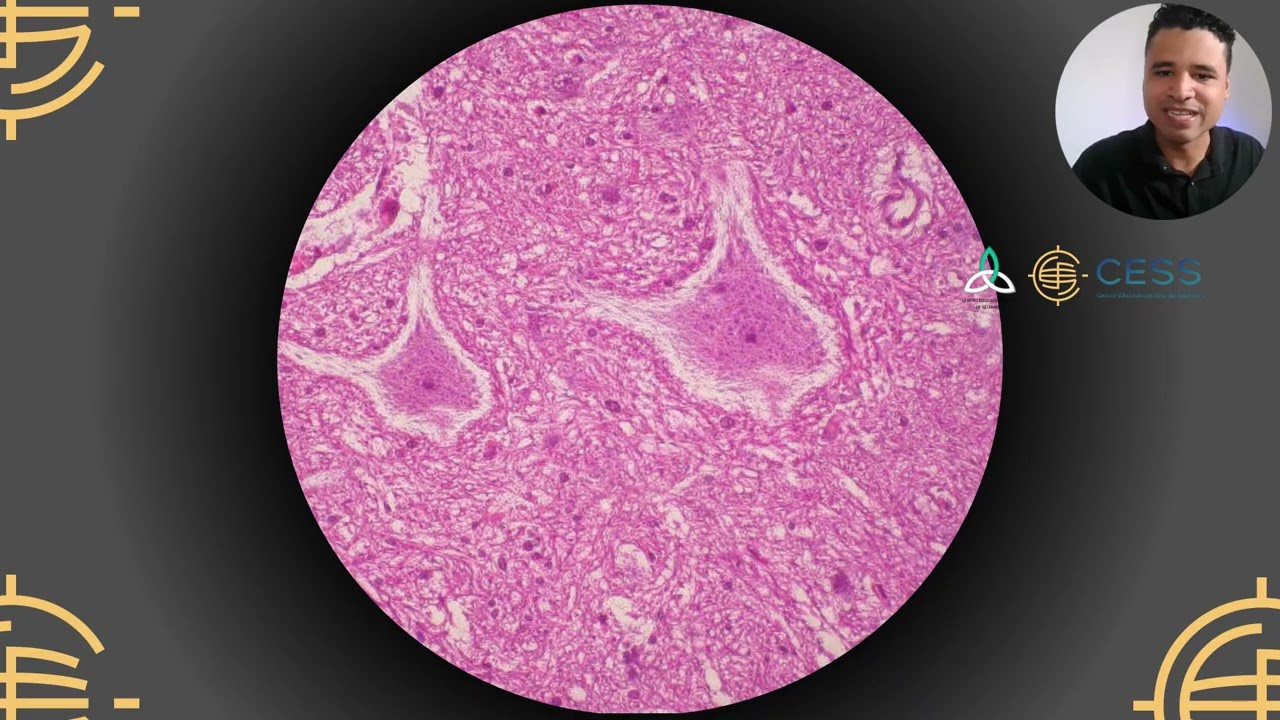
Added:Basic neuron anatomy, specifically the structural and functional differences between the soma, dendrites, and axons.
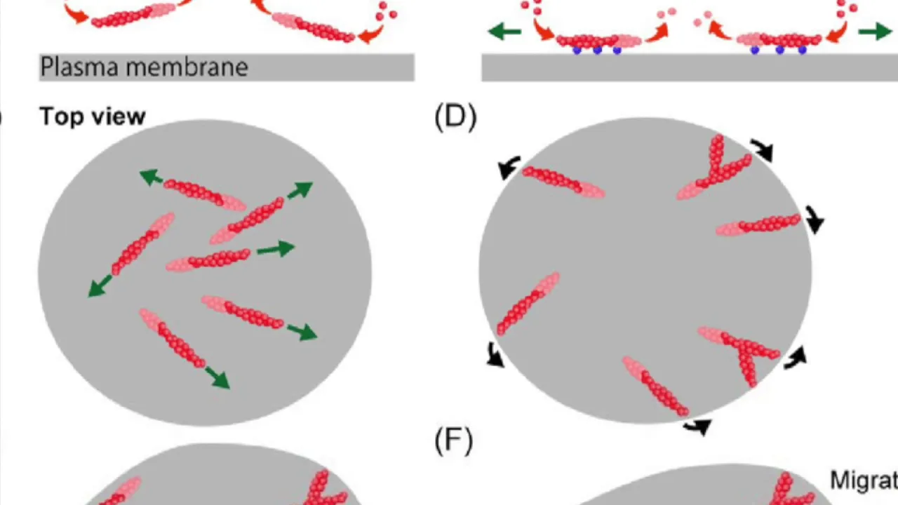
Fundamentals of cellular motility and cytoskeleton dynamics, particularly the roles of actin microfilaments and microtubules.
Basic principles of cell signaling, receptor-ligand interactions, and chemotaxis (how cells detect and move toward or away from chemical gradients).
An introductory understanding of what a synapse is and its role in neural communication.
Synaptogenesis: The precise molecular mechanisms by which a navigating growth cone transforms into a mature pre-synaptic terminal.
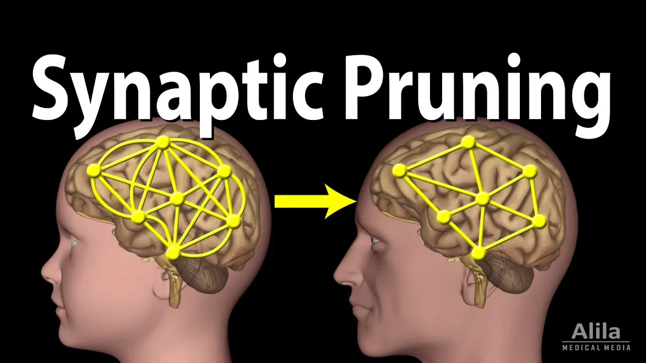
Activity-dependent neural pruning and circuit refinement, including the concept of critical developmental periods and Hebbian plasticity.
Axonal regeneration and the obstacles to repair in the adult central nervous system compared to the peripheral nervous system.
Neurodevelopmental disorders linked to axon guidance errors, such as corpus callosum agenesis, horizontal gaze palsy, and spectrum disorders.
17.4K views120likes9:59Original Release: 2010-03-09
our Sensations behaviors learning and memory are all functions of the brain and nervous system these brain activities are carried out by a complex system of billions of specialized cells called neurons brain information is transmitted along circuits of interconnected neurons electrical signals move between neurons along cellular processes called axons shown in green neuronal circuits are built during fetal life the tips of growing axons called growth cones in red navigate through developing tissues to their synaptic Targets this movie tells the story of the neuronal growth cone the Navigator of developing axons the Spanish neuroscientist Santiago Ramon kahal was the first person to see growth cones in fixed tissues however he imagined the Vitality of growth cone movements in living embryos growth cones protrude finger-like filopodia and flatten lamellopodia that extend and search their surroundings for clues that Mark the path to their targets a growth cone trip to its Target is like a road trip the growth cone must move through developing tissues and detect the roadways sign and exit ramps that lead to its destination first let's examine how an axon grows axon growth involves two fibrous components of the cell cytoskeleton microtubules in green and actin filaments in red microtubules are long Polymers of tubulin protein subunits microtubules support and Define neuronal shape encircling the cell body and extending along all neurites in order for an axon to grow microtubules in green must be constantly assembled and move down the elongating axon microtubules and axons continue to add tubulin summates in yellow as an axon grows at the growth cone the microt tual bundle in green spreads out and single microt tual enss penetrate a network of filaments in red which are polymers of actin protein subunits microtubules and aced filaments are Dynamic polymers their ends grow and Shrink by adding and losing subunits actin filaments disassemble completely but microt tual ends May shrink a bit and then regrow there is a third important component of the growth cone cytoskeleton in addition to microtubules in green and actin filaments in blue the motor protein mein in red lies within the actin filament Network myin Motors bind actin filaments and exert tensions to slide actin filaments rearranging growth cone components or pulling the growth cone forward thus the dynamic motility of a growth cone involves acant filament assembly and disassembly the bundling and linking of actin filaments the advance of M tubules in forces generated by my and Motors in addition to these cytoskeletal Dynamics a growth cone must adhere to its substrate in order to advance in the left image adhesion sites are black while on the Right image corresponding actin filament bundles are white microtubules green at the growth cone front do not Advance beyond the adhesion sites in red that are made by the extending growth cone margin in blue this Coincidence of microt tual ends AC in filament bundles and substrated adhesions reveals a key component of the mechanism by which growth cones Advance adhesion sites provide anchorage for M and motors to pull actin filaments AF and aligned advancing microtubules in green and other building blocks for axonal assembly by aligning the advance of microtubules in Associated components the locations of growth cone adhesions determine the path of growth cone migration this video shows microtubules in green advancing along bundles of actin filaments in red at the inherent front edge of a growth cone act an Assemble at the front advances the Leading Edge followed by adhesions to Anchor the Leading Edge and then microtubules Advance towards the adhesive sites remember the road turp analogy states that migrating growth cones detect the pathways Direction signs and exit ramps that allow navigation to their synaptic Targets in this de helping chicken leg incoming axons seen as thin black lines take a common path to exit ramps in which are arrows that lead to their specific targets growth cones must detect the extracellular guidance cues that Mark these Pathways proteins embedded in the plasma membrane are receptors for these guidance molecules when The receptors detect their specific guidance molecule a signal is transmitted in the growth cone to regulate the cytoskeleton adhesion molecules pave the pathways that growth cones follow while surfaces coated with repulsive molecules disrupt growth cone adhesion in our guard rails that channel growth cones along the proper pathway the top low mag view shows exal growth that follows a pattern surface of the adhesive molecule laminate the middle view shows neurons clustered on a patch of laminate while the lower view shows accents trekking along a l laminin path in this video filopodial antenni press forward from a growth cone to detect a laminin coated surface the growth cone turns and accelerates to reach its preferred pathway in this picture a growth cone hesitates at a border with a surface coated by a repulsive molecule while a podia in red tests the surface but the axon green turns to remain on its preferred pathway this video shows a growth cone turning at such a guard rail to remain on the preferred pathway in addition to surface bound molecules that Mark Pathways gradients of soluble attractive and repellent molecules tell growth cones what directions to move along the adhesive pathways these gradients of attractive or repellent guidance molecules trigger intracellular gradients of signals that either favor cytoskeletal advance for attractants or cytoskeletal Retreat or repellant in this video a growth cone turns towards a glass pipe pit that releases a soluble attractant growth cone spreads robustly toward WS the pipet again a growth cone turns towards a gradient of a soluble attractant actant filaments are preferentially formed towards the source of the attractant this local actin assembly creates a high actin filament density in the growth cone region that is closer to the source of the attractant cool colors blue and green indicate low actin density while hot colors red and orange mean High actin density in this video a growth cone reaches out to touch a bead that bears an attractant molecule microtubules and other axonal components rapidly shift towards the contact with the bead conversely in this video a growth cone rapidly turns away from its contact with a bead that's coated with a repellent molecule when a repellent is added to the median that bathe responsive neurons is in the right panel acting filaments and growth cones are disassembled and disrupted compared to the left thus positive and negative guidance molecules work in opposite ways to keep navigating growth cones on the proper Pathways to their synaptic Targets in conclusion the motility of exal growth cones is a fascinating aspect of neuronal development that is crucial to normal behaviors and is necessary to repair injured or diseased neural circuits
Up Next

5:01
Neurexin-Neuroligin Roles in Dendritogenesis
@cellvideoabstracts
3.4K views•2010-09-17

50:12
Bacterial Communication: Quorum Sensing and Biofilm Formation Explained
@JHUAAP
12.6K views•2011-06-28

5:35
Enteric Nervous System Explained: The Gut's Brain | Neurobiology Lecture
@alumniu6029
438 views•2018-09-12

7:08
Bacteriophages: Earth's Deadliest Killers and Future Antibiotics
@kurzgesagt
34.6M views•2018-05-13
Related Study Plans & Knowledge Roadmaps
Structured learning paths in Biology
biology
RNAi: Biogenesis, RISC & Gene Silencing
01Molecular Biology Foundations: DNA to Protein
02Introduction to RNA Interference (RNAi)
03Molecular Biogenesis of miRNA and siRNA
+ 2 more learning modules
10h5 modules
Explore Roadmap
biology
Mycorrhizae: Signaling, Biophysics & Ecology
01Foundations of Mycorrhizal Symbiosis
02Molecular Signaling and Root Colonization
03Biophysics and Bioenergetics of Nutrient Exchange
+ 1 more learning modules
11h4 modules
Explore Roadmap
biology
Circadian Clocks: TTFL, SCN & Periphery
01Foundations of Molecular Biology & Gene Expression
02Introduction to Chronobiology and Actograms
03The Core TTFL: Molecular Architecture of the Clock
+ 3 more learning modules
14h6 modules
Explore Roadmap
biology
Population Genetics: Math, Drift & Selection
01Genetic & Probability Foundations
02The Hardy-Weinberg Principle and Baseline Models
03Mathematical Modeling of Natural Selection
+ 3 more learning modules
18h6 modules
Explore Roadmap