

![Thorax 8 - Trachée, bronches, poumons, plèvre [Descriptive anatomy of the respiratory tract]](https://i.ytimg.com/vi/fOG_CXzk_Xc/maxresdefault.jpg)














![Body Fluid & Electrolytes [PART-II]](https://i.ytimg.com/vi/1eubrUCWHOE/hqdefault.jpg)



Gas exchange in the lungs occurs within alveoli, which are tiny balloon-like structures surrounded by capillaries; oxygen diffuses from the alveolar space (partial pressure ~105 mmHg) into the capillaries (partial pressure ~45 mmHg) while carbon dioxide diffuses in the opposite direction, driven by the pressure gradient across the thin respiratory membrane composed of epithelial cells, basement membrane, and endothelial cells.
Alveoli Structure and Gas Exchange Explained | Pulmonary Physiology
Added:Basic anatomy of the respiratory tract, including the pathway from the trachea to the bronchioles.
The physical principles of simple diffusion and how concentration gradients drive passive transport.
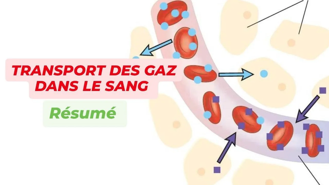
An introductory understanding of pulmonary circulation, specifically the role of capillaries and red blood cells.
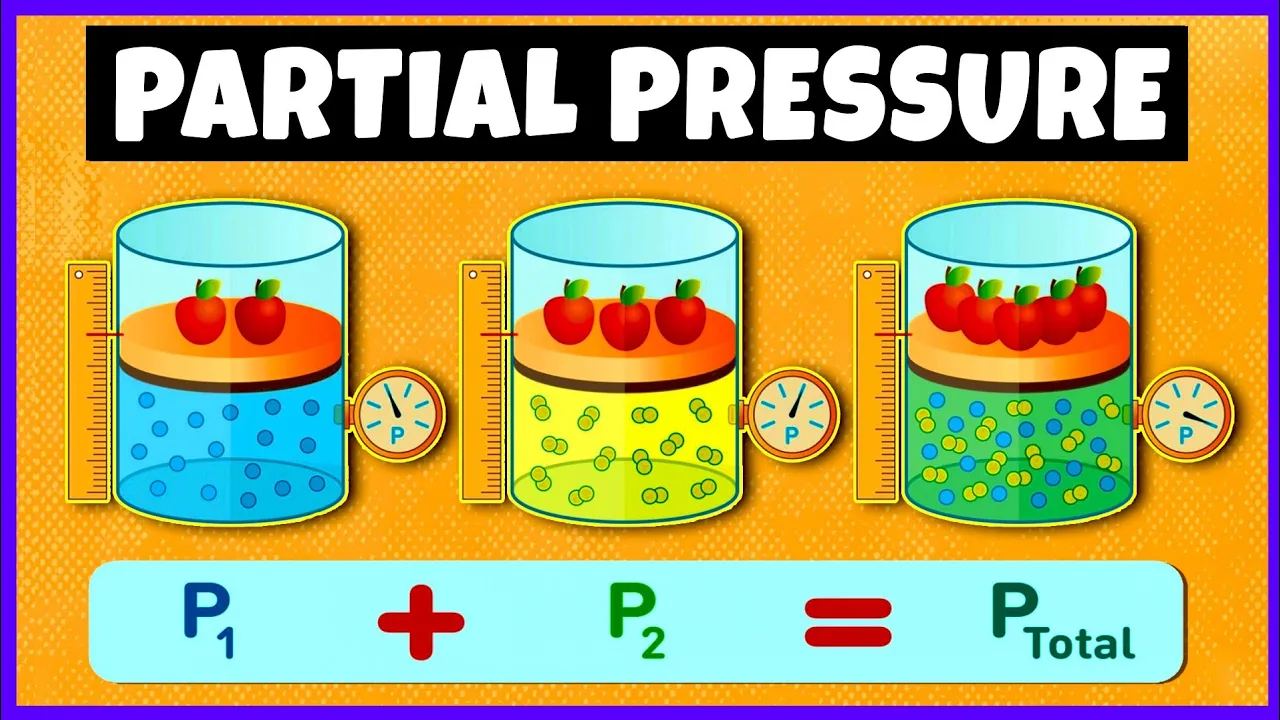
The concept of partial pressure of gases and how it contributes to total pressure (Dalton's Law).
The oxygen-hemoglobin dissociation curve and factors affecting oxygen binding affinity (Bohr and Haldane effects).
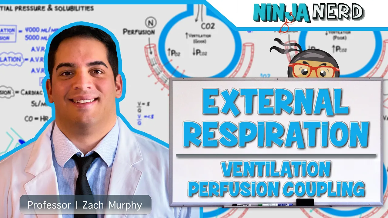
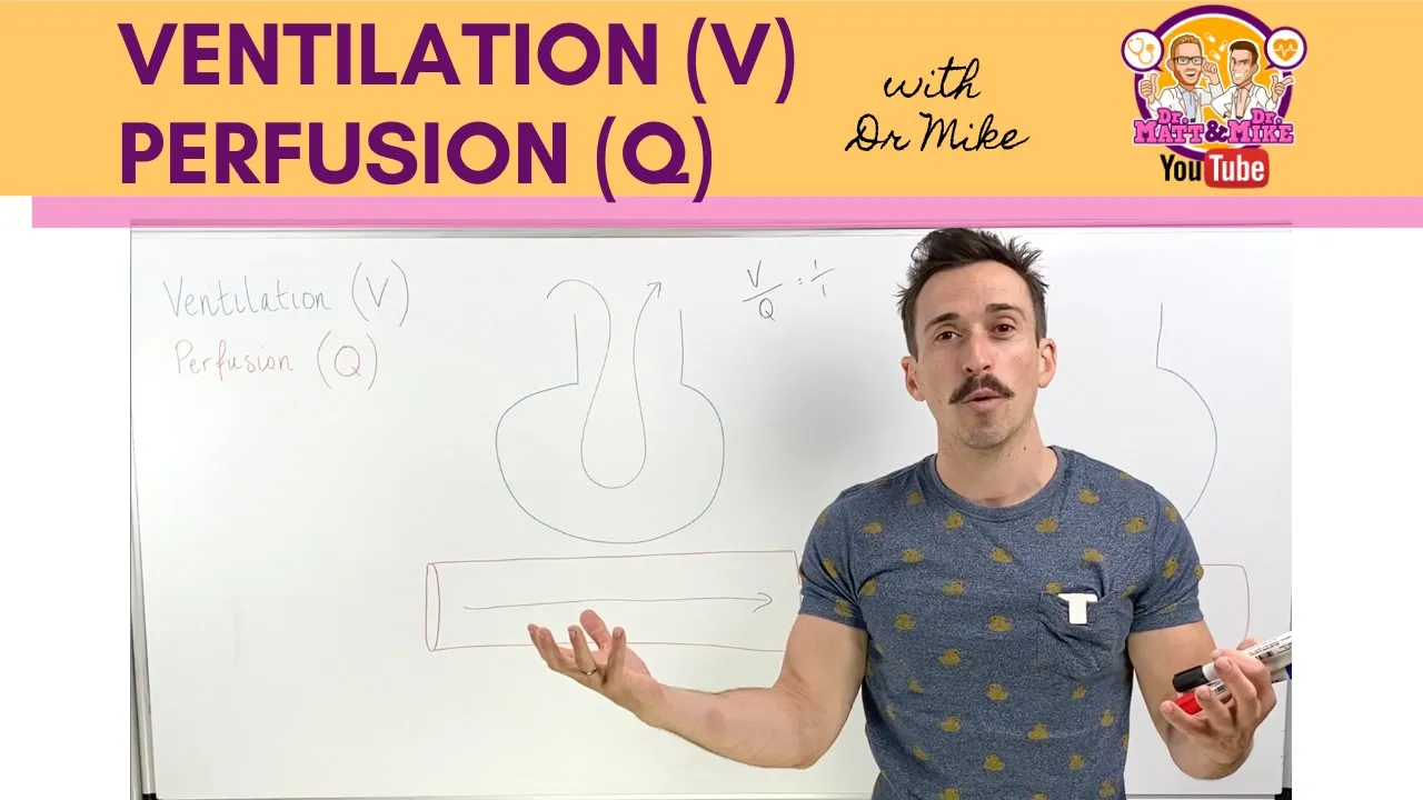
Ventilation-perfusion (V/Q) coupling and how the lungs match airflow with blood flow for optimal gas exchange.
Pathophysiological conditions that impair diffusion, such as emphysema, pulmonary fibrosis, and pneumonia.
The role of the respiratory system in maintaining systemic acid-base balance through the bicarbonate buffer system.
113.1K views1.2Klikes14:43Original Release: 2014-11-08
Inside our lungs, we have specialized structures called alvolar sacks. And these alvolar sacs contain many tiny balloonlike structures called alvoli.
And within the alvoli is where gas exchange actually takes place. Oxygen is exchanged for carbon dioxide. Now before we actually discuss how the process of gas exchange takes place within each individual alvoli, let's discuss what the structure of the alvolar sack is and what the individual alvolus actually looks like. Now recall in our discussion on the respiratory system we said that when we inhale when we breathe in air the air enters via the nose travels through the nasal cavity and then enters our fax and then connects with our larynx the voice box which then connects with the trachea our windpipe. Now the trachea ultimately bifurcates. It divides into two bronkey and each one of these broni subdivides into very tiny bronchioles that permeate through our lungs and at the end of each bronchule at the end of this very tiny air passageway are the alvolar sacs and this is shown by this diagram. So structure number two is the bronchial that is shown in brown. It basically extends all the way into this space number seven and space number seven is the alvolar sac space and this entire orange section is our alvolar sack that is described by number six. Now if we notice along our bronchule we also have these regions shown by red. So this portion this portion this portion and that is our smooth muscle that extends around our bronchule and it is capable of contracting and dilating that broncholle as needed. Now notice we have many of these individual tiny balloonlike structures shown by number one and those are our alvoli that is where gas exchange actually takes place.
So essentially this space number seven the alvolar sac space uh connects directly to the space within each one of these alvoli and that is known as the alvolar space. So if we examine each one of these alvol alvoli we basically get the following diagram and the space inside each one of these tiny alvoli looks something like this. That's the alvolar space. It's not the same as the alvolar sac space but they are connected to one another. And so the concentration of gas molecules inside the alvolar sac space number seven and the alvolar space number eight is exactly the same. Now before we actually take a look at the structure of the actual alvololis, let's discuss what this blue section is and what the red section is. So this blue uh section is our blood vessel. The pulmonary artery that actually brings deoxxygenated blood from the heart to our lungs. While the red blood vessel is our blood vessel called the pulmonary vein that brings oxygenated blood from each individual alvololis and to the heart of our body specifically to the left atrium of our body. So remember the pulmonary artery carries deoxxygenated blood away from the heart and to the lungs while the pulmonary vein carries oxygenated blood away from the lungs and to our heart. So we see that this entire section number six is the alvolar sack that contains many of these specialized balloonshaped structures we called alvoli. And within these alvoli is where gas exchange actually takes place. So we exchange oxygen for carbon dioxide. So remember oxygen is a very important molecule that is used by our individual cells in the process of cellular respiration to actually produce ATP the energy molecules used by the cell. And carbon dioxide is a waste product of cellular metabolism. And so we have to actually excrete it to the outside of our body. And this is what happens inside our lungs, specifically inside each alvoli. So now let's actually zoom in on one of these alvoli. And this is what a single alvolus actually looks like. So we have this connecting point this region here that connects number eight the alvolar space to number seven the alvolar sac space. And because we have this direct connection the concentration of our air molecules inside eight is the same as inside seven which is the same as our actually no it's not the same. So uh in seven and 8 we have the same exact concentration of gas molecules. Now notice that around the entire alvololis we basically have the system of blood vessels. So this blood vessel is our pulmonary arterial that brings deoxygenated blood and it loops around the entire alvololis until it gets to this section. And this is our capillary. It's the pulmonary capillary.
So number seven is the pulmonary capillary and number five is our pulmonary arterial. Now within the capillary we have exchange taking place.
Oxygen goes into the capillary and our carbon dioxide leaves the capillaries and goes into region number eight. And then our oxygenated blood travels via this blood vessel number six which is our pulmonary venule. It's a very small type of pulmonary vein. Now let's take a look at the actual membrane within which we have this diffusion of oxygen and carbon dioxide taking place. So notice we have two important types of cells within the alvololis. We have the cell labeled as number four. That is our alvolar cell type number two. And what this cell does is it produces and releases the pulmonary surfectant that is necessary to prevent the alvololis from actually collapsing when we exhale and to decrease the surface tension and therefore the pressure that is needed to actually inflate our alvololis.
Now uh the cells shown by these green cells number one. So if we zoom in on this small cross-section we get this blown up image. And so number one is our epithelial cells of the alvololis. These are the cells that line the wall of the alvololis. And the wall is shown by number two. That's the orange section.
And this consists of an extracellular matrix we call the basement membrane.
Now the basement membrane actually connects the epithelial cells of the alvololis to the endothelial cells of our blood vessels. So these cells shown in blue are the endothelial cells. So these cells are the endothelial cells of our pulmonary arterial and these cells are our art are the endothelial cells of our pulmonary venule.
Um okay so now that we know what the structure of our alvololis actually looks like let's discuss how gas exchange actually takes place and why oxygen is taken up by the capillaries but carbon dioxide is released by the capillaries. So how does gas exchange actually take place within each individual alvololis within our alvololis alvolar sack. So recall that the right ventricle of the heart pumps deoxxygenated blood into the pulmonary trunk which extends into the pulmonary arteries and these arteries bring deoxxygenated blood into the lungs. Now eventually the pulmonary arteries divide into smaller arteries and they ultimately divide into these pulmonary arterials that is shown by number five.
And these pulmonary arterials essentially circle around the alvoli until they connect with the pulmonary capillary. This section shown by number seven. So let's zoom in on this region that contains this capillary section here. So we basically get the following diagram. So we have the pulmonary arterial, we have the pulmonary capillary and we have the pulmonary venule. So our de oxygenated blood essentially travels along the pulmonary arterial until it gets to our capillary which is this section right here. Now de oxygenated blood has a relatively high concentration of carbon dioxide and a relatively low concentration of oxygen compared to the concentrations of these molecules gas molecules inside the alvolar space. So this region here is region number eight the alvolar space.
Now within the alvolar space we have a partial pressure of oxygen equaling to 105 mm of mercury while the partial pressure due to our carbon dioxide molecules that is 40 mm of mercury.
[snorts] Now inside the lumen of our pulmonary arterial these are the concentrations these are the partial pressures of these same gas molecules.
Notice the oxygen is 40 mm per mercury which is less than inside the alvolar space while the carbon dioxide has a higher concentration 45 mm per mercury inside the lumen of the arterial compared to our alvolar space. So we have a difference in pressure and whenever we have a difference in pressure we know we have a pressure gradient and these gas molecules will begin to move down their gradient from a high pressure to a low pressure. So as soon as the blood enters the capillary, we have this relatively thin wall that consists of the endothelium of the blood vessel, the capillary, the basement membrane as well as the epithelium of our alvololis. And this entire layer allows our diffusion of these gas molecules. And this layer that consists of these three different things is known as the respiratory membrane inside the capillary that allows diffusion to take place. And so carbon dioxide will diffuse down its pressure gradient from a high grad from a high pressure to a low pressure. And oxygen will also diffuse down its gradient but it will move from the outside to the inside of the capillary. also down its gradient from a valley of 105 to a valley of 45 mm of mercury. So this is exactly why our exchange takes place in the first place because there is a pressure gradient that exists between the space of the alvololis and the lumen of our capillary where the blood actually flows. Now by the time the blood actually ends up within our lumen of the uh pulmonary venule the concentration of carbon dioxide and oxygen will be the same inside the lumen as inside our alvolar space and that's exactly why the diffusion of these two gas molecules essentially stops and then our pulmonary venule connects with larger pulmonary arteries. ies and that carry the oxygenated blood into the left atrium of our heart. So once again the de oxygenated blood brought by the pulmonary arterial contains a relatively low partial pressure for oxygen and a relatively high partial pressure for carbon dioxide compared to the space inside our alvololis.
Therefore, due to this pressure difference, due to the existence of this pressure gradient, oxygen will diffuse into the capillary and carbon dioxide will diffuse out of the capillary down their pressure gradient. And this diffusion will continue until our partial pressure concentr uh the partial uh concentration or the partial pressure for oxygen is the same on the inside of the blood vessel as our inside space the space inside our alvololis. And the same is the same thing is true for the carbon dioxide. The diffusion of carbon dioxide will continue until the partial pressure inside our lumen of our blood vessel is the same as our alvolar space. So we see that inside each individual alvololis the reason that gas exchange takes place is because of the existence of a pressure gradient a difference in concentration between these two types of gas molecules found in the lumen and inside the alvolar Space.
Up Next
9:34
How Stressed Algae Curl Into Protective Cysts
@journeytomicro
70K views•2021-08-02

50:12
Bacterial Communication: Quorum Sensing and Biofilm Formation Explained
@JHUAAP
12.6K views•2011-06-28

7:47
Post-Translational Modifications Explained: Types & Functions
@AKLECTURES
136.8K views•2014-09-11

7:08
Bacteriophages: Earth's Deadliest Killers and Future Antibiotics
@kurzgesagt
34.6M views•2018-05-13
Related Study Plans & Knowledge Roadmaps
Structured learning paths in Biology
biology
RNAi: Biogenesis, RISC & Gene Silencing
01Molecular Biology Foundations: DNA to Protein
02Introduction to RNA Interference (RNAi)
03Molecular Biogenesis of miRNA and siRNA
+ 2 more learning modules
10h5 modules
Explore Roadmap
biology
Mycorrhizae: Signaling, Biophysics & Ecology
01Foundations of Mycorrhizal Symbiosis
02Molecular Signaling and Root Colonization
03Biophysics and Bioenergetics of Nutrient Exchange
+ 1 more learning modules
11h4 modules
Explore Roadmap
biology
Circadian Clocks: TTFL, SCN & Periphery
01Foundations of Molecular Biology & Gene Expression
02Introduction to Chronobiology and Actograms
03The Core TTFL: Molecular Architecture of the Clock
+ 3 more learning modules
14h6 modules
Explore Roadmap
biology
Population Genetics: Math, Drift & Selection
01Genetic & Probability Foundations
02The Hardy-Weinberg Principle and Baseline Models
03Mathematical Modeling of Natural Selection
+ 3 more learning modules
18h6 modules
Explore Roadmap