















The cerebrum contains three types of fiber pathways: association fibers (short U-fibers and long pathways like the superior longitudinal fasciculus connecting frontal and parietal lobes, and arcuate fasciculus connecting frontal and temporal lobes), projection fibers (corona radiata connecting cortex to subcortical structures), and commissural fibers (corpus callosum connecting hemispheres). These pathways support complex functions including language processing (dorsal stream via arcuate fasciculus for speech production and ventral stream via inferior fronto-occipital fasciculus for semantic processing) and visual processing (ventral stream for object recognition and dorsal stream for spatial navigation).
Fiber Pathways of the Cerebrum: Association, Projection, Commissural Tracts
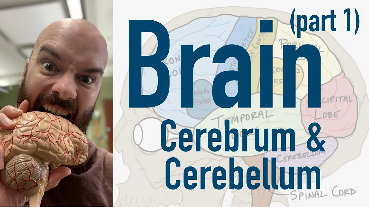
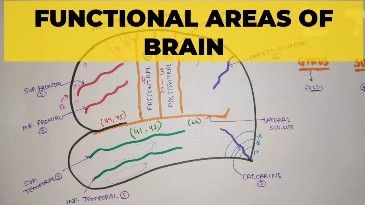
Added:Basic structural anatomy of the cerebrum, including the major lobes (frontal, parietal, temporal, occipital) and key cortical landmarks.
The anatomical and functional distinction between gray matter (neuronal cell bodies) and white matter (myelinated axons).
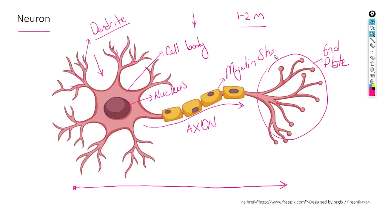
Fundamental neuron structure, specifically the role of axons and myelin in propagating electrical signals throughout the nervous system.
Anatomical directional terminology and spatial planes of the brain (e.g., coronal, sagittal, axial, ipsilateral, and contralateral).
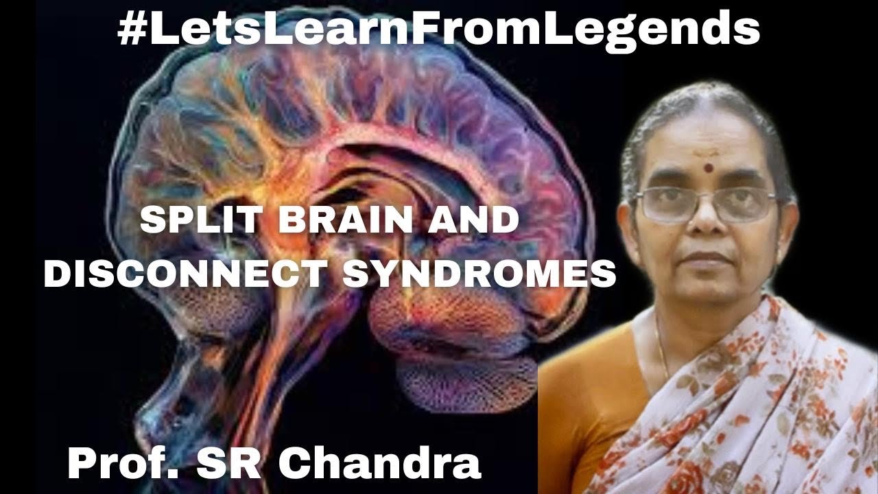
Clinical pathologies associated with tract disruption, such as disconnection syndromes (e.g., conduction aphasia or split-brain syndrome).
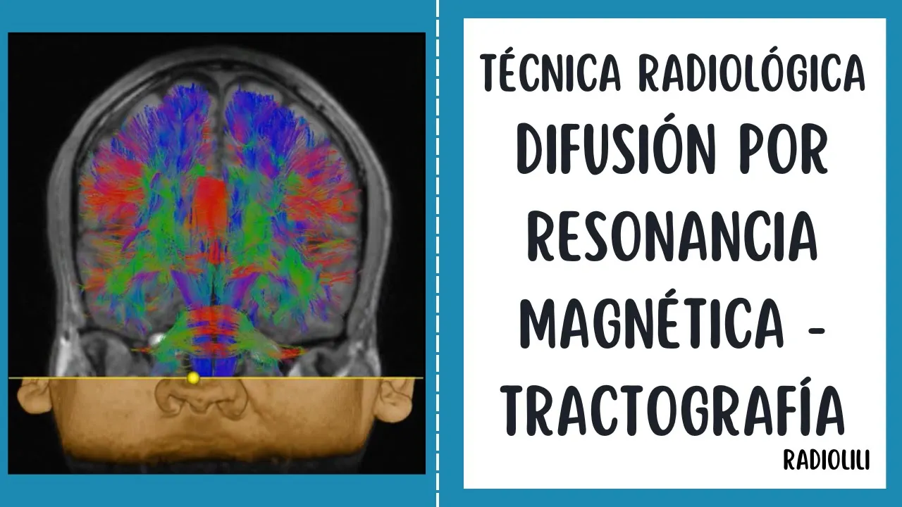
The principles and clinical applications of Diffusion Tensor Imaging (DTI) and tractography in mapping white matter pathways.
Neurosurgical planning and intraoperative subcortical mapping techniques used to preserve critical tract pathways during brain surgery.
Advanced cognitive neuroscience topics exploring how structural fiber pathways support functional connectivity networks like the Default Mode Network.
39.5K views741likes29:33Original Release: 2016-09-29
the cereum has gray and white matter the gray matter which is other layer of the cereum also called as the cortex is classified according to surface anatomy basically the frontal Lop is located in front of the central sulcus the occipital Lop is located behind an imaginary line drawn between the p oxyon lch and the top of the par occipital sulcus the paral l is located between the frontal and occipital LS the temporal Lop is the inferior part of the cereum the border between the peral and temporal lops is an imaginary line that curses from the end of the syan fusure to this imaginary line the white met of the cereum which underlies the gray matter and intervin between cortical gray matter and Asal ganglia contains three types of fibers the association fibers connect the different regions in the same hemisphere the projection fibers such as Corona radada connect the cortex with coral parts of the brain and spine cord the commcial fibers such as Corpus colossum connect the two hemispheres across the medium plane let's start from the association fibers which has the short and long types the short fibers also called as U fibers are located superficially just underlying the cortical gray matter and the long fibers are located at different position the arcade phicus is the frto Paro temporal long Association fibers that connects to VES area which consist of the middle and posterior parts of the superior and middle temporal gy to Bro area located in the posterior 2/3 of the inferior frontal jar this DTI demonstrates that the arcade fulus terminates in the precentral gyrus as well as broas area which is composed of the par Paris and triangularis in the inferior fontal gyus the arcade fulus is subdivided into a vental part which is blue one and a dorsal part which is orange one in the super silvian area the main long Association fiber pathway is the superior longitudinal fulus which which has three parts slf one dorsal pathway slf2 middle pathway and slf3 ventral pathway the slf one is positioned within the superior frontal gyus the slf2 within the middle frontal gyus and slf3 within the inferior frontal gyus the slf1 lies within the Superior Bank of the singl ccus and travels just above the singul to connect the superior paral and until terior single cortex the slf2 extends from the inferior paral L to the middle frontal gyus the sf3 curses within the front paral pulum and extends from the inferior paral L to the inferior frontal gyus the slf is considered as a higher order multisensory associative system the compartments of the superior longitudinal fulus and arcade facus can be seen on this DTI slf1 turquise is a dorsal pathway slf2 clear red is the middle pathway and slf3 yellow is the wal pathway in the super cilian area the wal blue and dorsal orange segments of the arcade phicus can be seen other long Association fiber pathway are the inferior front occipital fulus which is a front occipital connection the inferior longitudinal fasiculus which is a temporal occipital connection and the unate fasiculus which is a for temporal connection we are looking from above the function of the unate fasiculus is to link emotion and cognition as a oneic pathway in contrast to the singul or dorsal liic pathway additionally some resarch shows that the onate faul is involved in semantic processing of language are working memory and sound recognition language processing has a ventral semantic stream and a dorsal phological stream that connect B area and War area within the dorsal stream front the pr temporal regions are proposed as being involved in mapping aor speech sounds to articulatory representations the major pathway is arate forculus in contrast the one stream is proposed to be involved in mapping out your speech sounds to meaning the major fiber pathway in the one stream is the inferior front occipital fulus other fiber pathways are the extrem capsule onate fulus inferior longitudinal fulus and middle longitudinal fulus the Mage to the inferior front occipital fulus produces the semantic parasia in the dominant hemisphere visual processing has two Pathways like language processing visual information follows two main streams from the occipital La the ventral stream which is what pathway travels to the temporal L by means of the inferior longitudinal facus and relates to object identification and recognition the dorsal stream which is wire pathway travels to the paral which is the navigation center of the cereum by means of the occipital short Association fibers and the part of sf2 and relates to processing location of object in space the Corpus kosum is the major inpher Cal pathway that connects most NE cortical areas the Colossal fibers emanating from C caloras in one hem sare together above the lateral ventricle and enter the Corpus colossum to reach the other hemisphere they cross the midline by passing below the singul to form the roof of the lateral ventricle the colosal fibers arising from the Gen of the Corpus colossum turn in an anterior oblique direction to interconnect the frontal regions where they called to forceps minor at the level of the splenium fibers turn in a posterior obl direction to interconnect the paradoxal regions where they are called to force measure the Colossal fibers arising from the splenium named the toal fibers sweep downward to form the roof and lateral wall of the atrium temporal and oxital horns of the lateral ventricle other Comal pth are the anterior and posterior cures which Travers the midline the anterior commure resembles bicycle handlebars located immediately in front of The Columns of the forx and plays a role in visual processing together with the splenium of the Corpus kosum the anterior commure May compensate for congenitally absent Corpus colossum by integrating in mospheric visual information the mamary bodies and the Flor of the third ventricle can be seen here the posterior commiss is located on the dorsal aspect of the upper end of the cereal Aqueduct the anterior and posterior commissures can be seen in this medial wave the posterior commure originates from its nucleus located in front of the ocular Mo nucleus the posterior commure continues downward into the medial longitudinal alas culus the posterior commure is involved in popular light reflex the anterior crosses the midline at the base of the pan to connect to orbital frontal oipal and temporal Lopes especially amydala it has an anterior cruise that extends forward and a posterior cruise that extends laterally the posterior cruise is divided into temporal and occipital extensions for clinical perspective it was suggested that seizure activity can propagate to the cont lateral medial temporal L by way of the anterior commure so it's cutting is a part of the hemispherectomy procedure for control of intractable seizures after looking at the association and chal fiber Pathways let's move on to the projection fiber Pathways the front of paral projection fibers which have a vertical Direction can be called as the Corona radiata and the occipital projection fibers which have a horizontal Direction can be called a sedal strum the corona radiata is formed by the external and internal capsules above the upper level of the pan the Centrum SE is located above the level of the Corpus colossum and consist of the superior longitudinal fulus Association fibers Corona radada projection fibers and colossal chol fib we have seen all three types of fiber pathas of the cereum now we are going to look at all these fiber Pathways from the lateral to medial Direction in a stepwise manner as well as the central core and limic system functionally the frontal Lop is the conscious and to Center the perial Lop is navigation Center the occipital Lop is the vision center and the temporal Lop is the memory and sound center of the cereum the removal of the gray metal exposes the short Association fibers which interconnect knee body gyy the removal of the short Association fibers exposes the long Association fibers at a deeper position the most superficial long Association fibers are the superior longitudinal fulus which connects the frontal and perial lobs and AR fa which connects the frontal and the temporal lobs the removal of the archade fulus exposes the middle longitudinal facus and inferior longitudinal fulus the middle longitudinal fulus lies within the superior temporal gyus and the inferior longitudinal fulus lies within the inferior temporal gyus the middle longitudinal fulus connects the temporal Pole to the inferior parial especially the angular jars the middle longitudinal fulus is involved in location of sound in Space the inferior longitudinal fulus connects the temporal Pole to the dorsal lateral oxital L without reaching the visual cardex in this Dissection the frontal paral parum has been removed to expose the insula the central insular sulcus curses almost parallel with the central sulcus on the the convexity and divice the insular cortex into short and long insular gy the short insular gyri are located anterior to the central insular sulcus and the long insular gy are located posterior to the central insular sucus the insula is encircled by the limiting suai which are the superior inferior and anterior limiting susai the junction of the anterior and Superior limiting suai is called the anterior insular point and the junction of the superior and inferior limiting suide is called the poer nuar point these points and limiting suide are important during insular surgery an axial plane we are looking at the central core area located between the insular cortex laterally and ventricles medially the centrer core includes the extrem capsule clroom external capsule paman Globus palus carate nuus internal capsule and Thalamus the insular corex extrem capsule clustr external capsule paman globous palidos and internal capsule are positioned in the central core from the lateral to medial after removal of the insular cortex we can see the extrem capsule which is composed of the short Association fibers providing the connections among the insular gy and the opar the function of the extreme capsule is not clear but it has been suggested as a part of the semantic language stream in F Dissection the external capsule and claustrum were exposed by removing the extrom capsule the clroom and external capsule can be divided into dorsal and ventral parts the dorsal claustrum is here and the dorsal external capsule which is formed by cluster cortical fibers is here the ventral claustrum spits out into the ventral external capsule at the lemon insula to reach the Amala the cluster cortical fibers which is the dorsal external capsule CS from the dorsal clustr to the cortex between the supplementary motoria anteriorly and the pr Al L posteriorly the cluster Cal system has been related to the integration of visual somat sensory and mod informations just medial to the external capsule is the paman the paman is one of the Bas structures its main function is to regulate movements and learning another basal structure is the Globus palus which is located just medial to the paman in this picture the posterior half of the paman has been removed to expose the globous pilus and internal capsule the external and internal capsule come together to form the corona radiata above the pment a coronal section what we can see here is that the external and internal capsules come together above the pan to form the coronary rata also the Globes is situated medial to the the ventral external capsule is formed by the inferior front oipal fulus above and the unin fulus below at the lion insula the inferior front occipital fulus cses between the frontal and occipital LS the unate fulus connects the orbital frontal area to the temporal pole forming a hook shape removal of the PO and inferior front occipital phicus exposes the entire globous palus and anterior cure the Globus Palos sits just behind the anterior commure and plays in the regulation of voluntary movements the procedure now as pomy is used to treat some movement disorders the anterior commure courses below the wal aspect of the anterior LMP of the internal capsule in the hemisphere it moves cly and PES laterally through the ventral aspect of the globous padus in the grolia canal it cses downwards to the temporal pole just behind the UN fasiculus en llarge we previous Slide the anterior limp Geno and posterior limp of the internal capsule are observed the substantial inada is located in front and beneath the anterior commure here is the ventral clroom seates out in the UN fulus to reach the Amala an AAL section the anterior LMP of the internal capsule is located between the head of Cate nucleus and the lentiform nucleus which is composed of the paman and Globus palus the posterior LMP of the internal capsule is located between the lentiform nucleus and Thalamus the Geno is the junction of the anterior and posterior limbs a superior lateral wheel the Geno is located just behind the anterior commer at the same Coral level as the for Monro and posterior short insul gyus the removal of the anterior Lim of the internal capsule exposes the head of the c n here is a ventral claustrum which speds out into the onate fulus through which the Amala the onate fulus is is a for temporal long Association fiber pathway the unin fasiculus has a dorsal lateral part which goes to the orbital frontal area and a vent medial part which goes to the sepal area from the temporal pole clinically some DTI studies in patients with schizophrenia have found morphometric changes in the onate fasiculus at the same time disruption of the unate fulus during the entering medial temporal ectomy may be associated with psychosocial clinical Improvement because the onate fulus can no longer convey the pathological information from the temporal L to the orbital frontal cortex which is involved in decision making we are looking from above the dorsal lateral part of theate fulus which projects through lateral orbit frontal area and the vent medial part which projects through septal and medial over the frontal areas can be seen the C nus is an arched C shaped structure with head body and tail Parts it also forms the lateral wall of the lateral ventricle we are looking from above the CATE nucleus surrounds the lateral part of the thalamus and the St terminalis travels between the C nus and thalamus after removal of the thalamus we can see the position of the CATE nucleus in relation to the insular limiting suside the head of the C nucleus is positioned inside the insular area which is surrounded by the limiting suide the anterior insular point is positioned above the head of the card nucleus the body of the card nucleos sits above the superior limiting sulcus and passes just deep to the posterior ins point below the posterior ins point it's called the tail of the CATE nucleus and it travels behind the inferior limiting Circus the tail of the card nucleus crosses the inferior limiting circus before blending into the Amala among the Bas structures we have addressed the pan Globus pus and the cus now we are going to look at the rest of the Basel G structures we are looking from above the nucleus incumbant is located underneath the head of the C nucleus the subthalamic nucleos is located wial lateral to the right nucleos in the D posterior view of the dilic area the subthalamic nucleus is positioned V lateral to the red nucleus and dors medial to the internal capsule lateral wave the the substantia Negra is positioned on the ventral surface of the subthalamic nucleus the red nucleus is here the basal gangal structures has connections with cereal CTIC and brain stem let's see the projection fibers and their relationship with the lateral ventricle basically the front paral projection fibers can be called as the Corona radiata and the oxital projection fibers can be called as the sedal stratum the coronal radiative fibers run medial to the superior longitudinal fulus the Sagal stratum fibers run medial to the inferior font oipal fulus inferior longitudinal fulus and oipal extension of the anterior commer removal of the inferior front occipital fulus and occipital extension of the anterior commure exposes to sagittal stratum the Sagal stratum is composed of the oxy ponin fibers and oxamic fibers which is also called as the optic radiations the optic radiation fibers arise from the lateral genicot body and travel in the roof and lateral wall of the temporal horn and then lateral to The Atrium to reach the calar and sulcus notable the upper one thir of the atrium is free from the optic radiation fibers the optic radi iations are separated from the temporal horn and atrium of the lateral ventricle by the topedm The Atrium and occipital Horn of the lateral ventricle are covered laterally by the Sagal strum and topal fibers and medially by the forceps measure in the medial wall of the atrium we see the bulb of the Corpus colossum overlying the fibers of the forceps measure and the Cal AIS overlying the deep end of the calcarine ccus the anterior bundle of the optic radiations are called mayor's Loop and it extends up to the tip of the temporal horn the amydala forms the anterior wall of the temporal horn the hippocampus is located in the floor of the temporal horn removal of the thalamus exposes the third ventricle the stre terminalis arises from the sepal area which is located around the anterior Commission in the midline and Reps around the thalamus to blend into the amigdala the three amidar Talam extends from the sepal area to the habenula let's take a look at the subcortical white meter anatomy of the frontal L the lateral surface of the frontal L is bordered posteriorly by the Central ccus and superiorly by the superior hemispheric border the lower border of the lateral surface of the frontal L has an anterior part that faces the orbital roof and a posterior part the San border that faces the temporal L across the cvan fizure the subcortical white meter organization of the frontal L includes the projection chistol short and long Association fiber Pathways the superior longitudinal fulus 2 is situated within the middle frontal gyrus medial to the superior longitudinal fulus 2 are the corona radiat containing the frontal projection fibers in F Dissection the Colossal fibers at the level of the superior frontal gyrus the frontal horn and body of the lateral ventricle as well as the insular area at the level of the middle and inferior frontal gy have been exposed here is the anterior insular point which is an important Landmark for surgery the endal layer of the ventricle was opened the anterior insular Point corresponds to the frontal Horn of the lateral ventricle the cingulum which is a part of the lyic system runs medial to the Colossal fibers a similar dissection in another specimen the relationship between the Colossal fibers and cingulum can be seen the removal of the Centrum Sim exposes the Contra lateral frontal l the farther dissection of the central Cora exposes the inferior front occipital fulus and pan after seeing the frontal L let's take a look at the internal anatomy of the temporal l in order from the lateral to medial the temporal cordex and short Association fibers was removed to expose the long Association fibers the both middle and inferior longitudinal fauli originates from the temporal pole the middle longitudinal fulus cses within the superior temporal gyus while the inferior longitudinal fulus cses within the inferior temporal Gus fire Dissection the position of the unin fulus and inferior front oipal fulus can be seen here the removal of the unate fulus inferior front oal fulus and extensions of the anterior commure exposes the optic radiation fibers which arise from the lateral geniculate body and pass medial to the inferior limiting sulcus to form the roof and lateral wall of the temporal horn of the lateral ventricle after rising in the lateral geniculate body the optic gradiation fibers pass between the inferior limiting sulcus and tail of the C ncos to cover the roof and lateral wall of the temporal horn finally let's look at the limic system the limic system is a a group of interconnected cortical and subcortical areas that links viseral States and emotion to cognition and behavior the limic cortex is composed of the par hipocampal gyrus hippocampal formation which is hidden and singlet gyrus the parampal gyus is an elongated c-shaped gyus between the enus and ismost of the singular gyus where the lingal gyus merges into it laterally the collateral sulcus separated from the fusiform gyus this gyus forms a hook like expansion anteriorly called the ancus the paroc campus and ancus lie laterally and envelop the hippocampus the hippocampus plays a role in memory function the perample gyus is important for nursing behavior and attention the singul is a long Association fiber pathway which interconnects the par camp and single G medial wave the subcortical area of the lyic system basically includes the alaor system septile region hypothalamus and amydala the best known limic pathway that interconnects the limic cortical and subcortical areas is papy circuit to reveal fibers leave the hipocampal formation and proceed through the foric to reach the mamary body body from here a new pathway the mamic tract ascends up to the anterior talic nucleus the information in the anterior tmic nucleus continues to the singul gyus by means of the Talam singulate fibers which pass through the internal capsule from the singular gyus the information carries to the parampal gyus by the singul the parampal gyus projects to the hipocampal formation hence the circuit is format
Up Next

7:58
Broca's and Wernicke's Areas: Rethinking Brain's Language Centers
@SciShowPsych
63.9K views•2020-01-27

21:55
Deciphering Neural Circuits Controlling Anorexia in Mice | Richard Palmiter
@AllenInstitute
2.3K views•2011-10-10

4:10
Vagus Nerve (CN X): Anatomy, Nuclei & Functions Explained
@Alilamedicalmedia
305.2K views•2022-10-31

13:03
How Exercise Benefits Your Brain: Science Explained
@TED
11.4M views•2018-03-21
Related Study Plans & Knowledge Roadmaps
Structured learning paths in Neuroscience
neuroscience
Appetite Regulation: Brain Circuits & Hormones
01Foundations of Endocrinology and Brain Anatomy
02Introduction to Energy Homeostasis
03Peripheral Signals: Leptin, Ghrelin, and Insulin
+ 2 more learning modules
14h5 modules
Explore Roadmap
neuroscience
Neuroeconomics: Value, Risk & Brain Mechanisms
01Foundations of Neuroscience and Brain Anatomy
02Introduction to Neuroeconomics and Subjective Value
03Dopaminergic Prediction Errors & Reinforcement Learning
+ 3 more learning modules
18h6 modules
Explore Roadmap
neuroscience
Spatial Navigation: Cells, Grids & Brain Maps
01Foundations of Brain Anatomy and Memory
02Place Cells and Head-Direction Cells
03Grid Cells: The Brain's Coordinate System
+ 2 more learning modules
12h5 modules
Explore Roadmap
neuroscience
BCIs: Neural Decoding & Prosthetic Control
01Neuroscience Foundations: How the Brain Signals Intent
02Signal Acquisition: Recording the Brain's Activity
03Neural Decoding: Translating Brain Waves into Data
+ 2 more learning modules
25h5 modules
Explore Roadmap