Metastasis is the spread of cancer cells from one body region to another, occurring through a five-step cascade: (1) local invasion where tumor cells breach the basement membrane, (2) intravasation where cells enter blood or lymphatic vessels, (3) systemic transport where cells survive immune defenses and form tumor emboli, (4) arrest and extravasation where cells exit vessels into distant organ tissues, and (5) colonization where cells replicate and grow into new tumors; tumors are classified as carcinoma in situ when the basement membrane is intact versus invasive carcinoma when it is breached.
Metastatic Cancer: Mechanism of Metastasis & Tumor Staging
Added:Deep Dive
The fundamental differences between benign and malignant tumors, including the concepts of neoplasia and clonal expansion.

Neoplasia: Benign vs Malignant Tumors & Hallmarks of Cancer
278.3K
Neoplasia (neo = new, plasia = growth) is an abnormal mass of tissue growth exceeding and uncoordinated with normal tissues, persisting even after stimulus removal. It results from acquired mutations in a single cell and its clonal progeny, making all neoplasms clonal with identical genetic composition. Neoplasms are autonomous in proliferation but dependent on the body for oxygen and nutrients. Tumors consist of parenchyma (proliferating neoplastic cells) and stroma (connective tissue, blood vessels, immune cells). Four key differences distinguish benign from malignant tumors: differentiation/anaplasia (benign well-differentiated, malignant show pleomorphism and abnormal nuclei), growth rate (benign slow, malignant rapid with necrosis), local invasion (benign develop fibrous capsules, malignant invade tissues), and metastasis (only malignant spread to distant sites).
Click to watch →
![Biologia Molecular do Câncer [ Live com Certificado de Horas ]](https://i.ytimg.com/vi/imQUN3dPn1w/maxresdefault.jpg)
Biologia Molecular do Câncer [ Live com Certificado de Horas ]
1.6K
Tumor is a generic term for abnormal growth, neoplasia refers to disordered cell proliferation (which can be benign or malignant), and cancer specifically denotes malignant neoplasia. Benign neoplasias maintain cellular characteristics similar to their tissue of origin, show rare mitotic activity, remain encapsulated, do not invade adjacent tissues, and typically do not metastasize. Malignant neoplasias exhibit pleomorphism, high mitotic rates with atypical mitoses, lack encapsulation, actively invade surrounding tissues, and frequently metastasize. Benign tumors can often be completely removed surgically, while malignant tumors require wider margins due to invasive growth patterns.
Click to watch →

Chp6(Part2) Robbins Basic Patho | Characteristics of BENIGN & MALIGNANT Neoplasms |General Pathology
7.6K
This comprehensive lecture covers the three fundamental differences between benign and malignant tumors: (1) Differentiation and anaplasia - benign tumors are well-differentiated and resemble normal tissue, while malignant tumors range from well-differentiated to anaplastic (undifferentiated); (2) Local invasion - benign tumors grow as cohesive masses with capsules, while malignant tumors infiltrate and destroy surrounding tissues; (3) Metastasis - malignant tumors spread to discontinuous sites through lymphatic, hematogenous, or seeding pathways, while benign tumors never metastasize. Key microscopic features of malignancy include pleomorphism, nuclear abnormalities, atypical mitosis, and loss of polarity. Well-differentiated tumors retain functional capabilities, while malignant tumors may produce ectopic hormones. Dysplasia represents a pre-invasive stage that may regress. Understanding these distinctions is essential for accurate diagnosis and treatment planning.
Click to watch →
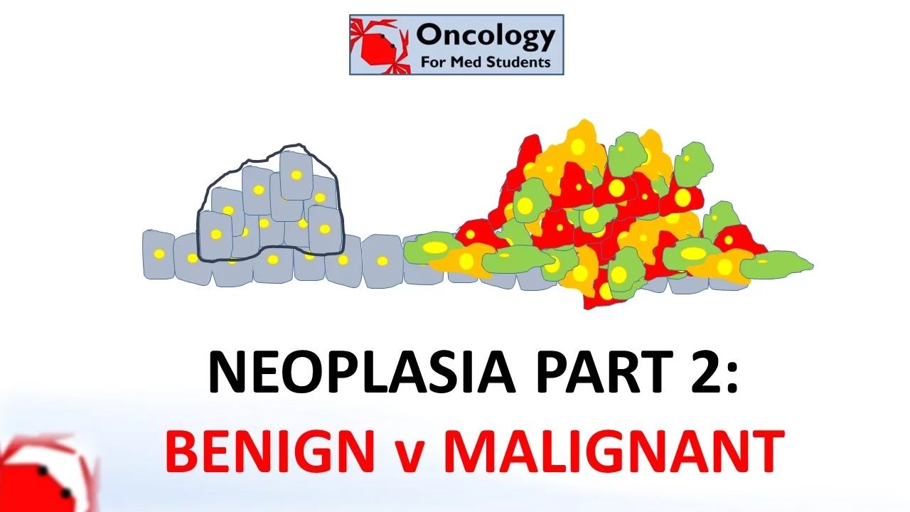
2. Neoplasia part 2: Differences between benign and malignant neoplasms
175.1K
Benign and malignant neoplasms differ in four key areas: (1) Differentiation - malignant tumors are less well-differentiated and appear more abnormal, while benign tumors maintain tissue-like appearance; (2) Rate of growth - malignant tumors generally grow faster than benign ones; (3) Local invasion - malignant tumors invade surrounding tissues and lack a fibrous capsule, making complete removal difficult; (4) Metastasis - malignant tumors can spread to distant sites through bloodstream, lymphatics, or body cavities, while benign tumors never metastasize. Even though benign tumors are usually harmless, they can cause problems when located in critical areas like the brain (due to pressure effects), when functioning as hormone-secreting adenomas, or when polyps may progress to malignancy.
Click to watch →
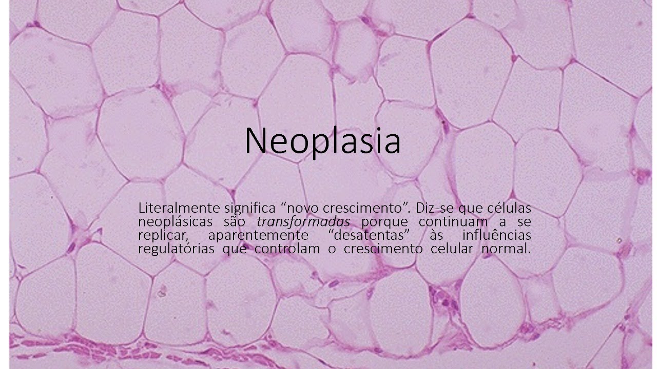
Neoplasia
6.6K
The main differences between benign and malignant neoplasias include: (1) Benign tumors are well-differentiated with cells that have specific functions, while malignant tumors are poorly differentiated with anaplastic cells; (2) Benign tumors are typically encapsulated, while malignant tumors invade surrounding tissues; (3) Benign tumors grow slowly and do not metastasize, while malignant tumors grow rapidly and can spread to distant sites.
Click to watch →
Deep Dive
Basic histology of epithelial tissues, specifically the structure and function of the basement membrane and the extracellular matrix (ECM).
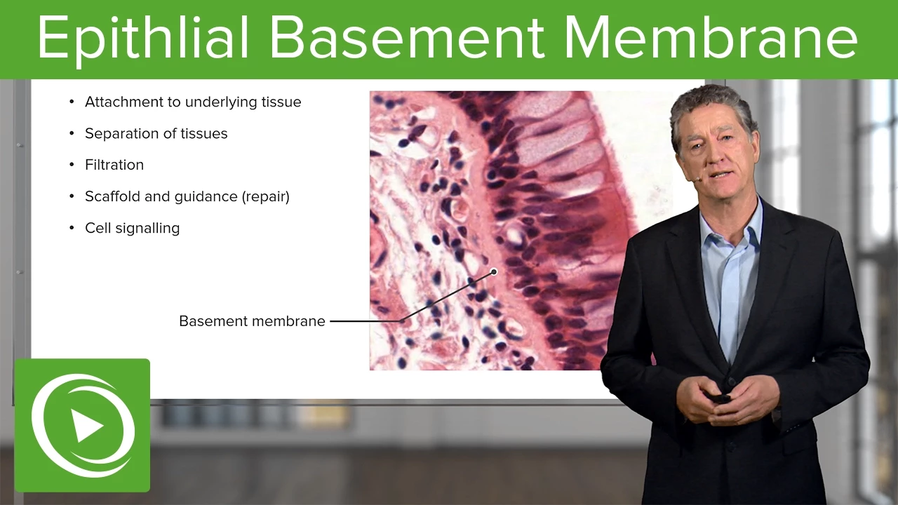
Epithlial Basement Membrane – Histology | Lecturio
53.5K
The basement membrane is a specialized extracellular matrix structure that supports epithelial tissues; it consists of two layers visible under electron microscopy—the basal lamina secreted by epithelial cells and the reticular lamina secreted by connective tissue cells—with hemidesmosomes anchoring epithelial cells to this membrane, enabling functions such as tissue separation, filtration, scaffold provision, and cell signaling.
Click to watch →

Tejido epitelial parte 1 de cuatro. Aldana Marcos Hernán
85.1K
The basement membrane (or basal lamina) is a specialized structure between epithelial tissue and connective tissue. It acts as a filter and provides attachment between the two tissue types. The basement membrane is produced by the epithelial cells themselves and contains complex molecules that help anchor the epithelium to the underlying connective tissue. Epithelial tissues perform various functions including absorption, secretion, protection, sensation, and storage.
Click to watch →

Tejido Epitelial (Parte 1) - Histología
38.3K
The basement membrane is a specialized extracellular matrix that anchors epithelial tissue to underlying connective tissue. It consists of: (1) Lamina lucida - electron-lucent layer immediately adjacent to the epithelial cell membrane; (2) Lamina densa - electron-dense layer containing collagen and other proteins; (3) Lamina reticularis - underlying connective tissue layer. The basement membrane is visible with special staining techniques (e.g., PAS stain) under light microscopy. It provides structural support, facilitates cell attachment, and serves as a barrier. In diseases like pemphigus vulgaris, autoantibodies attack hemidesmosomal components, causing separation of epithelium from the basement membrane and blister formation.
Click to watch →
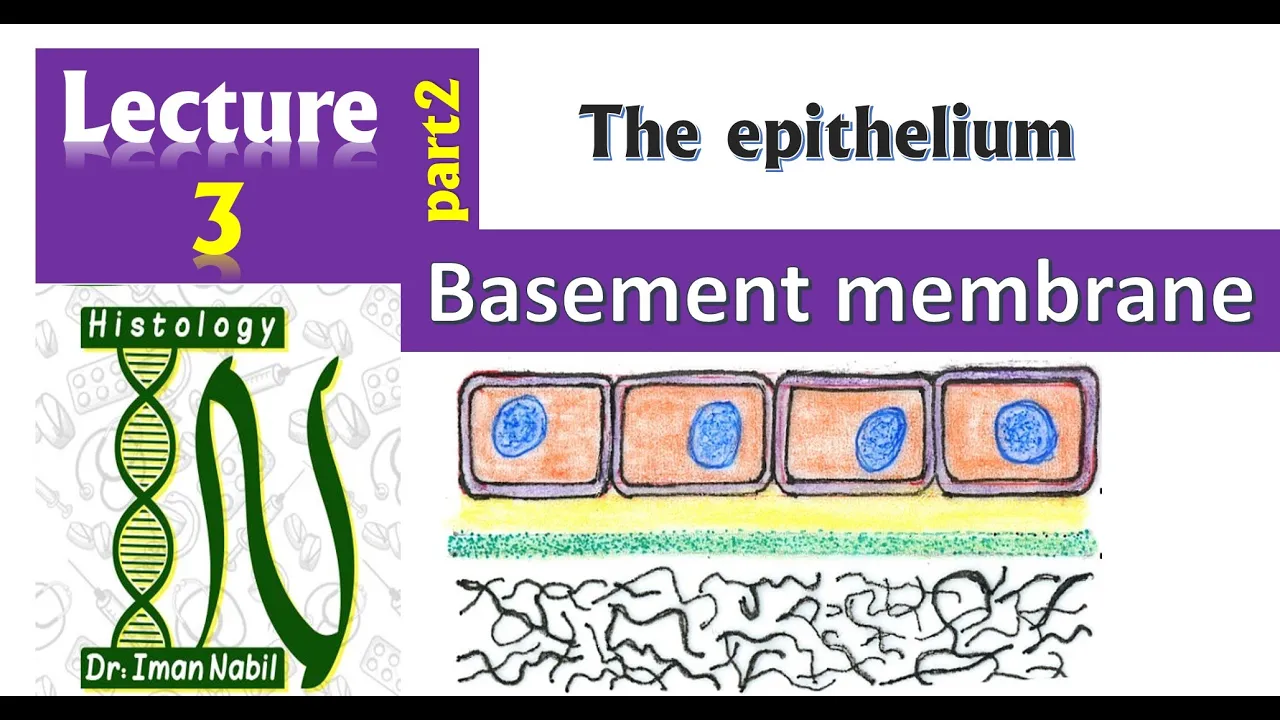
Structure of Basement membrane-2-Epithelium
47.2K
The basement membrane is a thin extracellular layer between epithelial cells and underlying connective tissue, consisting of three main components: the lamina basalis (containing type IV collagen, laminin, and proteoglycans like heparan sulfate with negative charge), the lamina reticularis (a network of type III collagen fibers called reticular fibers), and anchoring fibers made of type VII collagen that connect these layers; this structure enables selective filtration based on molecular size and charge, as seen in kidney function.
Click to watch →

Epithelial Tissue Class 11 | Structural Organisation in Animals | Class 11 Biology
183.4K
Epithelial tissue is avascular (lacks blood supply) and receives nutrients from underlying connective tissue through the basement membrane. The basement membrane is a non-cellular layer composed of mucopolysaccharides that anchors epithelial tissue to connective tissue and acts as a protective barrier. The epidermis (outer skin layer) consists of multiple epithelial cell layers with the outermost layer called stratum corneum. The dermis (connective tissue layer beneath epidermis) contains blood vessels that supply the avascular epidermis. This arrangement allows for protection while maintaining tissue viability through nutrient exchange.
Click to watch →
Deep Dive
The anatomy and physiology of the circulatory (cardiovascular) and lymphatic systems, which serve as the primary pathways for systemic cell transport.
What is the Lymphatic System? (Part 1: Anatomy and Physiology)
406.1K
The human body has two main circulatory systems for transporting fluid, cells, nutrients, and waste: the Cardiovascular (Blood) System and the Lymphatic System. These systems function like mass transit systems with passengers, routes, and destinations. Both systems consist of vessels (the train tracks and roadways) and passengers (cells, molecules, and fluid). These systems transport passengers from all over the body to their destinations.
Click to watch →

MSN - cardiovascular system (PR Yadav solution - #shaktibatch #vooglysir class 1010
6.5K
William Harvey discovered blood circulation. The body has four transport systems: circulatory (transports blood, oxygen, gases, nutrients, waste), urinary (excretory), lymphatic (immune defense and fluid balance), and endocrine (hormonal regulation). The lymphatic system includes lymph vessels, nodes, lacteals, spleen, thymus, and bone marrow. Lymph nodes trap antigens and contain macrophages that destroy them. B-lymphocytes mature in bone marrow for humoral immunity, while T-lymphocytes mature in the thymus for cellular immunity. The human heart has four chambers: right atrium, left atrium, right ventricle, and left ventricle. The apex is located 7-9 cm below the sternum, in the left fifth intercostal space at the midclavicular line. Heart valves are auscultated at specific locations: aortic valve (second intercostal space right sternal border), pulmonic valve (second intercostal space left sternal border), tricuspid valve (fifth intercostal space left sternal border), and mitral valve (fifth intercostal space midclavicular line). The plebostatic axis is at the intersection of the midclavicular line and fourth intercostal space, used as the zero point for manometer calibration for central venous pressure measurement.
Click to watch →

SISTEMA CIRCULATORIO / LINFATICO
141.1K
The circulatory system consists of the heart (a muscular organ with four chambers—two auricles and two ventricles—that pumps approximately 7 liters of blood daily through four valves) and blood vessels (arteries carrying oxygen-rich blood away from the heart and veins returning oxygen-depleted blood), while the lymphatic system transports immune cells and antibodies through lymphatic vessels and nodes to provide immune defense, with both systems requiring continuous movement to function properly.
Click to watch →
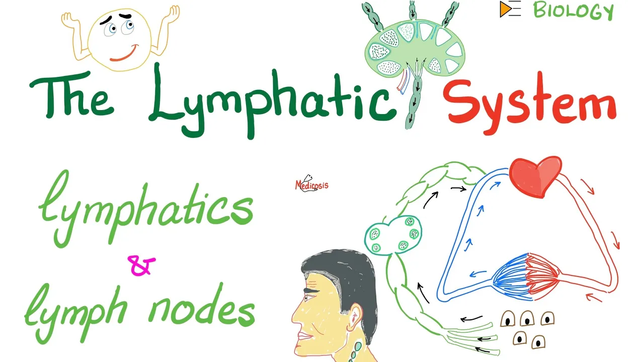
The Lymphatic System - Lymphatics and Lymph Nodes - Immunology - Biology
62.6K
The blood circulatory system is a closed loop starting from the heart's left ventricle, passing through arteries, arterioles, capillaries, venules, and veins, returning to the heart. Capillaries facilitate gas and nutrient exchange with cells. In contrast, the lymphatic system is an open system where lymph originates in interstitial fluid and terminates in veins. Approximately 10% of blood volume becomes lymph, collected by lymphatic capillaries and transported through afferent vessels to lymph nodes for filtration. This fundamental difference in system architecture explains why the lymphatic system serves as the primary route for immune surveillance and pathogen filtration.
Click to watch →

ANATOMİ FİNAL ÖZET KONU ANLATIMI TÜM ÜNİTELER #auzef #çocukgelişimi
16.2K
The cardiovascular system consists of the heart and blood vessels (arteries, veins, capillaries). The heart weighs 200-280g in females and 250-390g in males, with four chambers (two atria, two ventricles) and four valves. Blood contains plasma and cells: erythrocytes (oxygen transport), leukocytes (immune defense), and thrombocytes (clotting). Blood groups (A, B, AB, O) are determined by antigens on red blood cells. Blood pressure is measured as systolic (120 mmHg, heart contraction) and diastolic (80 mmHg, heart relaxation). Circulation includes pulmonary (right ventricle to lungs) and systemic (left ventricle to body) circuits. The lymphatic system (lymph vessels, nodes, spleen, thymus) supports immune function and fluid balance.
Click to watch →
Deep Dive
General mechanisms of cell-to-cell adhesion, including the roles of cell adhesion molecules like E-cadherin and integrins in maintaining tissue integrity.

Extracellular Matrix (ECM) CSIR NET Life Sciences Most Important Topic
490
Integrins are principal transmembrane receptors connecting cells to the extracellular matrix, consisting of alpha and beta subunits that span the plasma membrane. They function as linkers between ECM proteins and the actin cytoskeleton through adapter proteins talin and vinculin. Integrins exist in inactive and active conformational states: in the inactive state, subunits are folded together preventing matrix binding; in the active state, separation exposes binding sites for both ECM ligands and cytoskeletal linkers. Two activation mechanisms exist: outside-in signaling occurs when ECM binding induces conformational changes exposing intracellular sites; inside-out signaling occurs when intracellular signals promote talin binding to beta chains, blocking alpha-beta interactions and triggering activation. Cell-cell adhesion is mediated by cell adhesion molecules (CAMs) including selectins, cadherins, immunoglobulin superfamily members, and integrins. Syndecans represent membrane-anchored proteoglycans interacting with actin cytoskeleton and signaling molecules. These molecular systems collectively maintain tissue integrity, coordinate cellular responses to environmental cues, and enable dynamic processes including development, wound healing, and immune responses.
Click to watch →
BIOL 4610 Module 4.1 Part 2
114
Cellular interactions occur through transmembrane linker proteins that form either homophilic (identical proteins binding to each other) or heterophilic (different proteins binding) interactions; cadherins mediate calcium-dependent homophilic adhesion between cells, while integrins facilitate calcium-dependent heterophilic adhesion between cells and the extracellular matrix, with both types of junctions playing critical roles in tissue organization and immune responses such as leukocyte recruitment during inflammation.
Click to watch →

Cell Adhesion and cell to cell recognition
1.4K
Cell adhesion is the fundamental process by which cells attach to each other and form tissue architecture, mediated by cell adhesion molecules (CAMs) that include cadherins (calcium-dependent homophilic interactions), integrins (cell-matrix interactions), selectins (leukocyte homing), and immunoglobulin superfamily members; these CAMs cluster at specific junctions such as adherens junctions (cadherin-catenin-actin links), desmosomes (plakoglobin-plakophilin-intermediate filament links), tight junctions (claudin-based barriers), and gap junctions (connexin channels for direct cytoplasmic communication), each providing distinct structural and functional roles in tissue organization, mechanical strength, barrier formation, and intercellular signaling.
Click to watch →

Introduction #01 | Biologie cellulaire | 1er année universitaire (SNV)
15.3K
Cell adhesion molecules include cadherins and integrins. Cadherins are calcium-dependent adhesion proteins named after their tissue of discovery (E-cadherin in epithelial cells, N-cadherin in neurons, P-cadherin in placenta, V-cadherin in endothelial cells). Integrins are transmembrane proteins that connect cells to the extracellular matrix (collagen, fibronectin) and are involved in cell migration, adhesion, polarity, survival, proliferation, and differentiation.
Click to watch →
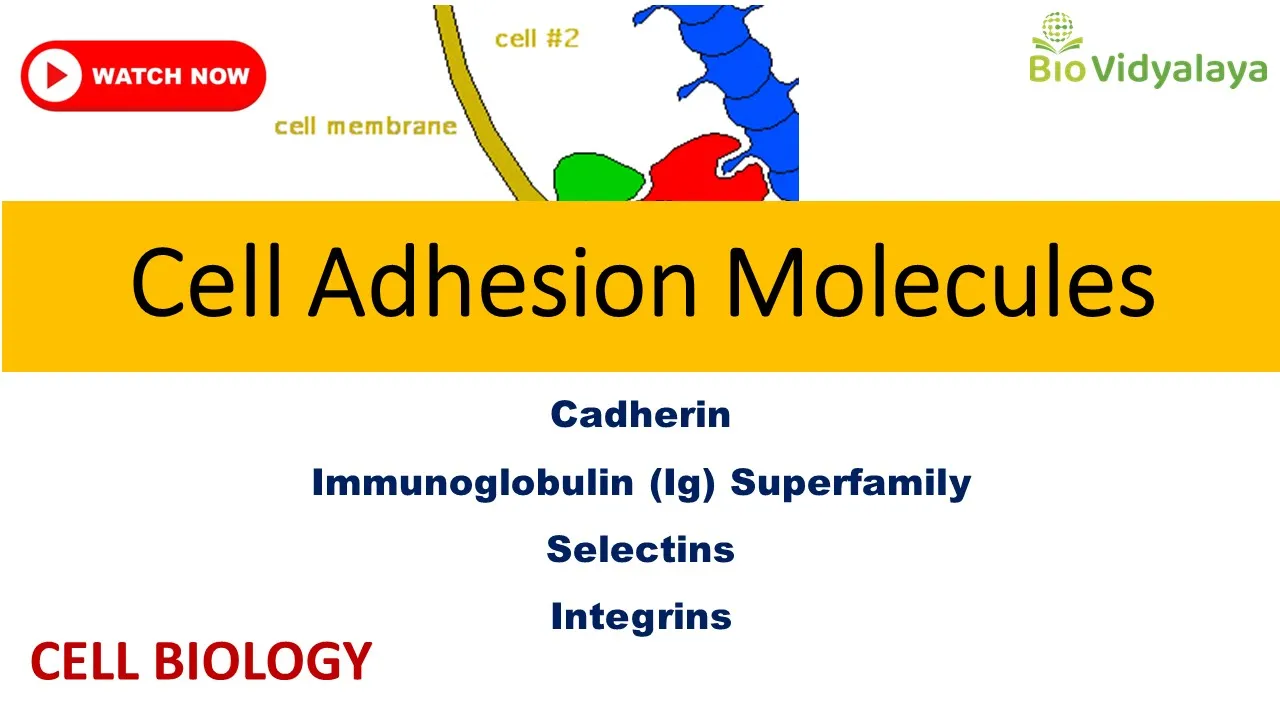
Cell Adhesion Molecules: Cadherin, Selectin, IgSF, Integrin | CELL BIOLOGY CSIR NET Lifescience
14.4K
Cell adhesion involves cell binding to extracellular matrix or other cells via cell adhesion molecules (CAMs)—transmembrane proteins with intracellular (cytoskeleton interaction), transmembrane, and extracellular domains. CAMs enable homophilic (identical CAMs) or heterophilic (different CAMs) interactions. Four major CAM families exist: cadherins, immunoglobulin superfamily, selectins, and integrins. Cadherins are calcium-dependent cell-cell adhesion molecules with extracellular cadherin domains containing calcium binding sites between domains. The C-terminal interacts with cytoskeleton via adapter proteins (beta-catenin, alpha-catenin, plakoglobin). Classical cadherins include E-cadherin (epithelial), N-cadherin (neural), and VE-cadherin (endothelial). Non-classical cadherins include desmosomal cadherins (desmocollin, desmoglein) forming desmosomes, and T-cadherin/LI-cadherin with GPI anchors. Protocadherins in the nervous system show homophilic binding and contribute to neural development.
Click to watch →
Prerequisite Knowledge
- Concept 01The fundamental differences between benign and malignant tumors, including the concepts of neoplasia and clonal expansion.
- Concept 02Basic histology of epithelial tissues, specifically the structure and function of the basement membrane and the extracellular matrix (ECM).
- Concept 03The anatomy and physiology of the circulatory (cardiovascular) and lymphatic systems, which serve as the primary pathways for systemic cell transport.
- Concept 04General mechanisms of cell-to-cell adhesion, including the roles of cell adhesion molecules like E-cadherin and integrins in maintaining tissue integrity.
Subsequent Learning
- Step 01The molecular biology of the Epithelial-to-Mesenchymal Transition (EMT) and how it confers migratory and invasive capabilities to carcinoma cells.
- Step 02Clinical application of the TNM (Tumor, Node, Metastasis) staging system and how staging dictates prognosis and guides oncological treatment protocols.
- Step 03Therapeutic strategies designed to target metastatic pathways, such as angiogenesis inhibitors and matrix metalloproteinase (MMP) inhibitors.
- Step 04The concept of 'organotropism' (the seed and soil hypothesis) and how the microenvironment of specific target organs attracts circulating tumor cells.
Metastasis Path
Invasion Steps
Final Colonization
Metastasis Path
0:00Playing Section
- 1
Defines metastasis and names three spread routes.
- 2
Explains venous invasion as most common hematogenous path.
The Early Dissemination and Parallel Progression Model
While the traditional linear cascade model views metastasis as a late-stage event occurring after a primary tumor has fully matured, the 'Early Dissemination and Parallel Progression' model offers a paradigm-shifting counterpoint. It proposes that cancer cells can disseminate to distant organs very early in tumor development, often before the primary tumor is clinically detectable or has undergone significant local invasion. These early-dispersed cells then evolve independently at secondary sites in parallel with the primary tumor. This challenges the classic assumption that metastatic capability is acquired late in tumor evolution and suggests that conventional anatomical staging (such as TNM) may fail to accurately predict or reflect the true timeline of systemic disease spread.
Deep Dive
The molecular biology of the Epithelial-to-Mesenchymal Transition (EMT) and how it confers migratory and invasive capabilities to carcinoma cells.

Principles of Oncology (Basics of Oncology) | Bailey 28th T&Ds |
5K
EMT is the process where epithelial cancer cells transform into mesenchymal cells to acquire migratory and invasive capabilities. Key regulators include SRC (sarcoma gene), Ras (intracellular signaling), integrins (cell-cell adhesion), cadherins/cam/camL (cellular connection molecules), Wnt/β-catenin pathway (colorectal cancer), and Notch pathway (basal cell carcinoma). This transition enables cells to access blood vessels for metastasis.
Click to watch →

El 90% de las Muertes por Cáncer Son por Esto: Invasión y Metástasis
180
Epithelial-mesenchymal transition (EMT) is a phenotypic switch where epithelial cells lose characteristics like E-cadherin, intercellular junctions, and apical polarity, while gaining mesenchymal features including N-cadherin, vimentin, migratory capacity, and apoptosis resistance. This process, normally involved in embryonic development and wound healing, is reactivated in cancer by transcription factors (Snail, Slug, Twist, ZEB1, ZEB2) and signaling pathways (TGF-β, WNT, Notch). EMT is typically incomplete, with metastatic cells often in hybrid states combining epithelial and mesenchymal features. EMT enables local invasion, intravasation, and confers drug resistance, and is reversible.
Click to watch →

Epithelial to Mesenchymal Transition | EMT | Molecular mechanism of EMT | significance of EMT
36.8K
Epithelial to Mesenchymal Transition (EMT) is a precisely regulated molecular process where epithelial cells lose their polarity, cell-cell adhesion, and become mesenchymal cells with enhanced migratory and invasive properties; this transition is characterized by loss of E-cadherin and cytokeratins, along with upregulation of N-cadherin, Snail, and Slug, and is triggered by transcription factors like ZEB1, Twist, and Snail through signaling pathways including TGF-beta and Notch/Delta, playing crucial roles in embryonic development (neural crest formation, mesoderm development), wound healing, and cancer metastasis.
Click to watch →

Lecture 11.1: Introduction to Cancer Biology - Part III
223
Epithelial-mesenchymal transition (EMT) is a key process in cancer metastasis where epithelial cells lose cell-cell adhesion (E-cadherin downregulation), acquire mesenchymal characteristics (increased motility, invasiveness), undergo morphological changes (loss of polarity), and degrade basement membrane and extracellular matrix. EMT molecular mechanisms involve transcription factors (SNAIL, TWIST, ZEB1) activated by signaling pathways (Wnt, TGF-β, RTK). Cancer cell migration patterns include single cell migration, collective migration, cell cluster migration, cord migration, and pushing growth. These patterns affect how cancer cells spread and metastasize.
Click to watch →

Dr. Robert Weinberg - "Cancer Stem Cells: A New Target in the Fight Against Cancer"
55.3K
Epithelial-mesenchymal transition (EMT) is an ancient developmental program (invented ~600-650 million years ago) where epithelial cells convert to motile mesenchymal cells. This program plays critical roles in embryonic development including neural crest formation and melanocyte migration. Carcinoma cells exploit EMT to acquire malignant traits: increased motility, invasiveness, metastatic potential, and therapy resistance. Research revealed that EMT does not simply create fibroblast-like cells but generates cells with stem cell characteristics, connecting EMT to cancer stem cell biology and explaining how tumors become aggressive and treatment-resistant.
Click to watch →
Deep Dive
Clinical application of the TNM (Tumor, Node, Metastasis) staging system and how staging dictates prognosis and guides oncological treatment protocols.

NORCET-2022 || MSN (Oncology) #3 || Important Questions || AIIMS || By Siddharth Sir
26.9K
The TNM classification system is used to stage cancer and determine prognosis, where T indicates tumor size (T0: no tumor, T1: ≤1cm, T2: >1-2cm, T3: >2-5cm, T4: >5cm), N indicates lymph node involvement (N0: none, N1: single node, N2: multiple nodes but not all, N3: all nodes involved), and M indicates metastasis (M0: none, M1: present). This staging system helps predict patient outcomes, with Stage I having 50-90% survival rates, Stage II having 20-30%, and Stage IV having approximately 5% survival rates.
Click to watch →

Рак молочной железы, виды рака, лечение и прогнозы выживаемости - врач-онколог Александр Гусляков
58.2K
TNM is the universal international classification system for staging any malignant process, developed by French scientist Denoix in 1942. T stands for tumor (size and extent), N for lymph node involvement, and M for metastasis. This standardized language allows oncologists worldwide to communicate about cancer staging regardless of nationality or language. The classification is assigned once at diagnosis and rarely changed during treatment unless there is significant disease progression. TNM staging determines treatment planning and prognosis for each patient.
Click to watch →
Breast Cancer
50.4K
Clinical examination positions: sitting semi-recumbent for inspection, supine for palpation. Arm positions: by side for mobility, raised above head for fixity to pectoralis major. Fixity to chest wall (ribs, intercostal muscles, serratus anterior) is checked by forward bending - if breast doesn't fall forward, tumor has infiltrated chest wall. Triple assessment includes clinical, radiological (mammography - always bilateral, CC and MLO views), and pathological (core biopsy preferred over FNAC for invasion/grade/receptor status). Multifocal (multiple lesions in one quadrant) vs multicentric (different quadrants, >4 cm apart). TNM staging: T1 <2 cm, T2 2-5 cm, T3 >5 cm. T4 includes T4A (chest wall), T4B (skin involvement - peau d'orange, satellite nodules, ulceration), T4C (both), T4D (inflammatory carcinoma). N1 (mobile axillary nodes), N2 (fixed), N3 (supraclavicular/infraclavicular/internal mammary). M0 (no metastasis), M1 (distant). Early breast cancer (Stage 1-2) treatment: modified radical mastectomy (removes breast, nipple complex, skin over tumor, and level 1-2 axillary lymph nodes) or breast conservation surgery (removes tumor with 1 cm margin plus radiotherapy). BCS is oncologically equivalent to mastectomy with better psychological quality of life. Contraindications for BCS: multicentricity, central/nipple complex tumors, BRCA mutation, persistently positive margins. Sentinel lymph node biopsy identifies first draining node using methylene blue or radioactive sulfur colloid. If negative, no axillary dissection needed. If positive, level 1-2 nodes are removed. Modified radical mastectomy approaches: Axillary clearance (retract pectoralis minor), Scapular (split pectoralis minor), Exin (retract without splitting). Level 3 dissection is intraoperative if level 2 nodes are hard. Boundaries: superiorly axillary vein, inferiorly latissimus dorsi pedicle, anteriorly pectoralis major, posteriorly latissimus dorsi, floor subscapularis. Structures to preserve: long thoracic nerve of Bell (damage causes winging), latissimus dorsi pedicle and nerve. Minimum 10 lymph nodes for adequate staging.
Click to watch →

Stages of Cancer: Tumor Staging and Grading TNM System Nursing NCLEX Review
353.2K
Cancer tumors are graded based on how abnormal cells appear under a microscope, with Grade I being well-differentiated (low grade, slow-growing) and Grade IV being undifferentiated (high grade, fast-spreading); cancer is staged using the TNM system (Tumor, Nodal involvement, Metastasis) which categorizes the primary tumor size and invasion (T), regional lymph node spread (N), and distant metastasis (M) to determine treatment approaches and prognosis.
Click to watch →

Breast Cancer | Clinical Medicine
67.4K
T staging: T1 (<2cm), T2 (2-5cm), T3 (>5cm), T4 (invasive to chest wall/skin). N staging: N0 (no node involvement), N1 (1-3 axillary nodes), N2 (≥4 axillary or any clavicular nodes), N3 (multiple nodes). M staging: M0 (no distant metastasis), M1 (distant metastasis).
Click to watch →
Deep Dive
Therapeutic strategies designed to target metastatic pathways, such as angiogenesis inhibitors and matrix metalloproteinase (MMP) inhibitors.
Metastasis Angiogenesis
915
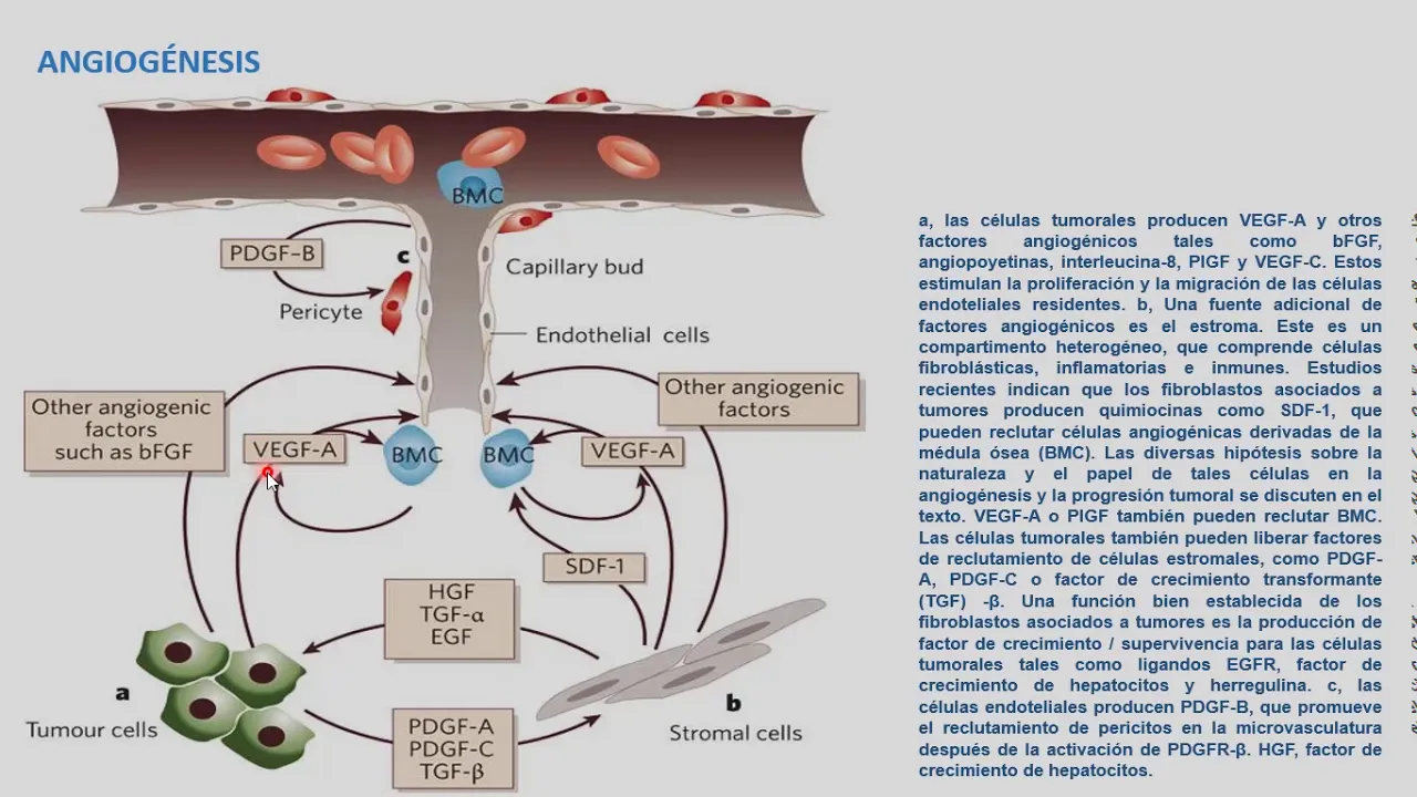
MMPs promote tumor progression through multiple mechanisms: releasing growth factors bound to the matrix, degrading the basement membrane to enable invasion, activating other MMPs, and activating integrins on cancer cells. The tumor microenvironment contains multiple cell types (tumor cells, stromal cells, immune cells) that contribute to MMP production. MMPs also help cancer cells evade immune recognition by degrading components of the extracellular matrix that would otherwise present tumor antigens to immune cells. MMP inhibitors have been developed as cancer drugs, but clinical efficacy has been limited. Alternative approaches target upstream regulators like HIF or NF-κB. MMPs represent important therapeutic targets for preventing tumor invasion and metastasis. The balance between MMPs and their inhibitors determines the extent of matrix remodeling and tumor progression.
Click to watch →
mod11lec62
2.5K
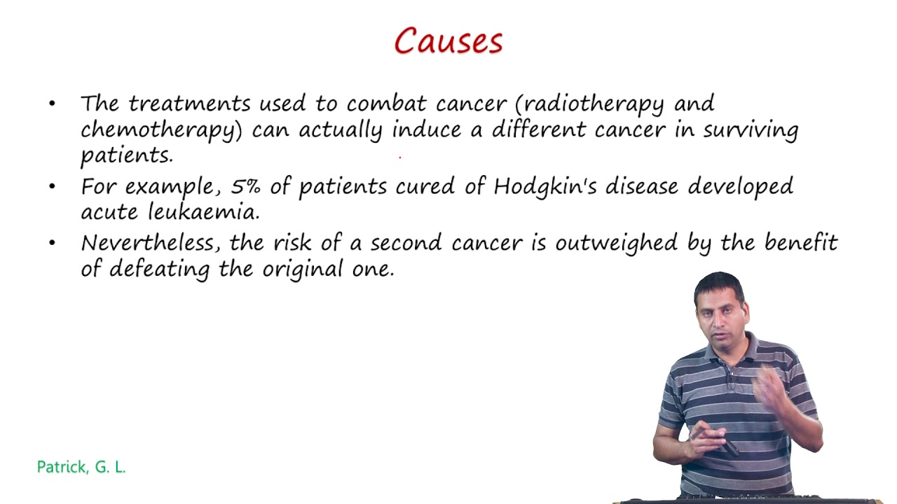
As tumors grow, cells become remote from blood supply, creating hypoxic conditions that trigger hypoxia-inducible factors (HIF). HIF activates genes releasing VEGF and FGF2, stimulating angiogenesis. Normally controlled by angiostatin and thrombospondin, this balance is disrupted in tumors, allowing increased blood supply and metastasis. Angiogenesis produces abnormal, disorganized, leaky blood vessels with integrins protecting new cells from apoptosis. Matrix metalloproteinases (MMPs) must break down basement membranes for angiogenesis to begin. Angiogenesis inhibitors are safer than cytotoxic agents but work best in combination. Drugs can be delivered via liposomes that exploit leaky tumor vasculature.
Click to watch →
Mechanisms of Angiogenesis Inhibitors in Metastatic Colorectal Cancer
4.2K
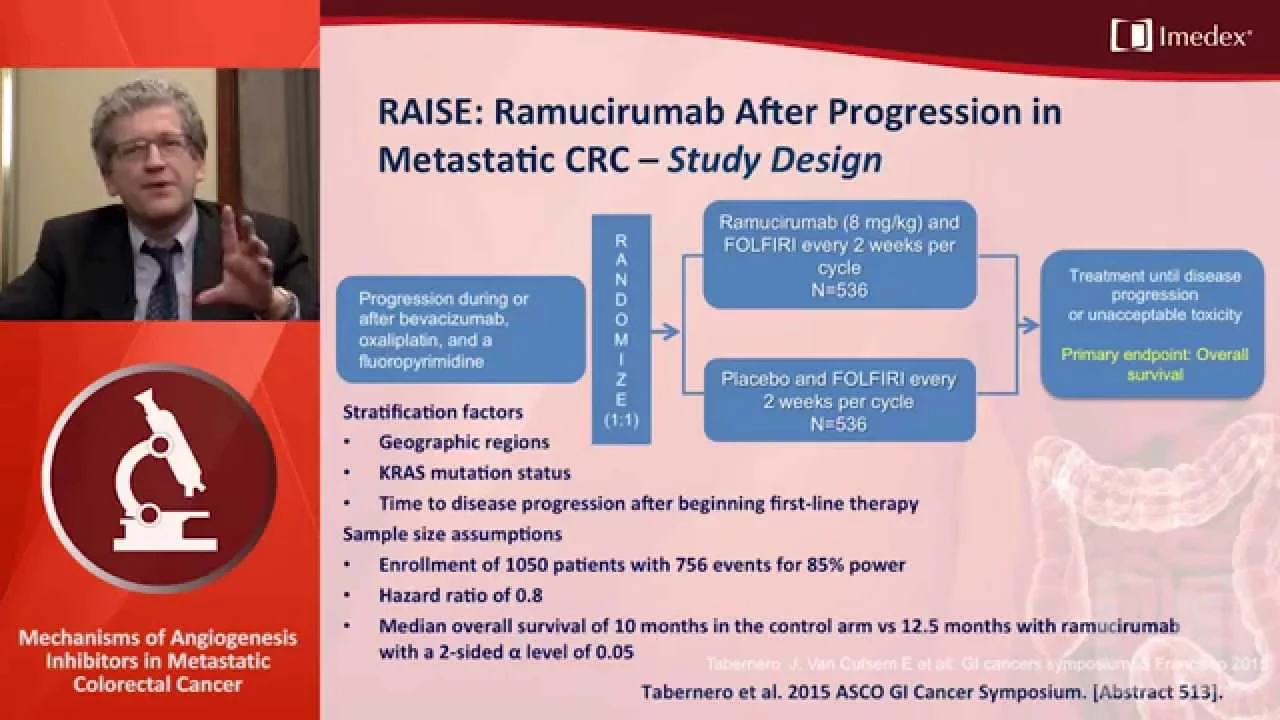
Angiogenesis inhibition in metastatic colorectal cancer targets the VEGF pathway to block tumor blood vessel formation; the process involves tumor hypoxia triggering VEGF production, which promotes chaotic vasculature, and antiangiogenic agents like bevacizumab, aflibercept, ramucirumab, and regorafenib work through different mechanisms (monoclonal antibodies, fusion proteins, tyrosine kinase inhibitors) to inhibit VEGF signaling, with clinical evidence showing improved survival and progression-free survival in both first-line and second-line treatment settings.
Click to watch →

Anticancer Drugs (Part-17 Final) Angiogenesis Inhibitors = Bevacizumab | Online Test Link
11.7K
Angiogenesis inhibitors are targeted anticancer drugs that block the formation of new blood vessels (angiogenesis), which tumors need to grow and spread; these drugs work by inhibiting vascular endothelial growth factor (VEGF) signaling, thereby cutting off the blood supply to tumors and preventing their growth and metastasis. A key example is bevacizumab, a monoclonal antibody that binds to VEGF and prevents it from activating its receptor, thus stopping the angiogenesis process. These targeted therapies offer advantages over conventional chemotherapy by specifically targeting cancer-associated pathways while minimizing damage to normal cells, though they can cause side effects such as increased risk of arterial thromboembolism and bleeding.
Click to watch →

PART 3: Clinical Utility of Angiogenesis Inhibitors in Metastatic Colorectal Cancer
261
Angiogenesis inhibitors targeting VEGF and FGFR pathways represent important therapeutic strategies in metastatic colorectal cancer, but their clinical application requires careful consideration of toxicity profiles, optimal dosing combinations, and the absence of definitive predictive biomarkers; while these agents offer significant clinical benefit, their integration into treatment algorithms must balance efficacy against manageable side effects, particularly when combining multiple targeted agents.
Click to watch →
Deep Dive
The concept of 'organotropism' (the seed and soil hypothesis) and how the microenvironment of specific target organs attracts circulating tumor cells.

Focal Adhesion Kinase Inhibition to Improve Losartan-Sunitinib Immunotherapy in Metastatic Osteo
311
The seed and soil hypothesis, introduced by Stephen Paget in the late 1800s, proposes that cancer metastasis depends on specific interactions between primary tumors ('seeds') and receptive target organs ('soil'). For many years discounted in favor of anatomical explanations, this concept has been validated by modern research showing that soluble factors from primary tumors circulate and prime distant organs to become receptive to metastatic cells. Tumor-derived exosomes—nano-sized particles carrying DNA, RNA, and proteins—function like hormones, traveling through the bloodstream to condition distant organs before metastatic cells arrive. This creates a receptive microenvironment that facilitates metastatic colonization, explaining why certain organs are preferentially targeted by specific cancers.
Click to watch →
Lecture 11.1: Introduction to Cancer Biology - Part III
223
Cancer cell dissemination routes include lymphatic spread (to lymph nodes) and hematogenous spread (through blood circulation). Circulating tumor cells (CTCs) can be detected in blood for prognosis. The seed and soil hypothesis of metastasis proposes that cancer cells (seeds) must find compatible microenvironments (soil) to grow. Different cancers preferentially metastasize to specific organs based on microenvironment compatibility. This explains why certain cancers commonly metastasize to specific organs (e.g., lung cancer to brain, breast cancer to bone).
Click to watch →
BIOLOGÍA MOLECULAR: Biología Molecular en la metástasis.
870
CTCs employ multiple survival strategies: clustering, platelet coating, and immunosuppressive signaling. Tumor cells secrete exosomes containing microRNAs that prepare pre-metastatic niches in distant organs. Organotropism explains why certain cancers metastasize to specific organs: lung cancer favors brain, bones, and adrenal glands; renal cell carcinoma targets lungs, liver, and bones; pancreatic cancer spreads to liver, stomach, and peritoneum. High mutational burden correlates with increased metastatic potential and responsiveness to targeted therapies.
Click to watch →

Seminario 23 Algunos mecanismos de carcinogénesis - Federico Waisberg
3.3K
Metastasis begins with lymphatic dissemination due to vessel permeability and exosome-mediated lymphatic development. Tumor-platelet interactions favor thrombotic events. Extravasation involves selectin-mediated initial adhesion, integrin-mediated stable adhesion, and diapedesis through vessel walls. Organotropism explains why breast cancer metastasizes to bone, colorectal to liver, and pancreatic to liver. The seed and soil theory proposes tumor-derived signals (like PTHrP activating osteoclasts) prepare favorable pre-metastatic niches. Exosomes carrying specific integrins (beta-1 alpha-6 for lung, alpha-v beta-5 for liver) provide molecular specificity for target organ selection. The pre-metastatic niche features high P38 activity, low ERK activity, fibroblasts with easily invaded extracellular matrix, and favorable leukocytes.
Click to watch →

How Does Cancer Spread To Other Parts of the Body and how to Prevent it? @mdanderson
168
The seed and soil hypothesis describes metastasis as interaction between tumor cells and distant organ microenvironments. Cells undergoing partial EMT (hybrid state) are most successful at establishing metastases. Metastatic cells can enter dormancy, surviving 10-15 years without dividing by resisting apoptosis. Organ tropism explains why cancers preferentially metastasize to specific organs: drainage patterns (blood vessel connections) and microenvironment suitability. Pancreatic cancer commonly metastasizes to the liver due to vascular drainage and the liver's growth factor-rich environment.
Click to watch →
Metastasis Path
Invasion Steps
Final Colonization
Metastasis Path
0:00- 1
Defines metastasis and names three spread routes.
- 2
Explains venous invasion as most common hematogenous path.
The Early Dissemination and Parallel Progression Model
While the traditional linear cascade model views metastasis as a late-stage event occurring after a primary tumor has fully matured, the 'Early Dissemination and Parallel Progression' model offers a paradigm-shifting counterpoint. It proposes that cancer cells can disseminate to distant organs very early in tumor development, often before the primary tumor is clinically detectable or has undergone significant local invasion. These early-dispersed cells then evolve independently at secondary sites in parallel with the primary tumor. This challenges the classic assumption that metastatic capability is acquired late in tumor evolution and suggests that conventional anatomical staging (such as TNM) may fail to accurately predict or reflect the true timeline of systemic disease spread.
The fundamental differences between benign and malignant tumors, including the concepts of neoplasia and clonal expansion.
Basic histology of epithelial tissues, specifically the structure and function of the basement membrane and the extracellular matrix (ECM).
The anatomy and physiology of the circulatory (cardiovascular) and lymphatic systems, which serve as the primary pathways for systemic cell transport.
General mechanisms of cell-to-cell adhesion, including the roles of cell adhesion molecules like E-cadherin and integrins in maintaining tissue integrity.
The molecular biology of the Epithelial-to-Mesenchymal Transition (EMT) and how it confers migratory and invasive capabilities to carcinoma cells.
Clinical application of the TNM (Tumor, Node, Metastasis) staging system and how staging dictates prognosis and guides oncological treatment protocols.
Therapeutic strategies designed to target metastatic pathways, such as angiogenesis inhibitors and matrix metalloproteinase (MMP) inhibitors.
The concept of 'organotropism' (the seed and soil hypothesis) and how the microenvironment of specific target organs attracts circulating tumor cells.
1.4K views116likes4:20@3dmedicalillustrationsOriginal Release: 2022-01-23
metastasis is the word used to describe a cluster of cancer cells in one area that arose from a cancer in another region of the body cancer that has spread in this way is called metastatic cancer metastatic cancer is named based on the site where the cancer began metastasis occurs by the following three roots transcholomic lymphatic spread and hematogenous spread the model in this video details the hematogenous or lymphatic root of metastasis you can see the blood vessels in this view that is the artery and the vein the veins have a thinner wall and a thin basement membrane as compared to an arterial wall making venous damage the commonest mode of hematogenous spread now you see the epithelial basement membrane which is the limiting structure defining whether the epithelial tumor is invasive or non-invasive the epithelial tumor as seen here is a mass of abnormal cells well vascularized with arteries and its branches some of this abnormal cells develop the special ability to invade the basement membrane this is the first step of metastasis the transformed cell that migrate within the tissue to the venules or lymphatic channels in the second step of metastasis the transformed malignant cell penetrate or invis intravasate into the vascular spaces or lymphatic spaces during intravasation the tumor cells invade across the basement membrane of the capillary or lymphatic channels the intravasated neoplastic cells now in the third step of metastasis are ready for their systemic dispersal to all tissues of the body and their success in metastasis requires that they survive the immune defense system formed by the lymphoid cells to invade the various defense and damaging events in the stage of vascular or lymphatic transport the cells form tumor ebola made of neoplastic cells platelets and fibrin in the fourth step the neoplastic cells that survive the hostile environments during transportation then arrest in in the capillary or lymphatic beds of distant organs or lymph nodes and engage in a second round of invasion a extravasation whereby neoplastic cells exit from the luminal spaces into the surrounding stromal or lymphoid tissue the fifth step is concerned with the progression of the colonizing tumor cells or micro metastasis to replicate in this new environment their neovascularization and progressive enlargement of the tumors that will threaten the survival of the host when the basement is intact that is called carcinoma in situ but when basement is not intact that is invasive carcinoma you
Up Next
26:44
Neoplasia: Benign vs Malignant Tumors & Hallmarks of Cancer
@medtoday
278.3K views•2020-11-20

48:47
Graphic Medicine: Comics for Collaborative Healthcare Communication
@nationalpatientadvocate
189 views•2023-12-04

10:28
Neuroanatomy: Central and Peripheral Nervous System Divisions Explained
@AKLECTURES
136.2K views•2014-09-20

3:05
Stages of Labor and Vaginal Birth | Childbirth Animation
@nucleusmedicalmedia
52.1M views•2017-08-18
Related Study Plans & Knowledge Roadmaps
Structured learning paths in Medicine
medicine
Abnormal LFTs: Diagnostic Workup & Pathology
01Liver Anatomy and Basic Physiology
02Introduction to Liver Function Tests (LFTs)
03Hepatocellular vs. Cholestatic Patterns
+ 3 more learning modules
12h6 modules
Explore Roadmap
medicine
Analyzing Blood Smears: RBC, WBC & Platelets
01Introduction to Blood Components & Smear Preparation
02Normal Morphology of Blood Cells
03Erythrocyte Pathologies & Morphological Variants
+ 2 more learning modules
12h5 modules
Explore Roadmap
medicine
Mechanical Ventilation: Waveforms & Alarms
01Anatomy and Physiology of Breathing
02Introduction to Mechanical Ventilation & Core Modes
03Ventilator Settings and Waveform Interpretation
+ 2 more learning modules
12h5 modules
Explore Roadmap
medicine
Neuro Exam: Localization & Stroke Syndromes
01Foundations of Neuroanatomy and Vascular Supply
02The Neurological Examination: Step-by-Step
03Principles of Neurological Localization
+ 3 more learning modules
18h6 modules
Explore Roadmap