















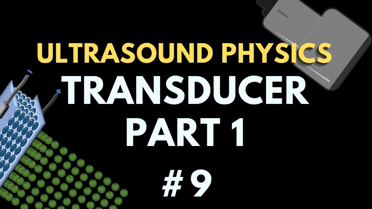







The piezoelectric effect is the conversion of mechanical energy into electrical energy, where compression of piezoelectric material (such as PZT crystals) generates an electrical current that can be measured by electrodes and converted into ultrasound images; conversely, the reverse piezoelectric effect converts electrical energy into mechanical energy, where applying an electric current causes the piezoelectric material to change shape, creating regions of compression and rarefaction that propagate as ultrasound waves through tissues. This bidirectional property enables ultrasound transducers to both generate ultrasound pulses for imaging and receive returning echoes from tissues.
Piezoelectric Effect and Reverse Piezoelectric Effect in Ultrasound
Added:Basic wave mechanics, including sound wave propagation, frequency, wavelength, and acoustic pressure.
Fundamentals of electromagnetism, specifically electric fields, electric charge polarization, and voltage.
Introductory solid-state physics, focusing on crystal lattice structures and non-centrosymmetric crystal symmetry.
The concept of energy transduction, particularly the conversion between mechanical energy and electrical energy.
The construction and design of ultrasound transducers, including matching layers, backing materials, and piezoelectric elements.
Principles of acoustic impedance and wave behavior (reflection, refraction, and attenuation) at tissue boundaries.
Ultrasound imaging modalities and signal processing, such as A-mode, B-mode, M-mode, and Doppler imaging.
Therapeutic applications of ultrasound, including High-Intensity Focused Ultrasound (HIFU) and lithotripsy.
52.1K views843likes10:54Original Release: 2023-04-03
we've spent the previous two talks looking at the components of the ultrasound transducer and we saw that the piezoelectric material was both responsible for generating ultrasound waves that propagate through tissues and receiving those returning Echoes and converting that mechanical energy into an electrical signal that we can then use to create our ultrasound image and I said at the time it was the result of the piezoelectric effect and the reverse piezoelectric effect that allowed us to both receive and generate those ultrasound pulses now we saw that if we apply an alternating current to alpisoelectric material that piezoelectric material will generate an ultrasound pulse that has a frequency that's equal to that alternating current and if we applied a short sharp burst of electric current to our piezoelectric material the piezoelectric material would resonate at a frequency that's dependent on the thickness of our piezoelectric material and the speed at which sound travels through that material now we're going to look at the piezoelectric effect in a little bit more detail in order to understand how we go about both receiving and generating those pulses now this may be a little bit of an addendum to our ultrasound physics module it's probably a little bit more depth than we need to know but there are a couple of Concepts that come up here that do come up in exams and understanding it in this depth helps us to explain those Concepts further so if we have a look at our ultrasound transducer here we can see that it has the ability to both receive these returning ultrasound pulses and generate ultrasound pulses now what are ultrasound pulses exactly they are regions of compression and rare reflection they're regions of localized pressure changes it's a mechanical energy that is moving through tissues a mechanical force moving through tissues and if we open up our ultrasound transducer it's this layer here the blue layer of piezoelectric material that is responsible for both receiving those mechanical forces and generating those mechanical forces now this transducer is a multi-element transducer we've got multiple crystals multiple transducer units that are separate from one another and we've got individual control over each one of those units we can get ultrasound machines where we have a single piezoelectric material transducer layer which is known as a single element transducer and going to look closely at the single element transducer when we look at our beam characteristics but for the most part in our ultrasound course we'll be looking at multi-element transducers so how exactly does this piezoelectric material both receive those incoming pulses and generate outgoing pulses well firstly this piezoelectric material is flanked by two electrodes there is an electrode on the patient's side and an electrode on the transducer side and these electrodes are responsible for either generating current within lpzt material or receiving current that's coming from our PCT material so let's start by looking at the piezoelectric effect now the piezoelectric effect is the conversion of mechanical energy into electrical energy and it's that electrical energy that we will convert into our ultrasound image the piezoelectric effect is responsible for those returning Echoes that mechanical energy that we then want to convert into an electrical energy so let's look at our piezoelectric material that's flanked by those two electrodes and we have this mechanical energy in the form of an ultrasound wave returning towards this electrode this region of compression then then compresses our pzt material our piezoelectric material that compression results in a current that can be measured by these electrodes and that current can then be converted into a pixel value a grayscale value that we will use on our ultrasound machine now the opposite is also true if we run a current through these electrodes that current will force the piezoelectric material to change shape now that change in shape is what is responsible for the mechanical energy propagation into the tissue as the piezoelectric material expands we get regions of compression here and as it then returns to its normal shape it forms a region of rare affection and it's those alternating regions of compression and rarefaction that result in the propagation of our ultrasound wave into the patient's tissue now the electric current conversion into a mechanical energy is Watson is the reverse piezoelectric effect so let's have a closer look at the actual chemical structure of alpisoelectric material now the most common material we use is what's known as a pzt crystal P standing for lead Z for zirconium and T4 titanium a lead zirconium titanate Crystal now in its basic form a pzt crystal has outer lead atoms that form these cuboid or rhombohedral shapes within that cuboid or rhombohedral shape is oxygen atoms that surround a central Titanium or zirconium atom now when we think of oxygen atoms interacting with other atoms let's take H2O water for example that oxygen shares its valence electrons with those two hydrogens and it's the sharing of those valence electrons that form the chemical bonds between the oxygen and the hydrogen now the electrons within that H2O molecule spend the most of their time by the oxygen atom relative to those hydrogen atoms so we can think of that oxygen atom as being relatively negatively charged compared to those hydrogen atoms now the same is also true here these oxygen atoms share valence electrons with that Central Titanium or zirconium and the electrons spend greater time around those oxygen atoms we can think of these oxygen atoms as being relatively negatively charged and the essential zirconium or titanium as being relatively positively charged now if we were to take the charge of all of these oxygen atoms and average them out we would see that that negative average would form in the center of this image here and the relative positive charge of the zirconium or titanium atom would match the average of these negative charge in the center of this pzt Crystal now if that zirconium was to shift or if the titanium was to shift our positive sensor here would not match our negative average sensor here we would have formed what is known as a dipole we will have a positive end to our pzt crystal and a negative and we formed a dipole here now how exactly do we go about forming these dipoles with nlpzt Crystal well it's a process known as polling of the crystals polarization of our PCT crystals how we do that is we heat up our pzt crystals to a temperature known as the curry temperature now the Curie temperature for pzt is about 350 degrees Celsius or 660 degrees Fahrenheit there is enough energy in that system there for our pzt Crystal to have a non-dipole structure now whenever we have a pzt crystal forming for the first time these dipoles are in random orientations when we heat it up to this Curie temperature we get the reduction of those dipoles then what we can do is apply an electric current over our pzt Crystal and cool it down slowly as we cool it down while maintaining that electric current we get all of our dipoles forming in the same direction we've got all our pzt crystals now having dipoles in similar orientations then the entire pzt Crystal can take on that dipole structure having a relatively positively charged and a relatively negatively charged end to that Crystal and this is the reason why we can't autoclave our ultrasound transducers to sterilize them when we autoclave something we would put it above the curry temperature and we would lose this dipole structure we would lose this orientation when the Piezo teeth crystals then go back below that Curie temperature these dipoles will again be in a random order and that pzt Crystal wouldn't function in the way we want it to function so let's have a look at this dipole and flip it on its side we flipped this pzt Crystal on its side and we've placed electrodes on either side of that pzt Crystal now we can think of this as a single molecule but in fact in our pzt Crystal we've got thousands of molecules with all the dipoles lined up a relative positive end and a relatively negative end now the piezoelectric effect provides a mechanical compression here as we compress this molecule we move that Titanium or zirconium into the center of those oxygen molecules we are effectively reducing that dipole that movement of Charge results in a current being formed within our electrodes that is the piezoelectric effect mechanical force resulting in a current formation so as we compress this molecule we get an electric current the reverse piezoelectric effect we can then run a current through these electrodes if we were to make this electrode negatively charged it would attract that zirconium or the titanium and pull it towards that negatively charged electrode we have then again compressed that PCT Crystal we've got the reverse piezoelectric effect causing compression here and ultimately rare a faction in our tissue and if we were to then switch that current we would create a compression within the tissue and alternating between those two will give us those regions of compression and rare refraction the reverse piezoelectric effect so that in a nutshell is the piezoelectric effect and the reverse piezoelectric effect now the diagrams I've used here are mainly for your understanding there are some nuances that I've left out that I don't feel are necessary for our Radiology Physics exam and don't add any further understanding into how we go about creating and receiving Ultra sound waves now in our next talk we are going to be looking at the various different ultrasound modes and how we can use the different modes to create different ultrasound images depending on what information we want clinically so I hope this has helped I hope you're studying for your ultrasound Physics Exam is going well and if you are studying for an exam I've linked a curated past paper question Bank below where I go through actual past paper questions in video format showing you how I would go about answering those questions so if you're interested in that go check it out otherwise I'll see you all in our next talk goodbye everybody
Up Next

16:01
Doppler Effect, Equation, and Angle Correction in Ultrasound Physics
@radiologytutorials
60.1K views•2023-04-19

12:53
21cm Hyperfine Transition in Neutral Hydrogen: Radio Astronomy Basics
@AaronRobertParsons
12.4K views•2011-10-13

24:46
Continuous vs Pulsed Wave Doppler Ultrasound: Key Differences Explained
@radiologytutorials
60.2K views•2023-04-24

27:14
Entropy and the Second Law of Thermodynamics Explained
@veritasium
27.5M views•2023-07-01
Related Study Plans & Knowledge Roadmaps
Structured learning paths in Physics
physics
Nuclear Physics (Decay, Fission & Reactors)
01Atomic Structure and the Binding Force
02Radioactive Decay and Half-Life
03Nuclear Fission Mechanics and Chain Reactions
+ 2 more learning modules
12h5 modules
Explore Roadmap
physics
Soft Matter (Polymers, Colloids & Rheology)
01Foundations of Soft Matter & Thermal Forces
02Polymer Physics and Macromolecules
03Colloids, Surfactants, and Self-Assembly
+ 2 more learning modules
38h5 modules
Explore Roadmap
physics
Analytical Mechanics: Lagrangian & Hamiltonian
01Mathematical Foundations: Calculus of Variations
02From Newtonian Mechanics to Constraint Dynamics
03The Principle of Least Action & Lagrangian Mechanics
+ 3 more learning modules
28h6 modules
Explore Roadmap
physics
General Relativity (Spacetime & Cosmology)
01Foundations of Relativity: Special Relativity
02The Geometry of Spacetime: Equivalence Principle & Curvature
03Einstein's Field Equations: The Mathematics of Gravity
+ 2 more learning modules
55h5 modules
Explore Roadmap