Medical Physics: Imaging, MRI & Radiotherapy
Learning Goal: Understanding Medical Physics: The Science of Medical Imaging, MRI, and Radiation Therapy. This curriculum explores how the fundamental principles of electromagnetism, nuclear decay, quantum spins, acoustics, and high-energy radiation are applied clinically to visualize anatomy, track metabolic processes, and treat cancer safely and effectively.
- Prerequisites: Basic high school level physics (algebra-based mechanics, waves, electromagnetism) and introductory biology (cell structure, DNA).
- Estimated Total Study Time: 15 hours
Module 1: Foundations of Radiation and Matter Interaction
Understand the core structure of the electromagnetic spectrum, the physics-based distinction between ionizing and non-ionizing radiation, and how high-energy photons interact with human tissues at the atomic level.
Recommended Videos
Why this video: This video directly bridges the gap highlighted in the review feedback. It establishes a clear, visual boundary on the electromagnetic spectrum separating ionizing radiation from non-ionizing radiation. It explains why non-ionizing waves (like microwaves) only stimulate kinetic/vibrational thermal states, whereas high-frequency radiation carries sufficient quantum energy per photon to alter atomic bonds directly.
Knowledge Checkpoint
- Differentiate between ionizing and non-ionizing radiation based on photon energy thresholds.
- Explain how low-energy electromagnetic waves cause heating without altering chemical structures.
Why this video: A deeper academic lecture analyzing the quantum-level mechanisms of attenuation. It breaks down the math and structural mechanics of four primary processes: coherent scattering, the photoelectric effect, Compton scattering, and pair production. This forms the analytical backbone of how radiological tissue contrast is generated.
Knowledge Checkpoint
- Define the photoelectric effect and its relationship to the atomic number () of absorbing tissues.
- Contrast coherent scattering (elastic) with Compton scattering (inelastic) regarding energy transfer.
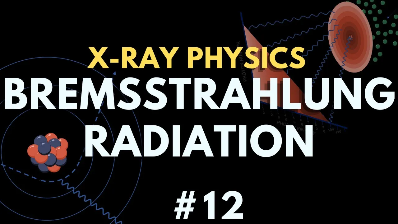
Why this video: Focuses explicitly on how diagnostic and therapeutic X-rays are physically engineered. It provides a detailed, animated breakdown of Bremsstrahlung (braking radiation) versus characteristic target interactions, explaining how electron deceleration near heavy atomic nuclei yields a continuous spectrum of diagnostic photons.
Knowledge Checkpoint
- Explain the physical mechanism behind the production of Bremsstrahlung radiation.
- Sketch or describe the continuous vs. discrete (characteristic) emission spectrum of a diagnostic X-ray tube.
Module 2: X-Ray Imaging and Computed Tomography (CT)
Learn the physical principles governing classic 2D X-ray projection radiography and the mechanical engineering that allows modern Computed Tomography (CT) systems to acquire thousands of projections to construct detailed 3D Hounsfield-mapped reconstructions.
Recommended Videos
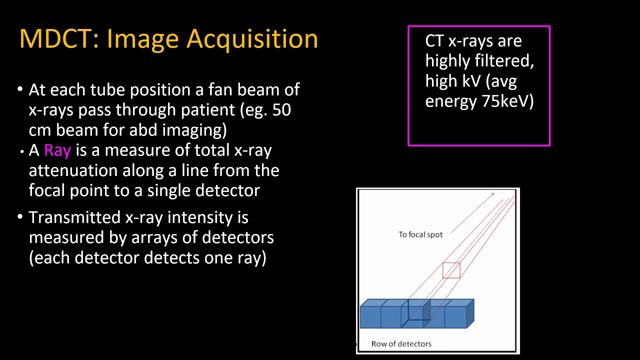
Why this video: An incredibly rigorous academic presentation covering the actual anatomy of a modern CT scanner. It covers key topics such as filtration (Bowtie filters for path-length compensation), collimation, and the physical characteristics of solid-state detector elements.
Knowledge Checkpoint
- State the function of a Bowtie filter in reducing peripheral patient dose and normalizing detector flux.
- Distinguish between pre-patient and post-patient collimation in CT imaging.
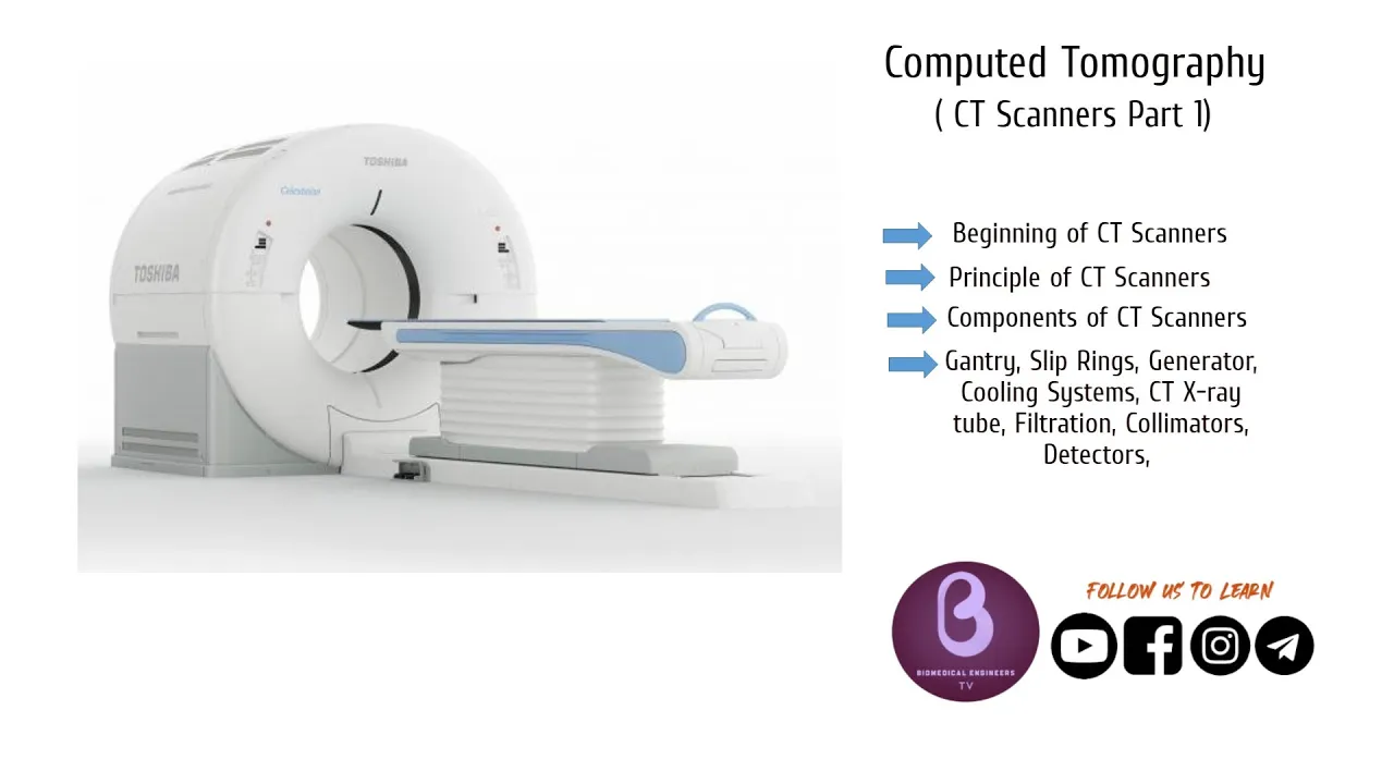
Why this video: Connects the historical timeline of CT developments (such as Godfrey Hounsfield's Nobel Prize-winning work) with modern clinical gantries. It offers a clear, dynamic look at modern helical scanning techniques.
Knowledge Checkpoint
- Explain the mechanical concept of "slip-ring" technology that enabled continuous rotational scanning.
- Define the scanning pitch in helical CT and its influence on patient dose and longitudinal resolution.
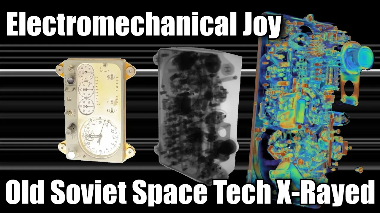
Why this video: Demystifies the mathematical concept of back-projection and voxel creation in an approachable yet scientifically accurate manner. It explains how hundreds of overlapping 2D attenuation profiles are reconstructed to resolve density values inside 3D volumes.
Knowledge Checkpoint
- Describe the process of converting multi-angle projection data (sinograms) back into spatial cross-sections.
- Define the Hounsfield Unit (HU) scale and state the nominal values for water, air, and dense cortical bone.
Module 3: Magnetic Resonance Imaging (MRI) Physics
Discover how strong static magnetic fields, radiofrequency excitation, and nuclear spin relaxation mechanisms are manipulated to produce detailed soft-tissue contrast without using ionizing radiation.
Recommended Videos
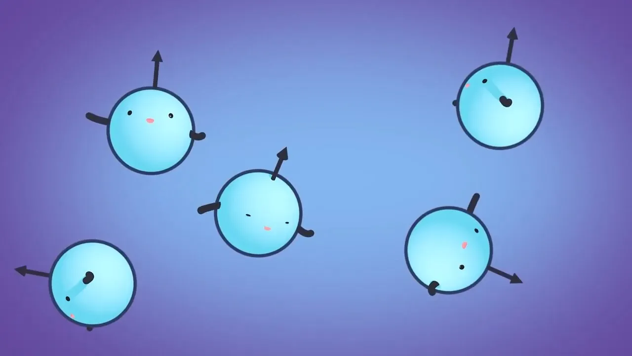
Why this video: Explains the foundational physics of MRI. It models hydrogen protons as microscopic magnetic dipoles, visualizes how they align with a static main field (), defines precession, and steps through how a 90-degree RF pulse tips net magnetization into the transverse plane.
Knowledge Checkpoint
- State the Larmor Equation and explain how precession frequency scales with external magnetic field strength.
- Describe what happens to net magnetization () during RF excitation at the resonant frequency.
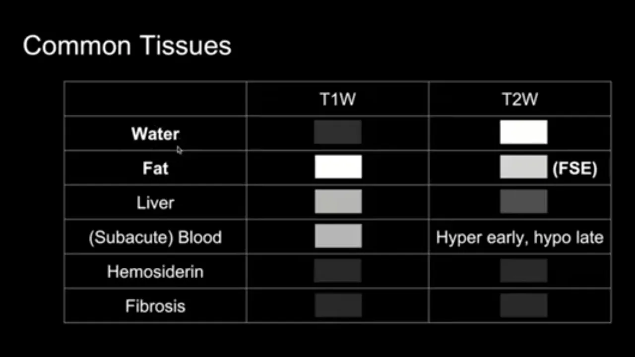
Why this video: An exceptionally clear visual breakdown of tissue-specific relaxation physics. It illustrates how energy exchange with the surrounding environment (T1/longitudinal recovery) differs from dephasing caused by local interactions (T2/transverse decay), showing how these values are exploited to color tissues differently.
Knowledge Checkpoint
- Compare T1 relaxation (spin-lattice) and T2 relaxation (spin-spin) in terms of energy loss and magnetic vector direction.
- Identify which physical parameter (T1 or T2) is characterized by a bright fluid appearance on a clinical MR slice.

Why this video: Delivers a deep, mathematically precise breakdown of longitudinal magnetization recovery. It outlines the thermal equilibrium kinetics of spins returning to , crucial for understanding advanced quantitative imaging.
Knowledge Checkpoint
- Write the mathematical function governing T1 exponential recovery and identify the point at which 63% recovery is achieved.
- Explain how molecular environment characteristics (such as the size and tumbling speed of molecules) dictate a tissue's local T1 value.
Module 4: Radioactive Decay and Nuclear Medicine (PET and SPECT)
Explore functional and molecular imaging. Learn about radioactive decay modes, the physical tracer principle, and how positron-electron annihilation and single-photon emissions are measured to record in vivo metabolic activity.
Recommended Videos
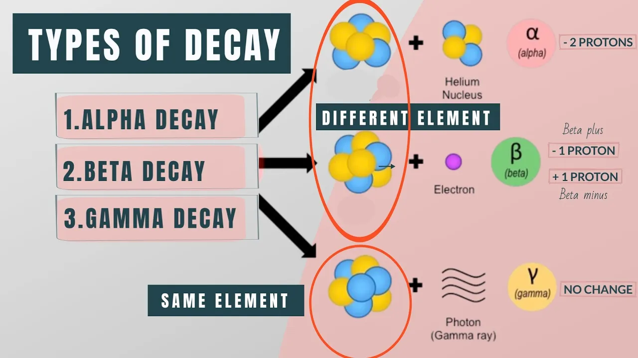
Why this video: Re-establishes the foundational nuclear physics required for functional imaging. It illustrates beta-plus (positron) decay and gamma-ray photon emissions, showing how unstable isotopes reach stable nuclear configurations.
Knowledge Checkpoint
- Explain the fundamental nuclear transition that occurs during beta-plus () decay.
- Distinguish between beta particles (charged) and gamma photons (uncharged) regarding range in human tissue.
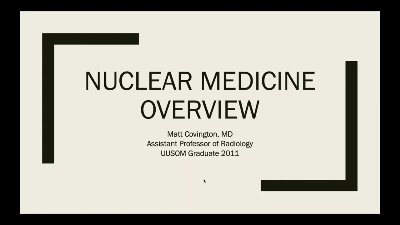
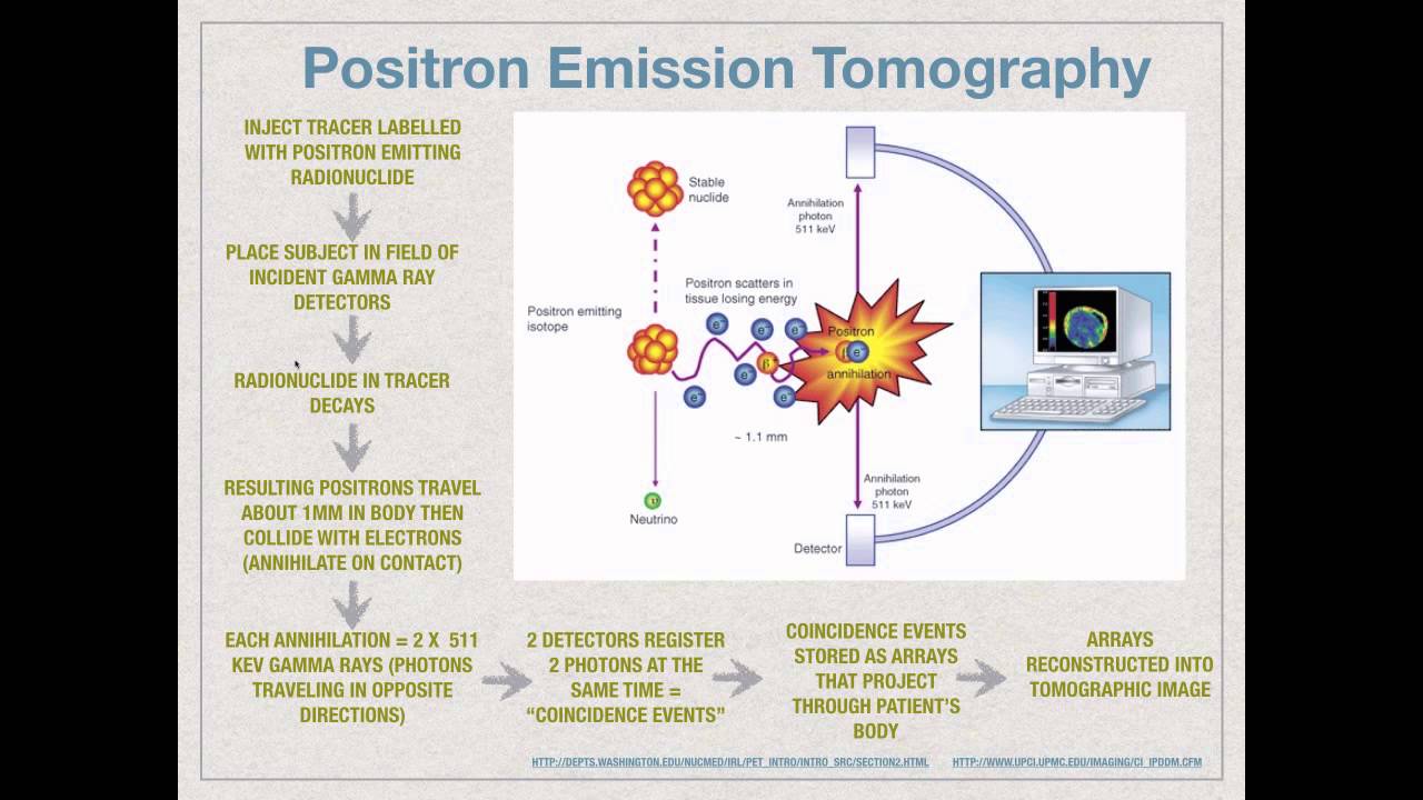
Why this video: This video addresses a major gap highlighted in the review feedback. It features an introductory-level, visual demonstration of positron-electron annihilation events within tissues. It shows how clinical PET scanners rely on these twin back-to-back 511 keV gamma rays.
Knowledge Checkpoint
- Describe the physical phenomenon of positron-electron annihilation.
- State the specific energy and angular relationship of the resulting photons.

Why this video: Expands on modern clinically paired systems (like PET/CT) and the concept of molecular targeting. It details how radioactive tracers track physiologic changes before anatomical abnormalities physically manifest.
Knowledge Checkpoint
- Explain the concept of coincidence detection in PET scanner ring electronics.
- Define why anatomical scans (CT/MRI) are often fused directly with metabolic functional scans (PET).
Why this video: Provides a comprehensive scientific comparison between Single Photon Emission Computed Tomography (SPECT) and Positron Emission Tomography (PET). This fulfills the review's request to clearly highlight the physical differences in imaging setup, detection, and isotopic physical properties.
Knowledge Checkpoint
- Contrast SPECT and PET in terms of the number of photons emitted per nuclear event and the use of physical collimators.
- Identify common radionuclides used in PET (e.g., Fluorine-18) versus those used in SPECT (e.g., Technetium-99m).
Module 5: Medical Ultrasound Physics
Study how high-frequency longitudinal acoustic pressure waves propagate through biological media, reflect at mechanical interfaces, and generate diagnostic anatomical and hemodynamic images via the piezoelectric effect.
Recommended Videos
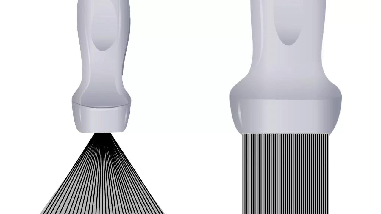
Why this video: Provides a complete primer on clinical acoustic physics. It defines sound as a mechanical wave, details the mathematical trade-off between penetration depth and spatial resolution across high and low frequencies, and explains common imaging modes (A-mode, B-mode, M-mode).
Knowledge Checkpoint
- Write the equation relating wave velocity, frequency, and wavelength in soft tissue.
- Describe the compromise between resolution and depth when selecting a high-frequency linear transducer versus a low-frequency curved array.

Why this video: Features an interactive crystal-lattice molecular animation explaining the direct and inverse piezoelectric effect. This is the primary physical process used to convert electrical energy into diagnostic acoustic energy, and vice-versa.
Knowledge Checkpoint
- Describe how physical deformation of an asymmetric crystal lattice (like quartz or PZT) generates an electrical potential difference.
- Explain how an AC voltage waveform is used to produce a therapeutic or diagnostic ultrasound pulse.
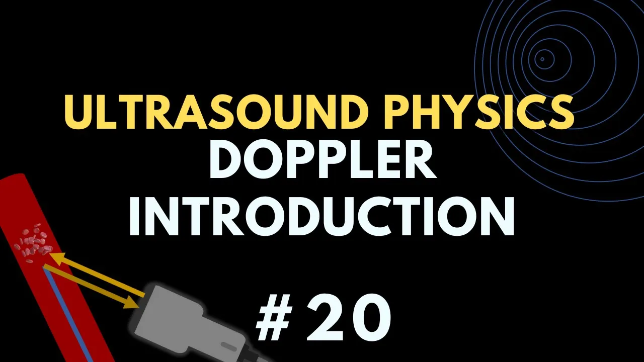
Why this video: Focuses on the math and physics of hemodynamic Doppler shift measurements. It breaks down the classic Doppler equation and demonstrates why angular alignment (the cosine factor) is essential for measuring real-time vascular blood flow velocities.
Knowledge Checkpoint
- State the Doppler equation and identify the variable that represents the direction of blood flow relative to the acoustic beam.
- Explain why a insonation angle yields a net Doppler shift value of zero, and state the clinically optimal angular range.
Module 6: Radiation Therapy and Dosimetry
Analyze the clinical deployment of high-energy Linear Accelerators (LINACs) to deliver tumor-cidal radiation doses. This module explores how treatment volumes are calculated, how internal brachytherapy compares to external beams, and the precise physical units used to define radiation safety limits.
Recommended Videos
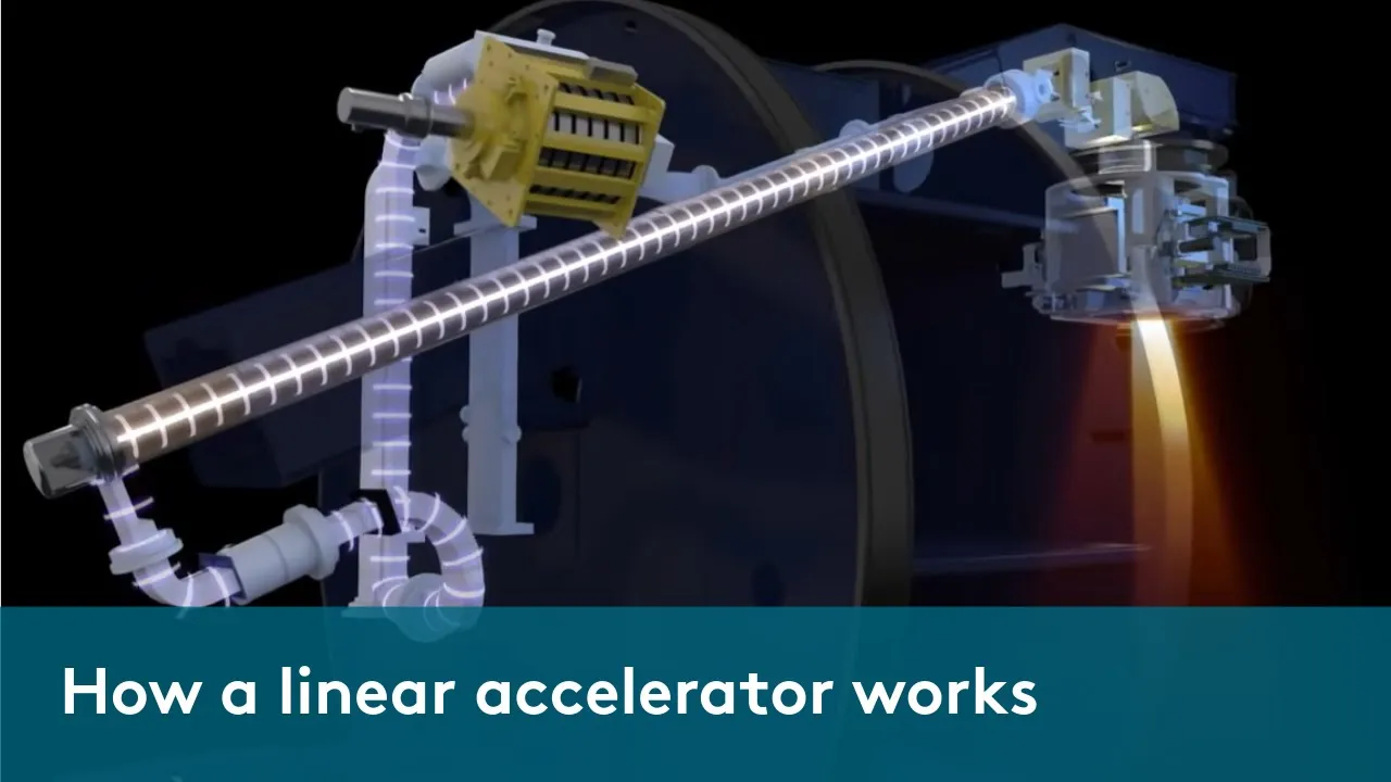
Why this video: Provides a complete mechanical and physical tour of a clinical LINAC. It tracks the path of electrons from the thermionic gun, through the microwave-driven waveguide, onto a heavy-metal target to generate high-energy (megavoltage) therapeutic X-ray beams. It also covers multi-leaf collimator (MLC) beam shaping.
Knowledge Checkpoint
- Trace the path of an electron inside a LINAC from generation to target impact.
- Explain how modern multi-leaf collimators (MLCs) dynamically shape treatment fields to conform to irregular 3D tumor shapes.
Why this video: A clear, concise comparison of radiation units. It details how the absolute physical energy absorbed in matter (the Gray) is adjusted to calculate the biological harm equivalent to human tissue (the Sievert) using radiation weighting factors.
Knowledge Checkpoint
- Define the Gray (Gy) in fundamental units of energy and mass.
- Calculate equivalent dose in Sieverts (Sv) given an absorbed dose in Grays and a radiation weighting factor ().

Why this video: Addresses the third key gap in the review feedback. It directly compares External Beam Radiation Therapy (delivered via megavoltage LINACs over multiple weeks) with Brachytherapy (involving direct implantation of radioactive isotopes/seeds internally). It compares their spatial dose profiles and treatment durations.
Knowledge Checkpoint
- Contrast External Beam Radiation Therapy (EBRT) and Brachytherapy in terms of the physical placement of the radiation source.
- Explain why brachytherapy offers highly localized doses that spare surrounding organs at risk (OARs), citing the inverse-square law.
Course Map
This map outlines the recommended progression through the material. Modules 1 and 3 are independent foundations. Modules 2, 4, and 6 directly build upon the concepts of radiation and matter interaction introduced in Module 1.
Key People Index
- Felix Bloch & Edward Purcell (Nobel Prize 1952): Discovered the physical phenomenon of Nuclear Magnetic Resonance (NMR) in bulk materials, establishing the foundational physics of proton manipulation and RF excitation.
- Godfrey Hounsfield (Nobel Prize 1979): Invented the computed axial tomography scanner, showing that multiple 2D X-ray projections could be mathematically reconstructed into a 3D digital matrix.
- Rolf Sievert (Swedish Physicist): Pioneered research in measuring biological effects of ionizing radiation. The SI unit for equivalent dose (Sievert, Sv) is named in his honor.
Final Self-Assessment
Test your understanding of the entire curriculum with this comprehensive self-assessment checklist.
- Can you define the critical quantum energy threshold that distinguishes ionizing from non-ionizing radiation?
- Can you explain why the photoelectric effect dominates low-energy diagnostic imaging and how it depends on a tissue's atomic number ()?
- Can you explain the physical origin of the continuous X-ray spectrum produced by Bremsstrahlung interaction?
- Can you define a Hounsfield Unit and state why water and air are calibrated to and respectively?
- Do you understand how slip-rings mechanically enable the helical acquisition of 3D CT projections?
- Can you state the Larmor Equation and calculate how proton precession changes if field strength () doubles?
- Can you contrast the molecular mechanics of T1 longitudinal relaxation (spin-lattice) with T2 transverse decay (spin-spin)?
- Can you explain why a positron-electron annihilation event must produce exactly two 511 keV photons moving apart?
- Can you contrast SPECT and PET imaging systems based on physical collimation and coincidence detection?
- Can you describe how both the direct and inverse piezoelectric effects are used inside a clinical ultrasound transducer?
- Can you write the Doppler shift equation and explain why angular correction is required when measuring vascular blood velocities?
- Can you define the physical difference between an absorbed radiation dose (measured in Grays) and an equivalent biological dose (measured in Sieverts)?
- Can you describe the clinical benefits of Brachytherapy compared to External Beam Radiation Therapy based on dose-gradient drop-off?