

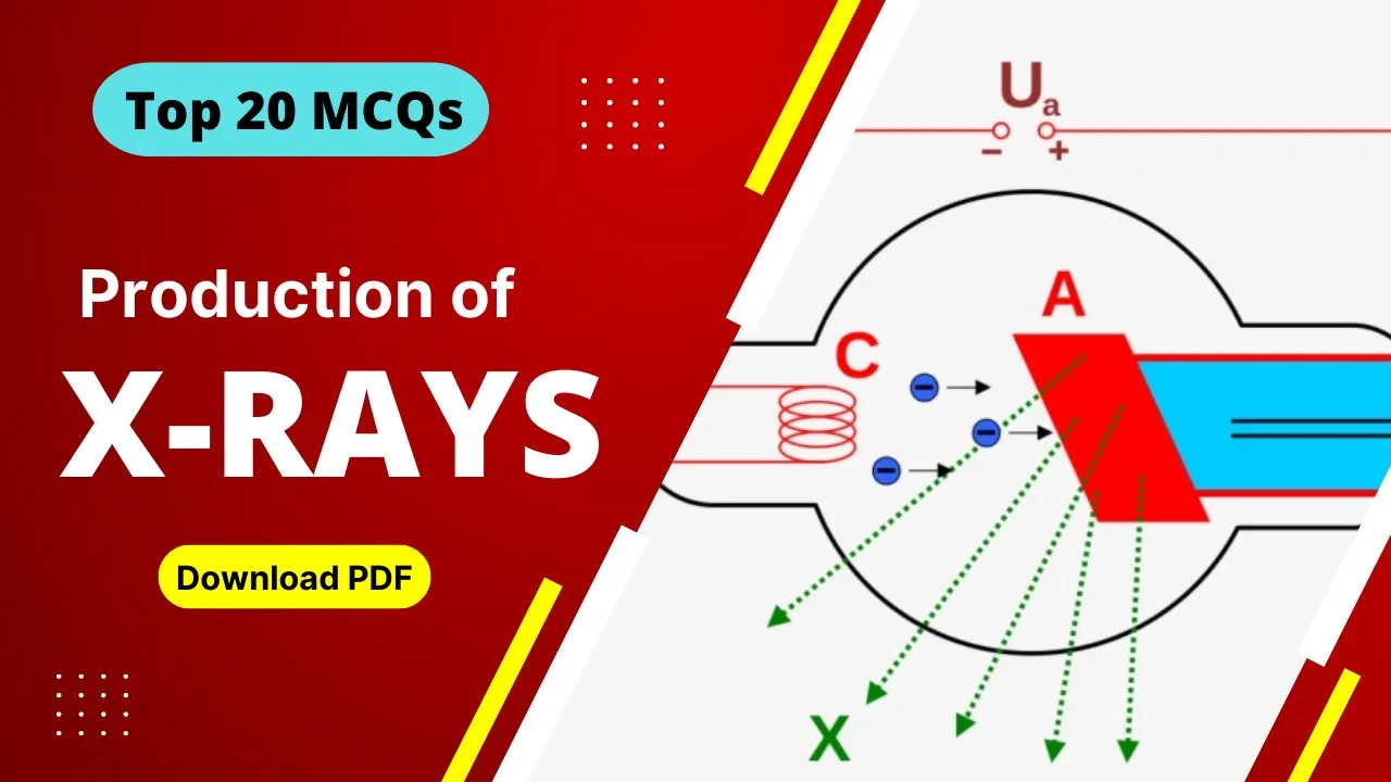
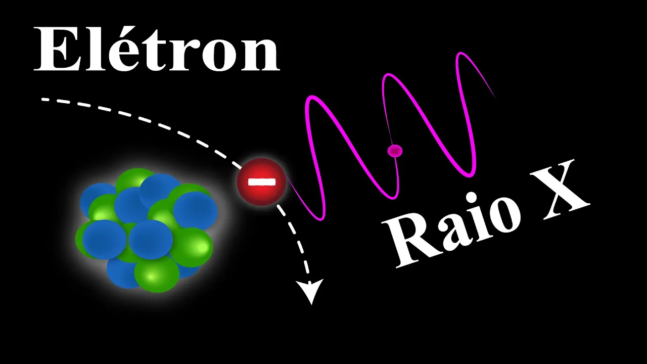
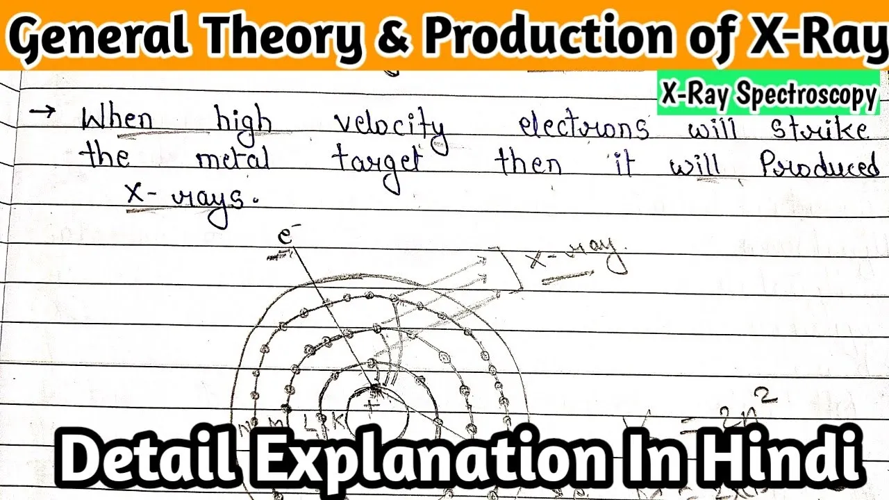
















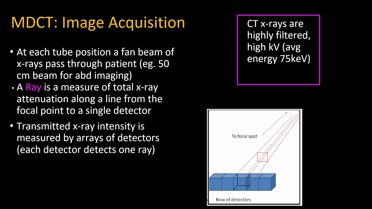






CT scanners, invented by Godfrey Hounsfield who won the Nobel Prize in 1979, use X-ray beams that rotate around the patient while detectors capture attenuation data from multiple angles to reconstruct cross-sectional images of the body; the system comprises a gantry housing the X-ray tube and detector array, slip rings for continuous rotation, a high-voltage generator producing 120-140 kV, cooling mechanisms, X-ray tubes with tungsten anodes, compensating filters to reduce patient dose and artifacts, collimators controlling slice thickness, and detector arrays that measure transmitted X-ray intensity to build 3D representations of scanned tissues.
Computed Tomography (CT) Scanners Explained: Principles & Components
Added:Basic physics of X-rays, including how electromagnetic radiation is produced and interacts with different matter.
Fundamentals of human anatomy and the concept of cross-sectional imaging planes (axial, sagittal, and coronal).
Elementary principles of digital signal processing, particularly how analog detector signals are digitized into image data.
Introduction to the concept of tissue attenuation and how different tissue densities (bone, air, water) absorb radiation.
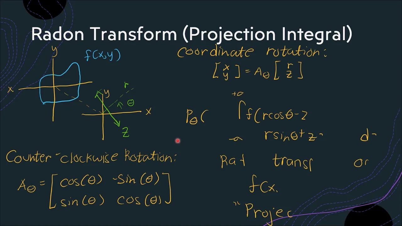
Mathematical concepts behind image reconstruction, such as the Radon Transform, Filtered Back Projection (FBP), and Iterative Reconstruction.
Advanced CT imaging technologies, including Multi-Detector CT (MDCT), Dual-Energy CT, and Cone-Beam CT (CBCT).
Radiation dosimetry, safety protocols, and dose optimization techniques to minimize patient exposure (e.g., the ALARA principle).
Identification and mitigation of common CT image artifacts, such as beam hardening, motion, and metal streaking.
Clinical applications of CT, including CT angiography, oncology staging, and the diagnostic use of contrast media.
90.7K views1.6Klikes10:46Original Release: 2021-07-03
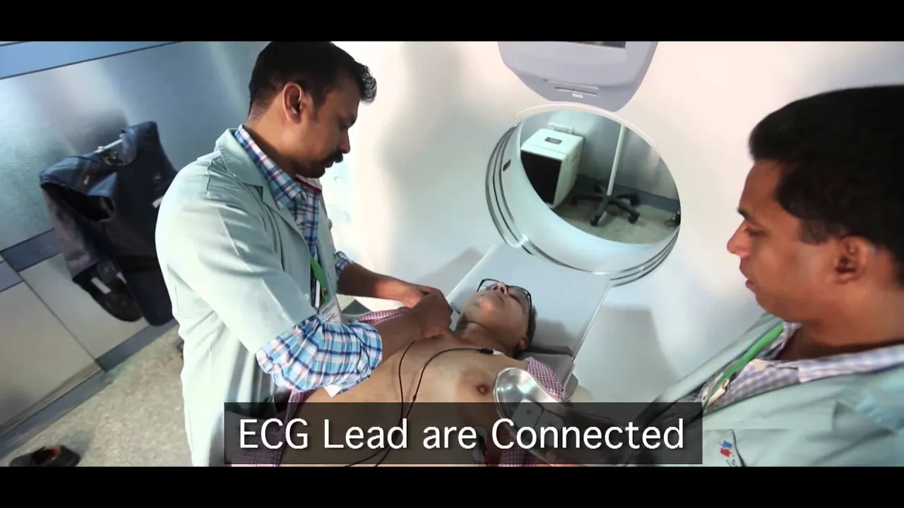
[Music] hi welcome to another video of biomedical engineers tv in this video we will look into computer tomography machines which is also known as a ct scanner let's look where it all began godfrey hounsfield a biomedical engineer contributed enormously towards the diagnosis of neurological and other disorders by virtue of his invention of the computed axial tomography scan for which he was awarded the nobel prize in 1979 working for the electrical and musical industry emi limited and in collaboration with two radiologists james ambrose and lewis creel he introduced the use of this machine in 1971 at the atkinson-morley's hospital in wimbledon he continued to improve the quality of the device and the human head was scanned for the first time in 1972 he continued his work on imaging of the human body was later concentrated on the next step in diagnostic radiology namely magnetic resonance imaging let's look into the principle of ct scanners ct is based on the fundamental principle that the density of the tissue passed by the x-ray beam can be measured from the calculation of the attenuation coefficient using this principle ct allows the reconstruction of the density of the body by two-dimensional section perpendicular to the axis of the acquisition system the ct x-ray tube typically with energy levels between 20 and 150 kev emits n photons monochromatic per unit of time the emitted x-rays form a beam which passes through the layer of biological material of thickness delta x a detector placed at the exit of the sample measures n positive delta and photons delta n smaller than 0 attenuation values of the x-ray beam are recorded and data used to build a 3d representation of the scanned object or tissue there are basically two processes of the absorption the photoelectric effect and the compton effect this phenomenon is represented by a single coefficient in the particular case of the ct the emitter of x-rays rotates around the patient and the detector placed in diametrically opposite sides pick up the images of a body section beam and detector move in synchrony unlike x-ray radiography the detectors of the ct scanner do not produce an image they measure the transmission of a thin beam one to ten millimeters of x-rays through a full scan of the body the image of that section is taken from different angles and this allows to retrieve the information on the depth in the third dimension let's learn about components of ct scanners ct scanners are composed of three important elements an x-ray tube a jan tree with a ring of x-ray sensitive detectors and a computer first we will look into a gentry the jantry is the donut-like or ring-shaped part of the ct scanner it houses many of the components necessary to produce and detect x-rays these components are mounted on a rotating scan frame components of the gantry are mounted onto a rotating scan frame gentries vary in total size as well as in the diameter of the opening or aperture the range size of aperture is typically 70 to 90 centimeters the gentry is designed to be tilted either forward or backward as needed to accommodate a variety of patients and examination protocols the degree to which the gentry can be tilted varies among systems but more or less 15 degrees to 30 degrees as usual the jantry also includes a laser light that is used to position the patient within the scanner control panels located on either side of the gentry opening allow the radiologic technologist to control the alignment lights gentry tilt and movement of the table in most scanners these functions may also be controlled via the operator's console a microphone is installed in the gentry to allow communication between the patient and the radiologic technologist throughout the scanning procedure the second component is slip rings old model design ct scanners used recoiling system cables to rotate the gentry frame this design limited the scan method to the step and shoot move and considerably limited the gentry rotation times newer systems use electromechanical devices called slip rings slip rings use a brush like apparatus to provide continuous electrical power and electronic communication across a rotating surface they permit the gentry frame to rotate continuously eliminating the need to straighten twisted system cables slip rings allow the gentry frame to rotate continuously making helical scan modes possible the third component is the generator high frequency generator is usually used in ct scanners the generators are designed to be small enough so that they can be located within the gentry highly stable three-phase generators have also been used but because these are standalone units near the jantry and require cables they have become obsolete generators produce high voltage and transmit it to the x-ray tube the power capacity of the generator is listed in kilowatts the power capacity of the generator determines the range of exposure techniques like kilovolts and ma settings available on a particular system ct generators produce high kilovolts generally 120 to 140 kilovolts to increase the intensity of the beam and thereby reduce patient dose in addition a higher kilovolt setting will help to reduce the heat load on the x-ray tube by allowing a lower ma setting and reducing the heat load on the x-ray tube will extend the life of the tube the fourth component is the cooling system cooling mechanisms are included in the gentry they can take many different forms such as blowers filters or devices that perform oil to air heat exchange cooling mechanisms are important because many components can be affected by temperature fluctuations the fifth component is the ct x-ray tube x-ray tubes produce the x-ray photos that create the ct image their design is a modification of a standard rotating anode tube such as the type used in angiography tungsten with an atomic number of 74 is often used for the anode target material because it produces a higher intensity x-ray beam this is because the intensity of x-ray production is approximately proportional to the atomic number of the target material ct scan tubes often contain more than one size of a focal spot 0.5 and 1 millimeter are the common size of a focal spot just as in standard x-ray tubes because of reduced penumbra small focal spot and computed tomography tubes produce sharper images like better spatial resolution but because they concentrate heat on a smaller area of the anode they cannot tolerate as much of the heat a very large amount of stress is placed on the ct scan tube scanning protocols often require multiple long exposures performed on numerous patients per day a ct scan tube must be designed to handle such stress the sixth component is filtration compensating filters are used to shape the x-ray beam they reduce the radiation dose to the patient and help to minimize image artifact as our teachers taught us that radiation emitted by a ct scan x-ray tube is polychromatic filtering the x-ray beam helps to reduce the range of x-ray energies that reach the patient by removing the long wavelength or soft x-rays these long wavelength x-rays are readily absorbed by the patient therefore they do not contribute to the ct image but do contribute to the radiation dose to the patient in addition creating a more uniform beam intensity improves the ct image by reducing artifacts that result from beam hardening filtering the x-ray beam helps to reduce the radiation dose taken by the patient and it also improves the image quality of the ct scanners the seventh component is collimators collimation restricts the x-ray beam to a specific area as a result it helps reduce scatter radiation the scatter radiation reduces image quality and increases the radiation dose to the patient reducing the scatter radiation improves contrast resolution and decreases patient dose collimation controls the slice thickness by narrowing or widening the x-ray beam the source collimator is located near the x-ray source and limits the amount of x-ray beam before it passes through the patient it is sometimes referred to as patient dose and determines how the dose is distributed across the slice thickness like the dose profile the source collimation resembles small shutters with an opening that adjusts dependent on the operator selection of slice thickness in mdct systems slice thickness is also influenced by the detector element configuration scanners vary in the choices of slice thickness available choices range from 0.5 to 10 millimeters and the last but not least component are the detectors the detectors are components of ct scan machines which collect information regarding the degree to which each anatomic structure attenuated the beam in conventional radiography we use a film screen system to record the attenuated information in ct we use detectors to collect the information the term detector refers to a single element or single type of detector used in a ct system the term detector array is used to describe the entire collection of detectors included in a ct scan system specifically the detector array compromises detector elements situated in an arc or a ring each of which measures the intensity of transmitted x-ray radiation along a beam projected from the x-ray source to that particular detector element also included in the array are elements referred to as reference detectors that help calibrate data and reduce artifacts detectors can be made from different substances each with their own advantage and disadvantages in the next video we will look into types of detectors in ct scanners types of ct scanners and their application thanks for watching biomedical engineers tv see you guys in the next part of this video [Music] [Music] [Music] you
Up Next
25:33
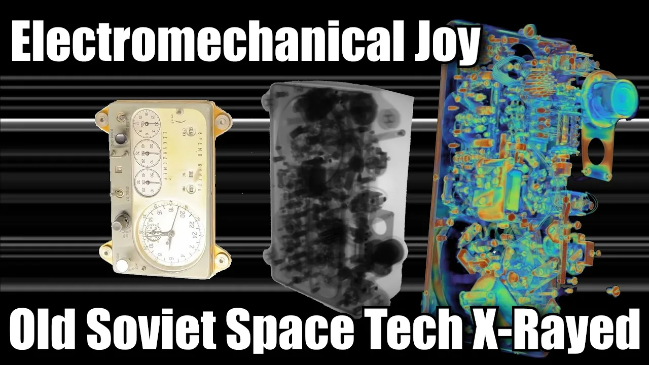
X-Ray CT Scanning of Heat Shield Tiles: Microstructure Analysis
@scottmanley
139.9K views•2025-06-10
30:32
Ultrasonic Transducers: Resonant Frequency Measurement and Horn Design
@imajeenyus42
229.9K views•2017-02-22

54:16
Polymer Environmental Degradation: Mechanisms & Stabilization
@iit
1.8K views•2012-07-10
33:39
How a Student's Question Saved a NYC Skyscraper from Collapse
@veritasium
22.8M views•2025-04-26
Related Study Plans & Knowledge Roadmaps
Structured learning paths in Engineering
engineering
Building Acoustic Levitators: Physics & Arduino
01The Physics of Sound and Standing Waves
02Piezoelectric Transducers and H-Bridge Drivers
03Arduino Programming: Generating 40 kHz Signals
+ 2 more learning modules
14h5 modules
Explore Roadmap
engineering
Building Peltier Cloud Chambers: Particle Tracks
01Cosmic Rays & Cloud Chamber Physics
02Thermoelectric Cooling (TEC) Fundamentals
03Custom Liquid-Cooling Loop Design
+ 2 more learning modules
18h5 modules
Explore Roadmap
engineering
Yagi-Uda Antennas: Modeling, Build & VNA Test
01Introduction to RF and Antenna Fundamentals
02Yagi-Uda Antenna Design Principles
03Antenna Modeling and Simulation in 4NEC2
+ 2 more learning modules
18h5 modules
Explore Roadmap
engineering
ECG Monitor Design: Op-Amps, Filters & Noise
01Electronics & Breadboard Basics
02Op-Amps and Instrumentation Amplifiers
03Bioelectricity and ECG Fundamentals
+ 2 more learning modules
18h5 modules
Explore Roadmap