






















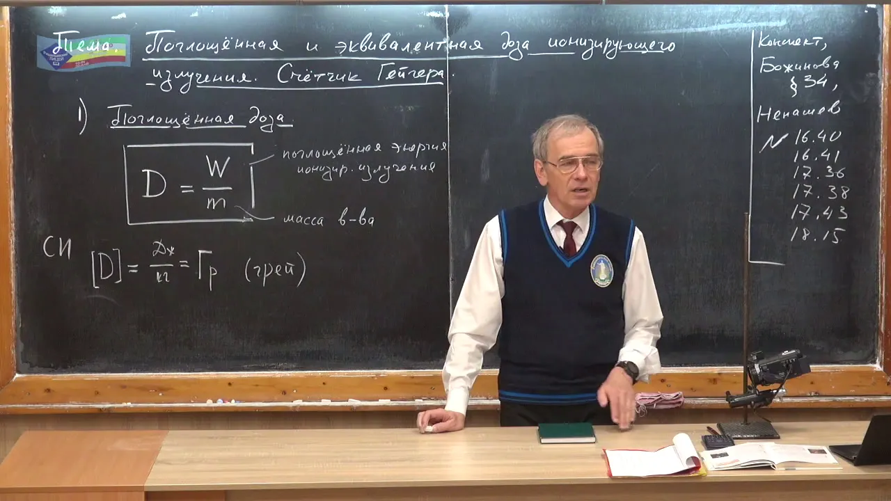



Radiation exposure is measured using two key units: the gray (Gy), which measures absorbed radiation dose as energy per kilogram of tissue, and the sievert (Sv), which adjusts for the different biological damage caused by various types of ionizing radiation (alpha, beta, gamma); for example, alpha particles require multiplying grays by 20 due to their high damage potential, while beta and gamma particles use a factor of 1, allowing people to understand and compare radiation risks from sources like medical scans or natural background exposure.
Radiation Units Explained: Gray, Sievert, and More
Added:The fundamental difference between ionizing and non-ionizing radiation, including the electromagnetic spectrum and particle radiation.
Basic atomic structure, isotopes, and the mechanisms of radioactive decay (alpha, beta, and gamma emission).
The concept of radioactivity or source strength (measured in Becquerels or Curies) versus radiation absorbed by a target.
The basic interaction of radiation with biological matter, specifically how ionizing radiation damages cellular structures and DNA.
The biological mechanisms of radiation-induced damage, distinguishing between deterministic effects (like radiation sickness) and stochastic effects (like cancer risk).
Radiation protection principles and safety standards, such as the ALARA (As Low As Reasonably Achievable) protocol, shielding, distance, and exposure time.
Medical physics and clinical dosimetry, specifically how radiation doses are planned and calculated for cancer radiation therapy and diagnostic imaging.
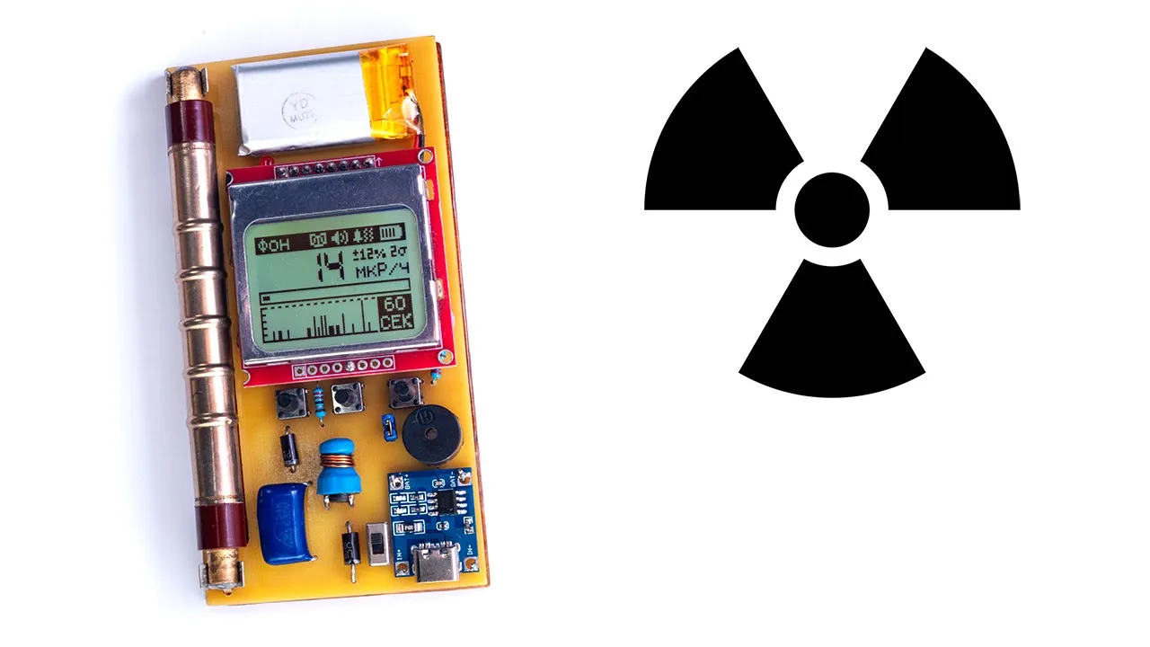
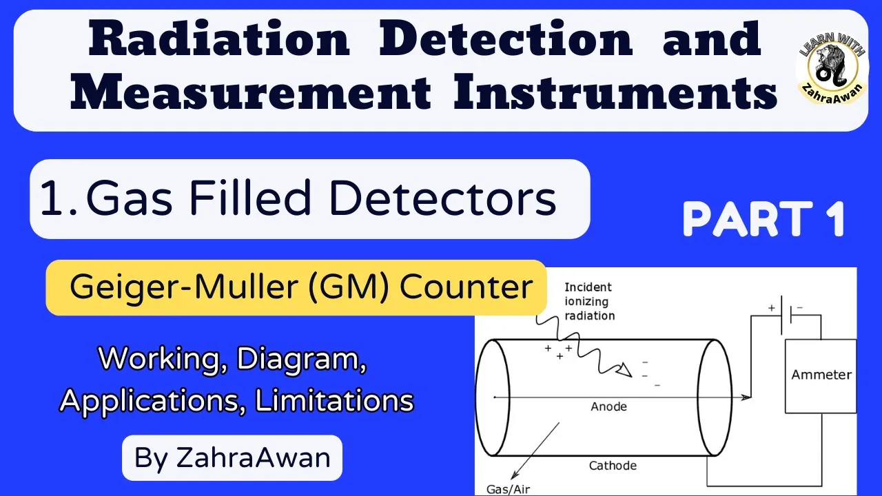
The operational principles of radiation detection and measurement devices, such as Geiger-Müller counters, scintillation detectors, and personal dosimeters.
Analysis of historical nuclear events (e.g., Chernobyl, Fukushima) and environmental radiation monitoring using established dose-limit guidelines.
342.1K views15.1Klikes6:05Original Release: 2019-09-12
[♪ INTRO] The term “radiation” is thrown around a lot.
Like, you might have heard that your cell phone gives off radiation.
Or maybe you just want to understand whether those x-rays your doc ordered are dangerous.
But we can’t have a really meaningful conversation about the relative dangers of radiation exposure without being clear about how much radiation we’re talking about.
And unfortunately, thanks to some accidents of history, the units we use to measure radiation and radiation exposure are… kind of a mess.
But there’s only two you really need to understand: the gray and the sievert.
To rewind a little, let’s start with that term radiation.
In physics, it refers to the energy carried by particles or waves, so technically, everything that reflects or emits light is “giving off radiation”.
But, generally, when people talk about harmful radiation exposure, they mean ionizing radiation.
That means particles with enough energy to rip electrons off of atoms, or, ionize them.
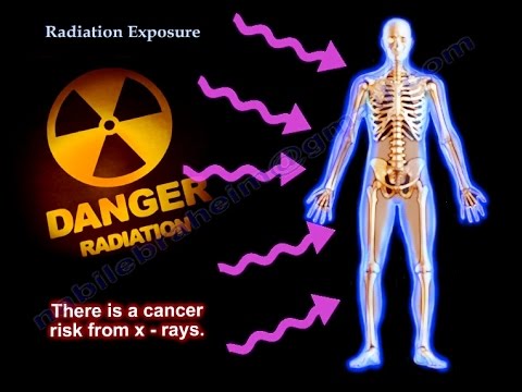
This is the radiation we care about being exposed to because it carries enough energy to break chemical bonds and cause mutations to DNA that increase your risk of cancer.
And to get this one out of the way real quick: cell phones don’t send signals using ionizing radiation—they use radio waves, which don’t have enough energy to damage cells.
The ionizing radiation we worry most about comes from nuclear decays— that’s when an atom breaks apart into smaller chunks, releasing other particles in the process.
And how easily these particles can ionize is, in part, determined by their energy, so that’s where we get to our first radiation units.
Particle energy is usually given in units called electronvolts, or eVs, and a typical radioactive particle may have an energy of about one megaelectronvolt, or MeV.
That’s a million eVs.
And while that might sound like a lot, it’s nothing compared to our everyday, standard unit of energy – the joule — the energy needed to lift 100 grams up by one meter — which has 6 trillion MeVs in it.
So one particle has a tiny amount of energy on a human scale.
Of course, radioactive sources don’t generally give off one particle at a time.
So we also have units to describe how many radioactive decays happen in a source per unit of time.
One becquerel, the standard unit for decay rates, means your source has one nuclear decay going on in it per second—basically, a tiny amount of activity.
Your body typically and safely emits several thousand becquerels all the time.
And there are older units that describe decay rates, too.
The curie, for example, was based on the decay of radium-226, but it’s now defined as 37 billion becquerels.
Somewhat related is the roentgen, which was in fashion for awhile— and recently popularized by the Chernobyl miniseries on HBO.
It focuses on the air instead of the radiation source.
Essentially, it quantifies how many electrons are being knocked off per cubic centimeter of air.
And if you’re standing next to something emitting one curie of radiation or in a room where a whole lot of electrons are being knocked off of air molecules, that’s probably not great for you.
But from a medical perspective, it’s not enough to know how many particles of radiation are in a room or are being emitted by something.
You need to know exactly how much radiation your body is absorbing: this is called the absorbed dose.
The standard unit for absorbed radiation dose is the gray.
A dose of one gray means that one kilogram of matter has absorbed one joule of radiation energy.
For instance, a CT scan in a hospital might expose you to seven milligrays, or seven thousandths of a gray.
Some people still use the rad— a historical unit now equivalent to 0.01 gray.
But whether you’re talking rads or grays, there’s an additional complication: some types of ionizing radiation do more damage to the body than others.
So the same amount of grays can do different amounts of damage, depending on the type of ionizing radiation.
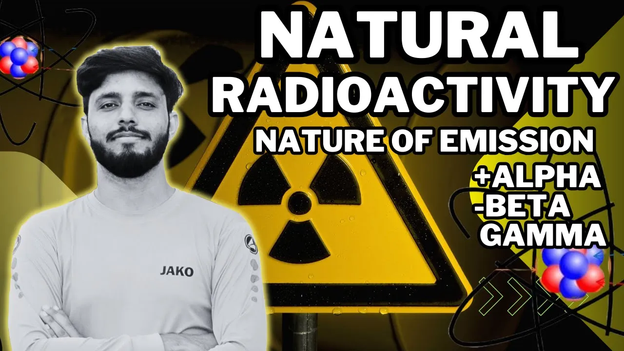
There are lots of types of ionizing radiation, but the three main ones are called alpha, beta, and gamma.
Each refers to a different type of particle ejected during nuclear decay.
Alpha particles are the heaviest, slowest, and most easily stopped, while gamma particles are the lightest, fastest, and hardest to contain.
And that’s what finally brings us to the sievert.
It modifies the gray to account for the different health risks associated with these particles.
To get sieverts, you multiply grays by a number specific to each type of radiation.
Most of the time, like for beta and gamma particles, the number you multiply by is just one.
But with some, like alpha particles, you multiple by twenty because alpha particles pack a real punch.
And that ‘twenty’ number isn’t arbitrary: it’s chosen based on the latest, constantly updated research into the effects of radiation on human health.
In that way, the sievert is a unit for human convenience: it takes the gray, which measures something exact and physical, and modifies it to tell humans how dangerous a type of radiation exposure might be when it interacts with our bodies.
You also may have heard people talk about rems when discussing exposure.
It’s the same idea, though an outdated version.
Rems is short for ‘roentgen equivalents in man’— one rem is now defined to be 0.01 sieverts.
But really, the sievert and the gray are the only units you need to remember to understand the overall impact of radiation exposure and make informed decisions about how you travel, receive healthcare, and… generally live your life.
Because the truth is there are lots of sources of radiation, including natural ones, and everyone is constantly exposed to some of it.
On average, people are exposed to a few millisieverts per year from their everyday lives.
The amount from medical scans varies depending on the size of the scan and the duration in the machine— from less than 0.01 millisieverts for a quick joint x-ray to about ten millisieverts for a full abdomen CT.
These raise your lifetime risk of cancer by one in a few million to one in two thousand, respectively.
And to put all that in perspective, the allowed limit for the average nuclear industry employee is 20 millisieverts per year.
Point is, while radiation exposure can be bad, it’s also something that happens to us every day.
And once you understand the lingo used to describe radiation, that fact doesn’t seem so scary.
Thanks for watching this episode of SciShow!
And a special thank you to our channel members.
You see, we’re only able to make free, educational science content like this episode because of the support of our viewers.
You can become a channel member by clicking that “join” button below.
And if you do, you’ll see that there are some perks to it— like, you get some special emojis you can use in chats.
But most of all, you get that warm, fuzzy feeling that tells you you’re supporting awesome science content on YouTube!
[♪ OUTRO]
Up Next

3:41
Introduction to Piezoelectric Effect: Generating Electricity from Crystals
@KeystoneScience
92.7K views•2016-12-21
16:48
MRI Basics: T1 vs T2 Relaxation and Tissue Contrast Explained
@navigatingradiology
101.8K views•2022-02-27

3:50
How Do Whales Avoid Deafening Themselves? Hearing Explained
@SciShow
187.4K views•2018-06-30

27:14
Entropy and the Second Law of Thermodynamics Explained
@veritasium
27.5M views•2023-07-01
Related Study Plans & Knowledge Roadmaps
Structured learning paths in Physics
physics
Medical Physics: Imaging, MRI & Radiotherapy
01Foundations of Radiation and Matter Interaction
02X-Ray Imaging and Computed Tomography (CT)
03Magnetic Resonance Imaging (MRI) Physics
+ 3 more learning modules
15h6 modules
Explore Roadmap
physics
Nuclear Physics (Decay, Fission & Reactors)
01Atomic Structure and the Binding Force
02Radioactive Decay and Half-Life
03Nuclear Fission Mechanics and Chain Reactions
+ 2 more learning modules
12h5 modules
Explore Roadmap
physics
Soft Matter (Polymers, Colloids & Rheology)
01Foundations of Soft Matter & Thermal Forces
02Polymer Physics and Macromolecules
03Colloids, Surfactants, and Self-Assembly
+ 2 more learning modules
38h5 modules
Explore Roadmap
physics
Analytical Mechanics: Lagrangian & Hamiltonian
01Mathematical Foundations: Calculus of Variations
02From Newtonian Mechanics to Constraint Dynamics
03The Principle of Least Action & Lagrangian Mechanics
+ 3 more learning modules
28h6 modules
Explore Roadmap