














![Internal Dosimetry: Quantitative Imaging (Iterative Image Reconstruction) (Part 2) [L46]](https://i.ytimg.com/vi_webp/ZAoLMVO0Ox0/maxresdefault.webp)





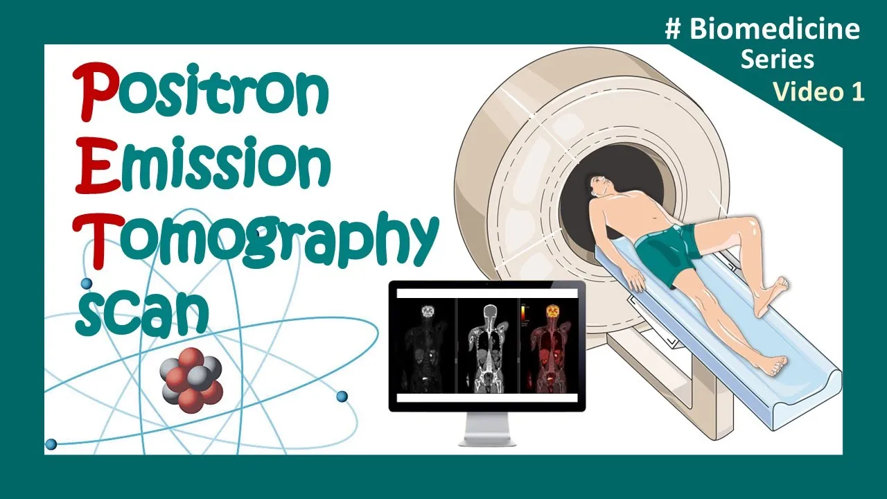
Nuclear medicine is a medical specialty that uses trace amounts of radioactive substances (radionuclides) to visualize and measure biological processes at the molecular level, distinguishing it from anatomical imaging techniques like X-rays, CT scans, and MRIs which only show physical structure; this tracer methodology allows doctors to observe metabolic activity, receptor binding, and cellular function to diagnose diseases such as cancer, heart conditions, and neurological disorders, while therapeutic applications use higher doses of targeted radionuclides to destroy cancer cells with minimal damage to healthy tissue.
Nuclear Medicine and Molecular Imaging Explained
Added:Basic atomic structure and the concept of radioactive isotopes, including half-life and decay modes (alpha, beta, and gamma).
Elementary human anatomy and physiology, specifically how organs metabolize compounds like glucose.
The fundamental difference between structural imaging (such as X-rays and CT scans) and functional/molecular imaging.
Basic principles of electromagnetism, particularly how high-energy photons like gamma rays interact with detectors.
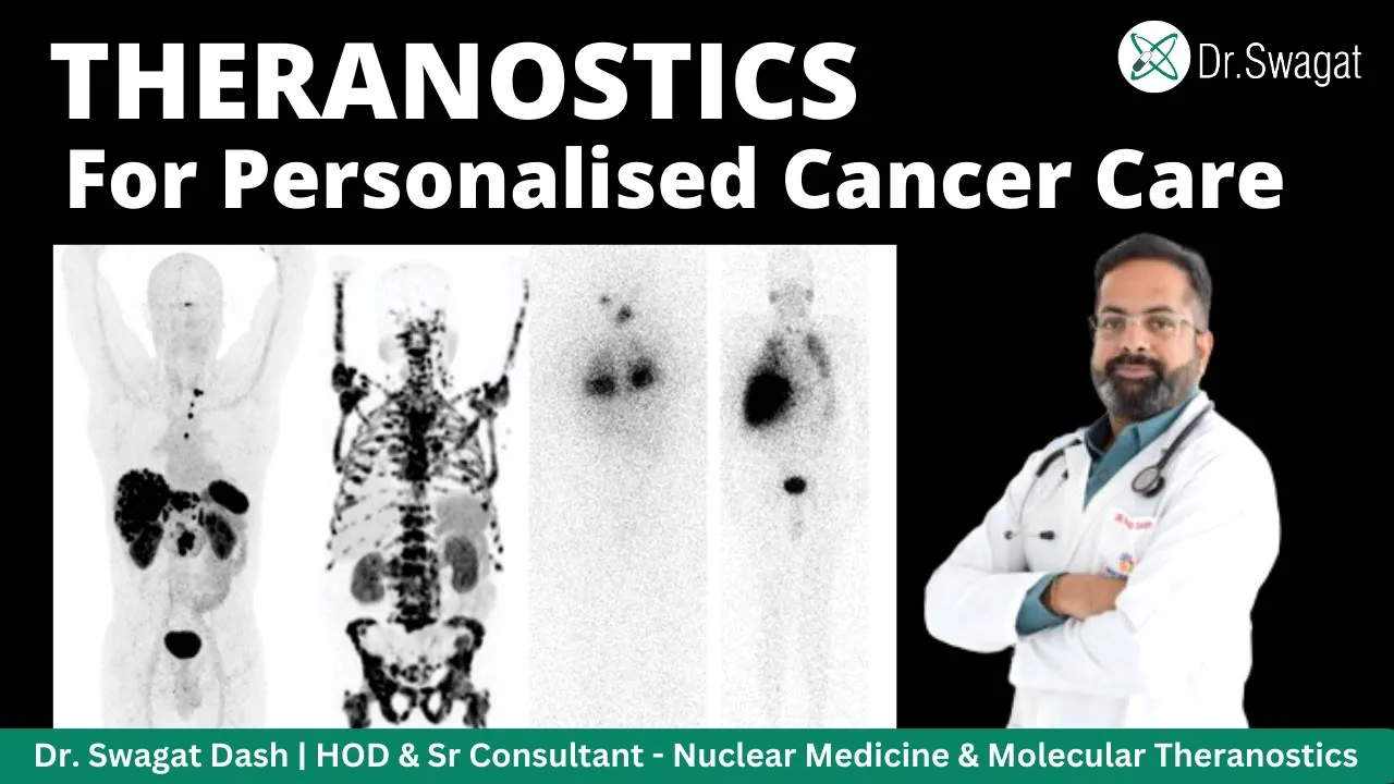
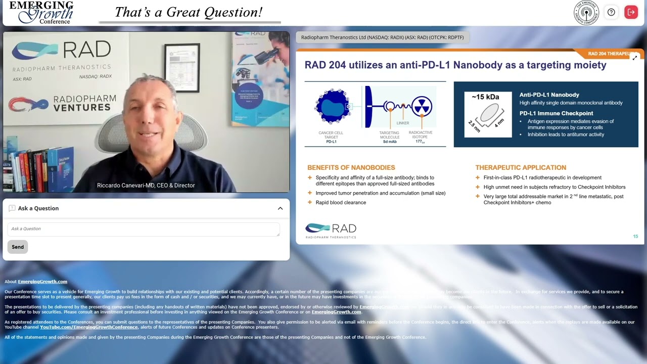
Theranostics: The emerging field that combines targeted diagnostic imaging with personalized radionuclide therapy (e.g., using Lutetium-177).
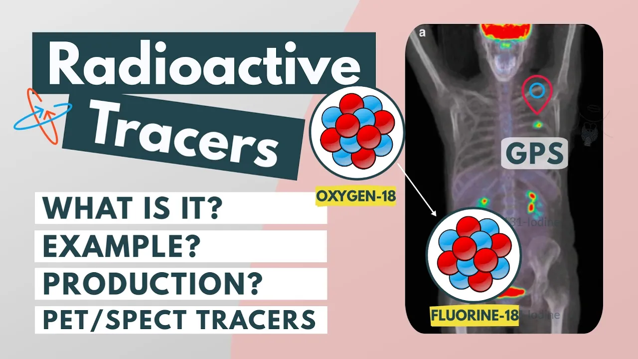
Radiochemistry and the synthesis of radiopharmaceuticals, including how cyclotrons produce short-lived isotopes like Fluorine-18.
Advanced mathematical image reconstruction, focusing on how PET/SPECT projection data is converted into 3D clinical images.
Radiation dosimetry and radiobiology: Calculating tissue-specific absorbed radiation doses and studying the biological safety limits of ionizing radiation.
70.1K views1.1Klikes46:42Original Release: 2019-08-16
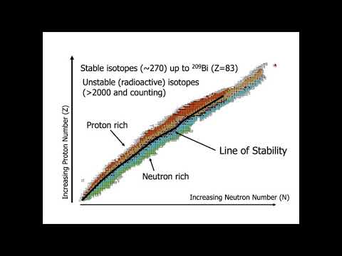
before I introduce our first speaker dr. Sunderland today really couldn't be possible without our support without the sponsors that we have that you'll see on the back of on the back of the schedule here and we have educational partners we have men's health Network we have NorCal Carson ette women heart and zero and then our industry partners advanced accelerator applications Blue Earth Diagnostics ITM and pro genex pharmaceuticals and I want to thank everybody for all the work and the participation and their help and in putting together this 8th patient education day so let's talk about dr. Sunderland University of Iowa serves as the director of the university's pet imaging center director of small animal em small animal Imaging Corps and associate professor professor of radiation oncology in the division of nuclear medicine associate professor of physics and astronomy he is also the vice chair of SN mmm eyes clinical trials Network I'd like to introduce dr. Sunderland thanks a lot um no I'm upset I I got rid of my second slide before I did which had the six pillars upon which the SN MMI is based and one of them is patient education patient advocacy so this is an important part of what the what the society does so my charge is actually just to do a very basic overview of nuclear medicine molecular imaging kind of how it works and what it does I've broken it up into four four parts just one slide on the difference between anatomical imaging and molecular and a nuclear medicine just to try and differentiate that from the get-go and then we'll talk about about 15 minutes on what nuclear medicine is and how it works kind of from a technical standpoint you'll forgive me I'm a physicist by training and so I have to do a little bit of that on the front end and then what is it used for we use it for Diagnostics and we're using it more and more in theory in the in the therapeutic or arena and so we'll get into that both what's approved and what's count by FDA and what's coming down the road and then a little bit on rate radiation safety considerations as we are in fact dealing with radiation so just starting on the front end here what you see in the upper left hand corner well in the left hand side are some anatomical imaging techniques you're familiar probably with most of them MRI in the top left that's functionally imaging imaging hydrogen or water density it gives you great soft tissue contrast beautiful anatomic anatomic images there's a great diagnostic CT down there which shows a nodule which can't really tell whether it's cancer enough you can see that little dot down down there you can see because of the pattern emphysema and what makes this particularly diagnostic is in the upper right hand upper right hand corner is the cigarette pack that's sitting there which helps the cancer diagnosis and geography by injecting dye in the veins you can actually in the artery of the vein maybe that's because the arteries you can actually look at the the arterial blood supply to the heart you can look for because of anatomy you can look for stenosis and back edges there ultrasound isn't just used Webster obstetrics it's used in cardiology is used in oncology and it's in and all of these are great and they have they have their uses but they're all fundamentally looking at anatomy whereas in the molecular imaging nuclear imaging biz we're largely looking at the biochemistry of the body the the anatomy is largely incidental what you see on the left of that slide is one of our FDG scans fluorodeoxyglucose and here what we're doing is we're imaging glucose metabolism because there's glucose metabolism all over the body you can kind of see the anatomy but that's that's secondary to the fact that we're imaging glucose metabolism you can see the uptake in the brain because we're thinking you can see the uptake in the heart because it's the muscle and it's pumping and they're using glucose for fuel but you can also see the tumor in the lung which would just be a nodule on a CT malignant not malignant don't know but because of the high uptake we can actually pretty pretty careful pretty confidently describe that as a as a malignant tumor you can see the kidney actually recognizes the radioactive glucose we injected is an imposter so the kidneys actually filtered out and you can it goes down to the bladder which you kind of see down there that's normal physiologic uptake what you see in the upper right is a DAT scan this is a SPECT scan where you inject something that goes into the the presynaptic dopaminergic system they use this for differential diagnosis of Parkinson's disease from other motion disorders and in the lower right hand corner you see some cardiac perfusion scans that look at blood flow to the heart to look for people who have cardiovascular disease these are just examples but in all of these cases we're looking at the function not so much the anatomy which is a it was a big difference so with that other way we're gonna get into how nuclear medicine works so why is it called nuclear medicine whereas a nuclear come from well in all cases in a nuclear man's medicine study were administering usually injecting but not exclusively a tiny quantity of drug and so in this case here's a molecule this is one of those sugar molecules this is Florida yaks d glucose and right there that's the f18 that's the radioactive tag so my kids have one of these is called a cell phone and they go around all over the country and I can track where they are because I follow where that phone is on my phone similarly that radioactive thing that's sitting there that's what we track and that's at what we image we don't image the molecule we image that radioactive nuclear flowing in this case fluorine-18 that's sitting there now when I say we're injecting a tiny amount I mean we're injecting a tiny amount that image that you saw of glucose metabolism about a millionth of a single grain of sugar that's the mass quantity of the drug okay the good news is there's almost no side effects from the drug there is radiation we have to worry about that but there are if I took a millionth of an of an Advil the headache that I have right now it wouldn't hit it at all right so we're at Sub pharmacologic doses here but then what we use is when that radioactive fluorine decays what we do is we get gamma rays and the gamma rays that come from the nucleus radioactively decaying which we'll talk about in a minute that's what we detect and that's how we get our images okay we're going to talk about then in much more grisly detail in moment so you're probably all familiar with the periodic table here right and so we do in in nuclei in the nuclear medicine business we use what Mother Nature gave to us so we have these elements right and you've all seen this but I think I always consider that the periodic table is a shallow two-dimensional representation of a three-dimensional space so if you actually look at carbon carbon six which is you can kind of see it going back there carbon carbon has six protons right that's by definition what carbon is and most of the carbon in our body is actually carbon-12 because it has six protons and six neutrons it's stable that's what we have in our body mostly there are some carbon 13 six protons gives carbon and seven neutrons which is 13 that's stable growing a small fraction of that right but the good news at least for us in in the biz is there's also carbon-14 that has six protons and eight neutrons that has too many Neutron for the nucleuses own good it's unstable it wants to get stable again and it does this by radioactive decay this has a half-life of about 6000 years we don't like to use it in people with a 6,000 year half-life but this is what you use for carbon dating right mother Nature gave this to us we have this we can actually and when we end up using it using it for carbon dating we can go the other way with carbon 11 this actually has six protons and five neutrons it has too many protons for its own good it ends up being unstable and it decays by positron decay it's actually so unstable it has a half-life of about 20 minutes but what we do in the nuclear medicine biz is we can take that carbon and we can attach it to a molecule whose physiology we want to trace and like in this case choline right we inject in choline is an fda-approved tracer to to image image prostate cancer and it's a little bit hard for me to point to but kind of in the in the lower left quadrant there there's a lymph node down there it's actually positive for disease and we can and on a CT and we have lymph nodes everywhere you see this you see this lymph node it's a lymph node it's the right size from lymph node along with all the other ones it's not diagnostic but that one has cancer and that helps them guide radiation therapy or surgery or something like that down the road so it's the c11 but it's Reid what's really important is the radiopharmaceutical they're just another real quick science slide the nice thing about radioactivity is frankly there's only three kinds we have to worry about if there's too many neutrons in the nucleus which is on the left it decays by beta decay that means it emits an electron from the nucleus which only goes about a millimeter if we have too many protons in the nucleus that's the middle one it decays by positron decay and when a positron gets submitted is of power so a positron is an electron except it's positively charged it's an antimatter electron it's a real I'm Anthony that means antimatter and what it does is it goes that same millimeter or so until it stops but when it stops it usually runs into an electron because there are electrons everywhere and when they get close to one another they annihilate that whole mass can neither be created nor destroyed thing right so that's wrong the electron has mass the positron has mass but after they meet they annihilate they're gone but in their place you get two gamma rays energy that are going off in opposite directions back to back that's what we detect when we do a PET scan and that energy you get that's the equals MC squared thing right the mem is mass the e is energy and then on the right side that we don't use so much for imaging but we use for therapy we have alpha decay where the nucleus emits this very massive it's like instead of emitting a ping-pong ball is emitting a bowling ball and that ends up being important when we talk about therapy a little bit from an imaging standpoint what's important is with the alphas and with the betas and frankly with the positrons as well see we can't detect the beta particle it's only going like a millimeter it's gonna stop in the body same thing with the positron but usually what happens is after it emits that that beta particle if the nucleus is in an excited state and it goes to the ground state when it does that it emits a gamma ray the gamma ray is a high-energy electromagnetic radiation and it can go through the body and this is what we're detecting when that nucleus decays by whatever it does it emits this gamma ray and the gamma ray can go through the body and we can detect it with our scanners this is my tribute to Bob Ross most of you even know Bob my even my kids know Bob Ross he's kind of a superstar now even for them so we we use the the these radionuclides that are available to us kind of as our palette we get like to choose the ones that that fit our needs for imaging or therapy this is just a list of ten there's probably about 60 or 70 useful radionuclides out there some of them have short half-lives like the imaging ones we'd like to inject the short lived ones for imaging because then they'll just go away by the next day they're pretty much gone for the therapeutic which is kind of up at the top if we're gonna inject therapeutic radionuclides we'd like to have them sit there bombarding the tumors for a really long time we'll get into that so having a longer half-life is a good thing and the kind of decay all these things enter into it we have to get very clever about how we use them but it's not just the radionuclide it's not just the half-life really it's all about the molecules we attach them to this is just three of several hundred that are out there most of them are on the research phase some of them are fda-approved and we use clinically here's here's three that are out there that all can represent different things there's that glucose one again that has fluorine 18 has a half-life of about two hours the case by positron decay actually all of these are positive chronometers the one that's in the middle is for our beta peer which we use for Alzheimer's disease what's interesting about this is it that's a great job of attaching to the amyloid in the brain for Alzheimer's disease and it's fda-approved and CMS doesn't reimburse for it and then on the right hand side we have gallium 6002 talk which is very much like net spot for those of you are familiar with it which is approved gallium 68 dota talk is not yet approved although we at the University of Iowa have submitted a new drug application and hopefully we're about two weeks away from from approval for for that so fingers crossed on that yes oh I'm sorry when I say CMS I mean Medicare right so so Medicare has chosen not to pay for those all time because we don't really have a treatment for it right so they say oh you diagnosed it so what that's there that's definitely I don't necessarily so bottom line here is so what we do is we have these radionuclides that are attached to these radio pharmaceuticals we inject them into patients and then we have to image them so if they're positron emitters we use the PET scanner that's designed to get both those so we have a ring of detectors it's designed to get both those gamma rays off that the detected at the same time if they just emit that single gamma ray then we use the SPECT scanner which is which is down below and so that's usually the beta emitters we use we use respect scans for and the standard plane or gamma camera images we generally use the same suspect scanner which is down below the procedure is pretty simple in general we administrator the radiopharmaceutical ninety ninety-five percent of the time it's by injection but we do have other other modes of administration so I'm going to show you this this is a some friends of mine work with the company in China and they built a pet scanner that's two meters long so you can get the whole patient in there at the same time and this is one of the very first images that they see if I can get my very first images they injected FDG in the leg and these this is one second images you can see it going up the leg into the lungs into the brain we're about 45 seconds in it's beginning to distribute into the heart into the liver etc we're and now it's kind of going to minutes you can see it going into the brain as it's being metabolized you can see the kidney has actually filtered out and the bladder is filling up you can see all this all this overtime first time we've ever been able to see what an injection does all at once let alone what what FDG does so we can get we can get full pharmacokinetics of all of these things now with this with this new new device PET scanner but two meters long very cool bottom line is we do inject this stuff there's an uptake period that uptake period could be anywhere from a few minutes to maybe an hour or two when you put put them in the scanner the scan typically takes fifteen to thirty minutes actually these days with new scanners that's usually down to 10 to 20 in order to get a whole body scan so that's how we do it now the question is how do we get the images just a couple of really quick slides here once again we have our glucose molecule that actually has the has the unstable nucleus when it decays once again we have that positron that goes off in about a millimeter we get the annihilation and then we have those rings of detectors which actually try and detect both those photons at the same time at the same time means within about five billions of a second of one another we can actually kind of trace back to where the event occurred because two points define a straight line we don't just detect one of these we detect tens of millions of these events and from that we can reconstruct these images so in pet we're looking at two photons at once inspect so this is that scan this is the one that looks for Parkinson's disease it has iodine 123 which has been a bit of a longer half-life but when it emits that that that beta particle but then the important thing is it emits that single gamma there's two detectors one on the top and one on the bottom it detects if it's going up or going down we actually detect it and those detectors actually rotate around the patient over time there was about a half reservoir half revolution and from that we can reconstruct what the the distribution is in the brain or really wherever or the heart or whatever is we're looking and that's as much as I'm going to go into how those scanners work I just want to repeat again that nuclear medicine is always tracer methodology the quantities the mass quantities that we inject are really really small for the most part although FDA doesn't like us to say it there's really no side effects associated with these quote-unquote drugs because the mass quantity is so small that doesn't mean we don't have to worry about the radiation dose it doesn't mean you don't have to worry about who there might be you might get a bruise that the infect it that at the injection site but the drug itself the mass is so small we really don't worry about that too much and fda realized they really make us check it but they realize that these are really low risk drugs from that standpoint it's not like you're giving a therapeutic drug for these for these diagnostic and masses the mass is really small okay so now that's how how this all works let's go to some of the applications I'm going to talk about the diagnostic ones first the current ones and then some of the unapproved ones that are coming down the pike each of them this isn't by no means an exhaustive list it's just a sampling I think I go through about ten or so and I'm going to go through a couple of therapeutic applications and then on into safety so all of these slides coming up right now have the same format you've seen the FDG ones I'm not going to go and go into too much detail here but I'm going to go with a format so in the upper left hand corner is the molecular structure I know you can't see it very well and frankly I don't care what I want you to realize is they're just all different so whatever the application is the molecule has to be tuned to the physiology that we want to trace I have a modality whether it's pet respect or standard nuclear medicine I kind of have anything to have an example what it's used for what its availability is and the regulatory and reimbursement situation and then almost most important is the localization mechanism why it does what we think it does so this we've talked about Florida oxy glucose it's used for a wide array of solid solid cancers solid tumors but it's not used very often at least for prostate cancer or neuroendocrine tumors those are relatively indolent they don't usually grow very fast and as a result the glucose uptake they don't require a whole lot of energy so they show up very well so we have to be a little it's discriminating how it uses and how we use it it turns out that most cancers use glucose like crazy which is why they light up like little Christmas lights kind of all over the place because they've actually got something wrong with a metabolic Abba lism that makes them burn burn glucose it's approved it's available just about everywhere in the in the USA this is net spot gallium 68 donut 8 it's a much more complicated mountain molecule gallium 68 has a half-life of about an hour it's used specifically for neuro endocrine tumors why is that because neuroendocrine tumors overexpressed somatostatin receptors commonly found there and so they've been clever enough to define a drug molecule that seeks this out and binds to it and actually sticks and pretty much won't let go okay and that's what this this net spot this gallium 68 donut 8 does it is approved it is reimbursed although you have to have to get pre-approval by insurance companies it is not quite ubiquitous as far as availability is concerned and I think this is a modification of something from from Josh's website where he has the sites where this is produced his doesn't have little circles I have little circles that about a 1-hour radius around these because it only has a half-life of about an hour and you don't make much of it when you do a synthesis so these are the places you have to be kind of put within one of those circles in order to get this because it's not widely distributed okay but the good news it is is it is available okay so on to prostate cancer so I got a couple of so I showed choline before that's one approved agent another approved daven agent is is f-18 FAA CBC which has the trade name axiom --n it's for prostate cancer it's a this is an amino acid and it targets a transporter that that is overexpressed in prostate cancer and here you can see in in this image the little dot there in the pelvis which is where it's spread too which is very helpful for the for the physician to either do surgery or radiation therapy to make sure it targets it this one I made a point of showing the the CT background and those color ones right so sometimes the PET scans are so specific you can't see the background Anatomy so the surgeon doesn't know where to go because there's no landmarks so most PET scanners really all PET scanners these days you can get a CT done at the same time so you have the anatomical backdrop in which to project the PET scan so you know exactly where you're you're looking this once again is available just about all over the country because it's transportable you can go you know on the order of three hours outside the reimbursement situation is good but pre-approval is is required cardiac perfusion so I have two examples here both pet and SPECT they do pretty much the same thing perfusion means how much blood is going to different parts of the heart the left ventricle is the important one here this is what pumps it all the way through that's a big muscle of muscular part of the heart and it's kind of shaped like a bullet and so where you see those round Donuts that's like looking credit cross-sections of the bullet and what you like to see is like what you see that sestamibi SPECT scan on the right the rubidium on the left the rubidium on the right and what you like to see is that nice round doughnut they're the ones below it that look like like this is a vertical slice through it the other one on the bottom is a horizontal slice once again those are normal not normal is what you see down below the apex is the tip of the bullet and if you can see under rest conditions at the tip it's pretty much thinned out that means there's probably a stenosis not a whole lot of blood getting there and under stress conditions they get on the treadmill and they inject it you can see there's almost no blood getting there because the stenosis is so much when people have have angina they have pain heart pain when they exercise this is what's happening that heart that heart muscle is not getting the perfusion they can see the extent of it in the impact of stress on it with these cardiac perfusion I mentioned this brain imaging before drug companies were all hot on this because they found a way to image the amyloid three companies went as far as to get these approved none of these are reimbursed all of them are approved they are available they're being used extensively in clinical trials largely to look at treatment treatment drugs because they'd like to know whether somebody actually has Alzheimer's disease or not before they actually try the drugs this is an example of kind of how it works I should go back and say the ones on the right I'm sorry I'm sorry on the left is what normal uptake is that's a white matter uptake but when when you start developing amyloid plaques on the cortex which is the outside of the brain that's what you see on the right hand side they all they look a little bit different but in all cases you see that the amyloid spreading to the to the outside where this ends up being important is in a series like this these are for different patients the one on the left is normal the two middle ones are the important ones MCI stands for mild mild cognitive impairment that means you're a little bit forgetful if we get what you said five or ten minutes ago but there's a lot of causes for that and it could be early on these two middle patients have the same symptoms one of them doesn't have Alzheimer's disease the other one does and and you can see the mild cognitive impairment amyloid burden is pretty much the same as Fletch Alzheimer's disease s the ad on the far right so differentiating it is actually pretty important but CMS and the Medicare people don't seem to think it's that important yet I only do this one this is a bread and butter nuclear medicine scan this is this is actually not SPECT this is just plain er I I bring this up because I mentioned different kinds of administration so the ventilation scan is done with xenon 133 which is radioactive gas you inhale this so that's what you see on the left-hand side that's just see where the air is actually getting in the lungs because sometimes sometimes those are not normal what you see on the right they use this for identifying pulmonary embolisms those should be kind of uniform lung kind of black on either side this is actually looking at where the blood goes because it's not just where the air goes the air has to has to go through go through and then get into the blood where where you see those in homogeneity is where it's light in there that's where the Bloods not getting where it's supposed to this is a standard nuclear medicine scan done with two different two different drugs one xenon one technetium-99 labeled I mentioned and showed this Parkinson's disease one this that scan this is an approved agent iodine 123 inspect and and this is taken up and stored in the vesicles of presynaptic brain cells which are generally sitting in the caudate and the putamen which is those others comma-shaped things on the left that's normal the putamen begins to the cells begin to die and then in in in Parkinson's disease and so you can see on the right hand side the tails have kind of gone away and that's a telltale sign that the patient actually does have Parkinson's you can also see it's a lil a symmetric and so you can see one is a little bit more diseased than the and the other okay that's just some basic approved ones here are some not fda-approved but coming this is a very exciting another exciting prostate cancer agent gallium 68 PSMA there's a lot of excitement over this the choline is good the acumen is good but the PSMA is seems to be able to detect much smaller lymph nodes sooner at least that's what the literature is looking like there is a phase 3 study that was done completed between the University of California San Francisco and UCLA they are literally a couple weeks away from submitting their new drug application we hope that that they get this in approval is likely but not certainly because that's up to FDA with any within about a year widespread availability would be maybe three years or so that's because not everybody can do it if I if we want to do in Iowa we would have to submit an a NDA that's a whole nother application and that goes to the office of generic drugs and takes them about two years to approve those so it'll be available likely nearby we can go the expanded access IND route but we'll just have to see on that but it's got to get approved first but it's but it's it's quite exciting it's a good agent frankly it's likely a stopgap solution because there is another f18 labeled PSMA this f-18 DCC f py l which is in phase 3 clinical trials right now this does pretty much the same thing as the gallium 68 PSMA but you can make a lot more of it in with the two-hour half-life you can distribute it a lot further so for widespread public availability this if I look into my crystal ball will likely be what most places use two to three years down the road assuming the phase three clinical trial is successful assuming they submit the NDA in assuming FDA accepts it so but that's an exciting very exciting area as well that first one the gallium is academically sponsored that the pyl compound is industry and the neuro endocrine space is is not is not static at all there's this OBS OBS 202 which is for neuro endocrine tumors it works a lot like net spot it works a lot like dota talk except it's in an antagonist which which at least it's hypothesized it has some advantages both from an imaging and a therapy standpoint it too is not approved yet just phase two and it's been and that's really just starting so it's a better way I want to move on to therapeutic applications just for a couple minutes and to this we need to begin to understand a little bit about radiation and not just what it can do for us but the harmful effects and I'm going to get into the and get into the dose doses here in a minute but we do know that very large doses of radiation can kill cells or the very least damage the DNA so they don't go on but if a human gets very large doses of radiation all at once and five 500,000 millirem and we'll talk about what a milligram is in in a couple minutes it can cause acute harm and even death we know this from Hiroshima and Nagasaki these kinds of things that's been been been studied extensively so we know that radiation is dangerous at particularly in very large doses however we can use these toxic effects of radiation to our advantage if very large doses of beams of radiation are being aimed very specifically and carefully at tumors the cancer cells we can selectively treat this scanner know the cancer now this is not molecular molecular imaging or nuclear imaging right now I'm just kind of going outside of this to show it external beam radiation therapy does well here we have a CT scan of somebody going through their lungs and you Candice can see circled there's a tumor there and what you do in radiation therapy is you shoot a beam of radiation which is sub-lethal from one direction right and it goes on through still out of radiation but then you do it from another angle and then they overlap and where they overlap you now have twice that dose and you do it from another angle another and so what you do is you you get a lot of dose to the tumor and you get less to the outside and that's targeting the radiation to the tumor but not not exclusively you still your rating and everything else outside of it wouldn't it be nice if you could just get their radiation just to the tumor and not this other stuff so that's what targeted radionuclide therapy is all about and those of you who are PRRT and some of these prostate cancer trials it all seems quite new is really quite old in in fact Saul Hertz back in 1936 had the idea to treat thyroid cancer with radioactive iodine and he treated his first patient in 1941 you can actually ingest it so we talked about inhaling we talked about injections you can actually take a pill that has a radioactive iodine-131 goes in the system and the body just moves it to the thyroid and pretty much nowhere else and you can obliterate the thyroid and more importantly of thyroid cancer tens of thousands of people have been treated with this over over the years so this targeted radionuclide therapy is is is not new but what we're getting what's happening now is where we're using it a lot more specifically because because of three things one is we're getting better and better at characterizing at a molecular level the different kinds of cancer the kinds of chemicals that are specifically on the surface of melanoma or on the surface of prostate cancer and we target drugs to this then we're getting really good at designing custom drugs there will I go specifically to those targets then lastly and sometimes this is the hardest step then we actually have to take those drugs but we have to attach the radioactive atom to it so we can actually get it to so we can actually follow it but we're getting very good at these things and so we're doing it for four neuroendocrine tumors we're doing it for prostate cancer those are pretty far along but there are a lot of companies who are developing things for non-small-cell lung cancer for melanoma for all these cancers and keep watching the space because a very exciting very exciting area not just for you guys but for companies because this is a very lucrative thing and I can let me go back and just say I mean the good news about about these things it goes really specifically to the tumors right you don't have all this kind of crosstalk it does go to other other other organs and all but not nearly so not nearly so much so the more targeted you can be the better off you are and so this is just an example this is with this is with an annette spot scan where somebody has has tumor pretty much pretty much everywhere you know and wouldn't it be nice and this is just with the gallium right short short half-life so the idea here is we take that same dota toc that's the chemical that binds to the somatostatin receptors you take that same camp but you instead of attaching this gallium 68 you attach lutetium 177 it has a half-life of about a week so you inject it into the patient it goat now it gives off a gamma-ray so we can image it that's why you have an image right and we didn't just inject a little we injected a lot we injected it out about a hundred times more it's a hundred times more and it's got a half-life which is a hundred times as long so it's like little machine guns shooting the tumors for weeks until it decays away because it stays bound and so if you can do that and it happens happens to be successful I mean there's no other way to treat this right then you go through and and in some cases in the precaution you in the literature they always show you the really good ones right there so not not not very often do you have kind of complete responses like this but it can be it can be pretty dramatic sometimes right and so this is that this is the promise this is what we're what we're looking for now I'm just so that you know so the imaging part is the diagnostic the therapeutic part the targeted radiography is the therapeutic which you might hear the word theranostics the Thera is for the therapy what we do for the lutetium the gnostic is the diagnostic that we do with that with the gallium in this case and the therapeutic part is just targeted radionuclide therapy the good news is we haven't recently have have a recent approval lutetium 177 doe rotate luna Thera it was approved just once again here's another good example of here's a patient who has a lot of cancer you treat them and afterwards the cancer largely goes away this is not necessarily a cure this reduces the tumor burden and hopefully substantially but it can it's it's remarkable and you keep on doing this over time and you can add a lot to it this is results from the trial a lot of you have probably seen this the Green Line is the people but it's progression free survival that doesn't mean the cancer goes away it just means it's not getting any worse right and so the Green Line is people who have received a luna Thera and the grey line is people who just kind of had the standard treatment which is not really much treatment at all and you see that you know after two years almost all of the the minimally treated people had had progressed but about 60% of the patient people who had been treating their cancer hadn't gotten any worse in some cases gotten better so it's pretty dramatic you don't usually see plots like this with treatments if you look at these other chemotherapy drugs they're not anywhere near as good as this so this is what what's kind of quite exciting they're doing the same thing with this with the lutetium PSMA now this one's in this one's in clinical trials so this is not approved right this was the image of the year I think last year at the SN mi annual meeting this was from a group actually groups down in Australia these are six different cases and on the left-hand side in all cases is the for treatment and on the right is the after treatment they have artificially kind of shaded where the tumor is in red so you can more easily see the before and after in some cases complete response in other cases dramatic dramatic responses once again the shows shows great promise as I said this is in Phase three trials right now and hopefully at least the accrual will be done probably within about a year they have follow-up for a couple of years to look at survival benefit and all and so so hopefully in a few years we'll we'll have some good news on this front but it's it's a very promising now I mentioned alpha particles at the very beginning you know shooting bowling balls and stuff and also as I've been showing these great responses here this this is a PSMA this is a prostate cancer patient with with horrible horrible metastatic disease they tried to lutetium 177 PSMA and it didn't get any better in fact it got worse and if you look at the PSA levels down here down at the bottom you can see it actually gets worse but what they did was they took the same PSMA molecule that that targets the surface and they attached actinium 225 which is an alpha emitter and they gave the patient this two times and they did respond and they responded dramatically and you can see after a third you know the cancer is almost in almost completely gone at least visually here right so there's a lot of enthusiasm about alphas you got to be careful about alphas because we don't really fully understand the dosimetry as I mentioned kind of shooting this bowling ball so you really have to worry about toxicity issues with this kind of thing and we're really just in the beginning phases but there's a lot of enthusiasm you know remember we're years out from doing this clinically but but there's a lot of excitement in this area in a lot of work just a few few minutes on radiation safety and and we are dealing with radioactivity here a lot of times in literature you'll see the unit millisievert for personal purposes of this talk I want to use the millirem it's not the standard unit but I like it because we get about one Miller rim which is from from background radiation we get about one milligram of radiation dose per day so that's an easy thing to just kind of remember in the back way so by walking around we got 1 mil rim okay now where do we get it from we get it from cosmic rays we get it from radioactivity the earth there's naturally-occurring radioactivity in our body and about half of it we get from radon that we breathe that's kind of in the basement of our our homes usually but one milligram per day so that's what we all get just kind of in the background I mentioned before we get large doses of radiation that can it can be toxic to us I will and kill us we do know and that's at the 500,000 says 500,000 times we get in a day if you get that all at once at about a 50% chance that that's going to kill you okay we know that if we go down about a factor of 10 to 50 thousand milligrams all at once that's the threshold at which we can start to detect maybe we get a few more cancers than we otherwise would okay that's where the data actually shows yeah we really we really don't want to go there so none of the diagnostic tests want to be in a Cape place where where we can we can we can be there most of the tests are way below that and I'll show you some some data on that we do conservatively assume and although there's little evidence that even a small amount of radiation dose is bad for you we don't know that a small amount of radiation it's bad for you but we conservatively assume that that is the case that's all diagnostic radiology and nuclear medicine is based upon that so it's prudent for Public Safety to assume that even that even small doses of radiation of bad for you but there's no real good evidence for that frankly so also benchmark I as a radiation worker I can get five thousand millirem per year and I'm within within bounds that's because we've never noticed we have never seen any ill effects of radiation at five thousand milligrams per year so we're in in this safety zone okay and I get and our technologists who treat they usually get they don't get quite up to the limit but they're on the order of a thousand or two thousand annually that they get in in in administering these patients there are their drugs so on the left hand side every day dose now I flew out here I got five milligram because I'm so the atmosphere does a good job of filtering you know cosmic rays I get about five milligram just flying out here from the east coast or the middle so get five times mat ground the 310 millirem per year is the average exposure and 365 days in a year that's where I got the about one per day okay and there's the five thousand per year now if we look in the right hand side we have the standard medical imaging radiation doses so chest x-ray do we have pneumonia or not about ten milligram it's about ten days a background radiation not that big deal remember a technologist can get on the order of five thousand per year and not worry about it right if we go down to the nuclear medicine stuff most of standard nuclear medicine about two to four hundred milligram that's about a year's worth you get in in that and for a pet-ct study you're you're you're between a thousand let's say two thousand which is still below what our technologists get right and and the bottom line is for all of these you're getting it done for a reason right so in the medical imaging biz this is risk benefit risk benefit and so and so I don't put a lot of stock in these but this is for perspective sake and hopefully this will be put up on the website and you may have to take some of this with a grain of salt but if you look at at the life expectancy effect of things like living in poverty you're talking about you know 10 years of your life if you do if you look statistically you know that if you're if you're thirty pounds overweight you know that's a that's a that's a three year effect radiation workers down at that five thousand millirem statistically we don't know this you know maybe twenty five days but you're getting down I mean a couple of two cups of coffee and I'm way more than that that's that's what I guess why I need already need to worry about and we have that we have the lifetime fatal risk from everyday activity anyway these are all Israel here and and what they estimate for like PET scans or bone scans or things like this these are just worst-case estimates there's no firm evidence behind they think that's the problem with these things they make it sound like we know we don't know where linear linearly kind of projecting backwards from these high doses so so we don't know how much radiation is too much radiation for a diagnostic test the answer is any more than it's necessary right each image imaging procedure takes a certain amount of radiation to perform it appropriately using too much leads to unnecessary radiation goes to the patient too little may not provide enough information each imaging procedure is optimized for the medical task at hand and I'm going to go a little off script here and so this is my personal opinion it's nobody else's when people in particular get PET scans it's usually life-threatening conditions you want the scan to be of diagnostic quality if people start worrying too much about radiation dose and I think the pendulum is kind of swung too much that way you run the risk that somebody's gonna say you I want to spare your radiation dose I'm only gonna give you half the dose but if if the image quality isn't good because of it and you don't get the proper diagnosis that's way more harm than getting a little bit more radiation so so you really want the test you know the test to be done right so when you take a picture with your with your with your cell phone and kind of dark you know you can go into Photoshop and you can try and but if the information isn't there and the information isn't there so you need the right exposure and we work very hard to do that but but as far as I'm concerned it's all about the image quality right and because because you have in general life-threatening disease you want to make sure you get it right what is the image image community doing there's an image gently campaign for pediatric imaging there's image wisely for dose optimization for adult imaging the society takes this very seriously we're tweaking we're doing both looking there are six or seven dosimetry sessions tomorrow alone where scientists are looking into this both birth or therapeutic standpoint and and diagnostic so bottom line is SN mi has a webpage on this that takes you to these these different programs and and I think they're doing it they're going a good job of kind of optimizing it for all of the different procedures that are out there and I think with that I'll stop thank you
Up Next
111:08
Nuclear Medicine Basics: What Radiologists & Clinicians Should Know
@universityofutahdepartment8630
10K views•2020-04-22

14:39
Coagulation Cascade: Understanding Blood Clotting (Part 1)
@khanacademymedicine
1.3M views•2014-08-18

10:28
Neuroanatomy: Central and Peripheral Nervous System Divisions Explained
@AKLECTURES
136.2K views•2014-09-20

3:05
Stages of Labor and Vaginal Birth | Childbirth Animation
@nucleusmedicalmedia
52.1M views•2017-08-18
Related Study Plans & Knowledge Roadmaps
Structured learning paths in Medicine
medicine
Abnormal LFTs: Diagnostic Workup & Pathology
01Liver Anatomy and Basic Physiology
02Introduction to Liver Function Tests (LFTs)
03Hepatocellular vs. Cholestatic Patterns
+ 3 more learning modules
12h6 modules
Explore Roadmap
medicine
Analyzing Blood Smears: RBC, WBC & Platelets
01Introduction to Blood Components & Smear Preparation
02Normal Morphology of Blood Cells
03Erythrocyte Pathologies & Morphological Variants
+ 2 more learning modules
12h5 modules
Explore Roadmap
medicine
Mechanical Ventilation: Waveforms & Alarms
01Anatomy and Physiology of Breathing
02Introduction to Mechanical Ventilation & Core Modes
03Ventilator Settings and Waveform Interpretation
+ 2 more learning modules
12h5 modules
Explore Roadmap
medicine
Neuro Exam: Localization & Stroke Syndromes
01Foundations of Neuroanatomy and Vascular Supply
02The Neurological Examination: Step-by-Step
03Principles of Neurological Localization
+ 3 more learning modules
18h6 modules
Explore Roadmap