











![[TACFA EDN médecine 24/25] Conférence 20 bis : Radiologie n°4](https://i.ytimg.com/vi/eYdPJcKATbU/maxresdefault.jpg)

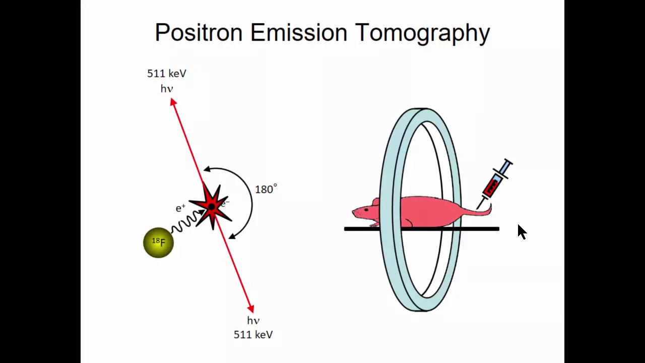
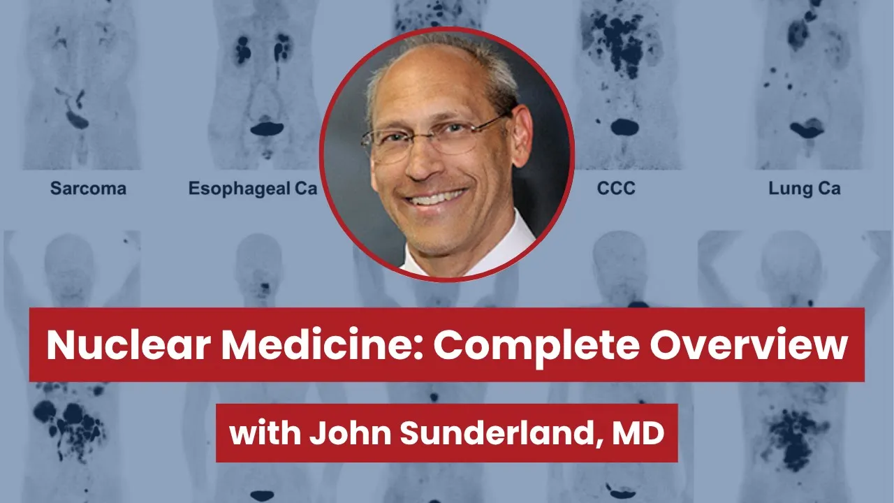
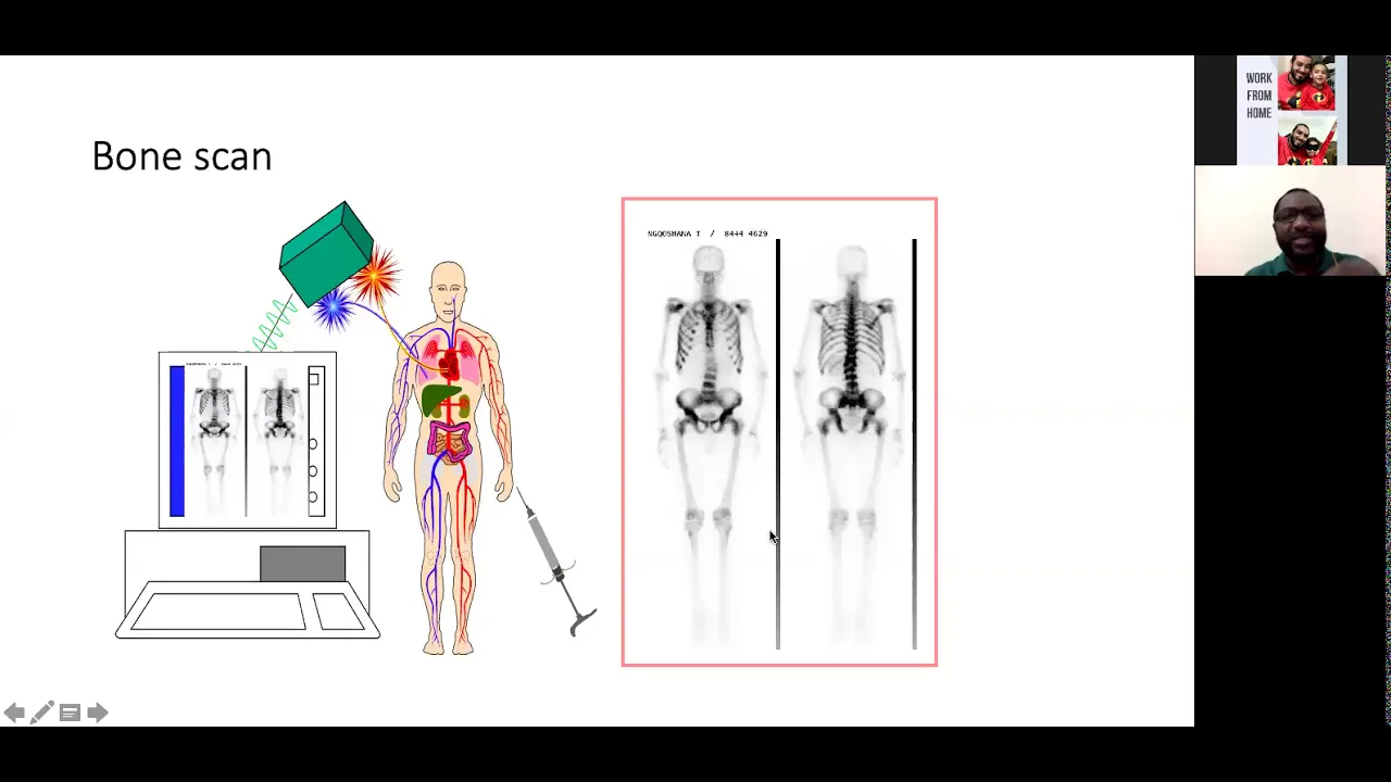
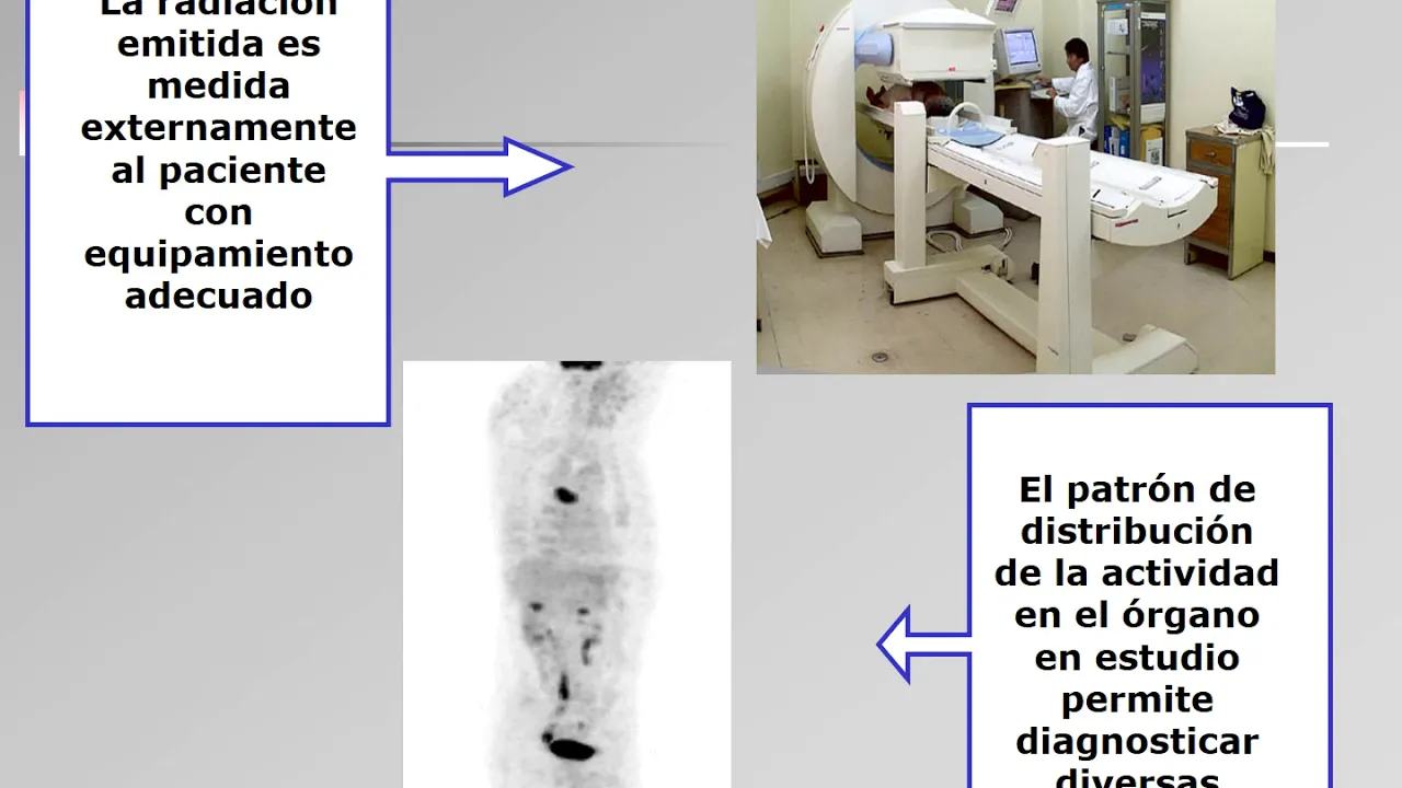
Nuclear medicine is a medical specialty that uses internalized radiation (via IV injection, ingestion, or other routes) combined with specialized imaging technology to diagnose and treat diseases, particularly cancers and metabolic disorders, by exploiting the unique physiological targeting properties of radiopharmaceuticals that accumulate in specific tissues or processes.
Nuclear Medicine Basics: What Radiologists & Clinicians Should Know
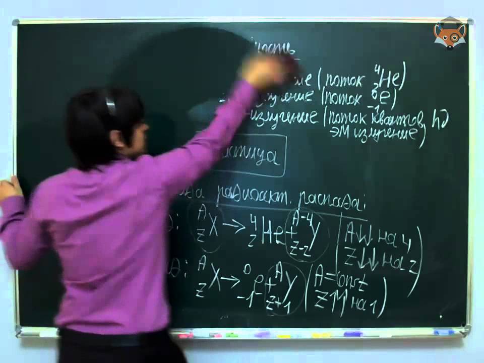
Added:Basic atomic physics and radioactivity, including isotopes, half-lives, and radioactive decay modes (alpha, beta, and gamma emissions).
The conceptual difference between anatomical imaging (such as CT and MRI) and functional or molecular imaging.
Fundamentals of human physiology and cellular metabolism, as radiopharmaceuticals are designed to target specific biological pathways.
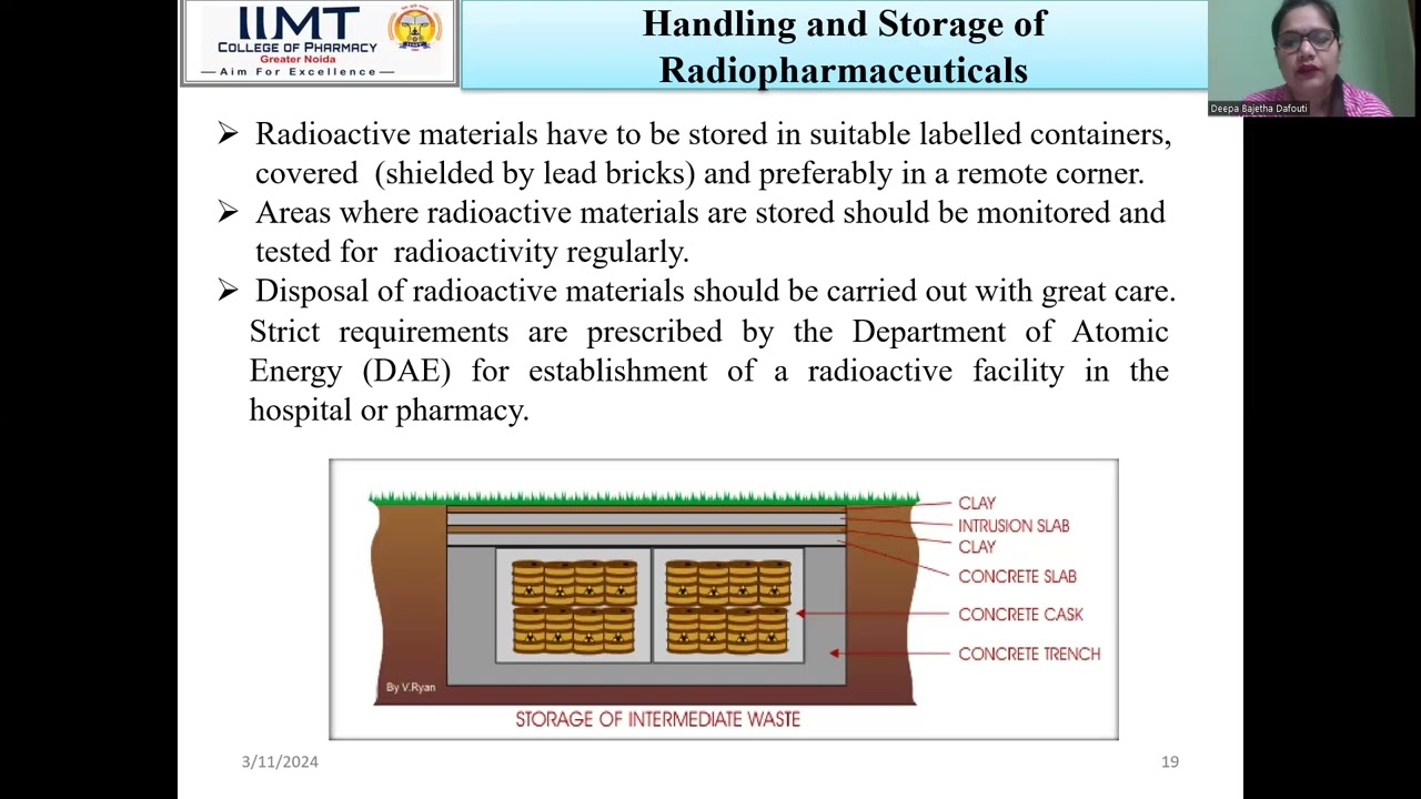
General radiation safety principles, including the biological effects of ionizing radiation and the ALARA (As Low As Reasonably Achievable) concept.
Hybrid imaging modalities, specifically the integration and clinical utility of PET/CT, PET/MRI, and SPECT/CT systems.
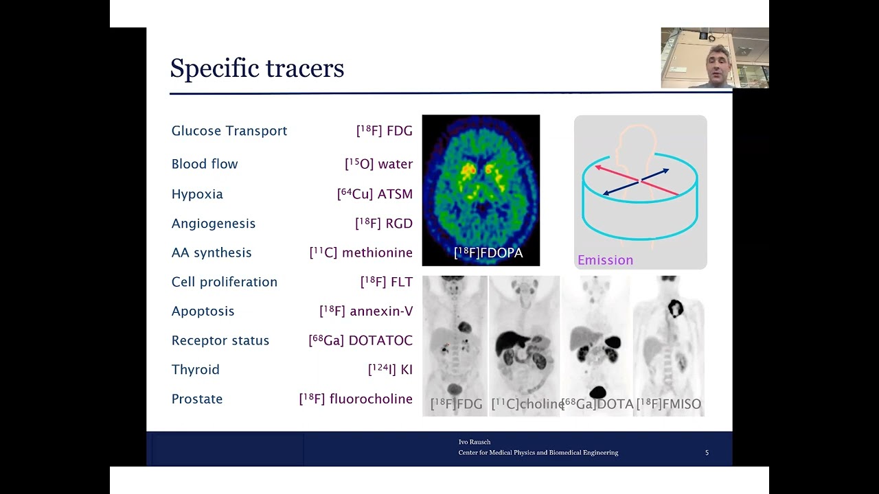
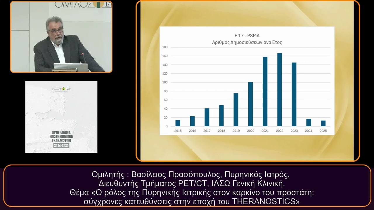
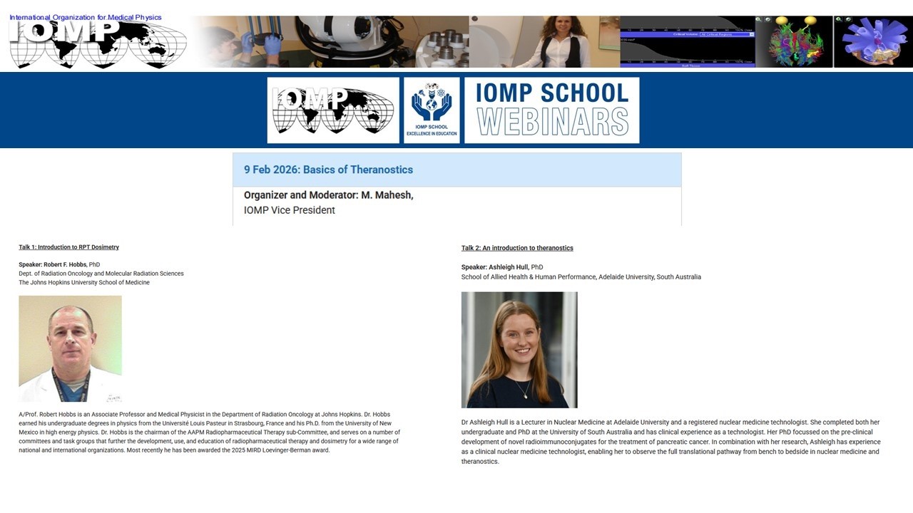
The clinical paradigm of Theranostics, utilizing specific molecular targets for both diagnostic imaging and targeted radionuclide therapy (e.g., Lu-177 PSMA).
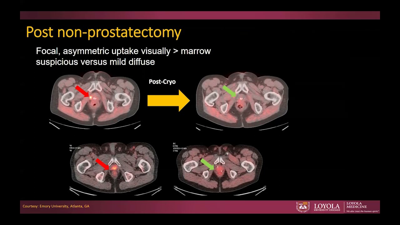
Clinical image interpretation protocols, focusing on distinguishing normal physiological tracer biodistribution from pathological uptake and identifying common artifacts.
Radiopharmacy operations, covering the synthesis, sterile preparation, and quality control of PET and SPECT radiotracers.
10K views201likes1:51:08Original Release: 2020-04-22
we'll check that if you if any of you see the quiz questions come and it would be helpful to me if you let me know that they made it and with that let's let's just go ahead and get into this okay so first what is nuclear medicine this is a question that's a little more complicated than you may suspect and then we'll talk about some selected nuclear medicine exams that I think are important for people to understand that are not going into radiology and things that might help you when you're interns and just general nuclear medicine study and knowledge that I think would benefit any physician and then I'll also talk about some nuclear medicine therapies because nuclear medicine doctors are mostly diagnosticians but they are also treating physicians okay just reading some of these other comments okay so what is nuclear medicine and sorry I need to arrange my screen a little differently here I yes I'm struggling for real estate on my display because I'm using a laptop and here we go okay so nuclear medicine is actually its own specialty and it is also part of radiology so that's a little bit confusing but historically and even today you can find nuclear medicine programs that are completely separate from radiology so it is its own feel that has its own Medical Board the American Board of nuclear medicine and if you choose you can still go through the pure nuclear medicine route however when nuclear medicine started merging with radiology and that mostly happened when we started having PET CT because initially it was only pet there is no CT and then we realized we confuse the pet images on a CT image and now you have a PET CT and that goes along with all of the information that you can see on a CT scan and at that point radiology started getting more involved and we're at the point now where within the last few years there has been discussion on whether the American Board of radiology should just completely incorporate nuclear radiology I don't want to say takeover but that's the way it appears to be to nuclear medicine doctors is that this could be seen as a takeover of nuclear medicine by radiology there was a vote by the ABN M to see if they would dissolve and just fall under the umbrella of the American Board of radiology and they opted not to do that so as it stands we still have two separate medical boards and to be a practicing nuclear medicine doctor you can go through either pathway I first see in the future that more of the nuclear medicine practitioners in the country will be radiologists because just by sheer number there's way more nuclear so the kind of the term that is often uses nuclear radiologists versus nuclear medicine but even then is interchangeable but there are just a lot more radiologists out there and the training programs for radiology are a lot bigger so by sheer numbers there will be many more radiologists that are doing all the nuclear medicine but not all of them and there are certain programs Stanford is one washio and st. Louis is another where a lot of the practicing nuclear medicine doctors and you know attending physicians are not radiologists they are pure nuclear medicine and they do a good job but just to be aware that there are two separate things here and what I'm really trying to do is have two hours to present an overview of an entire specialty area in medicine so it's a tall order when radiologists do nuclear medicine most of them will not do any training beyond residency but there are nuclear medicine fellowship pathways that you can do after a radiology residency and there are also programs where you do a combined radiology nuclear medicine residency and that's what I did and I think that was a great option to be able to go through residency and also get subspecialty decertification nuclear medicine so essentially during my four years of radiology I spent 16 months on nuclear medicine and the rest on a radiology but if I wanted to I could also do a fellowship after but I think that combined pathway can make a lot of sense and then after rhetta's radiology residency I did a women's imaging fellowship as well but that was my path so what makes new pure medicine different than radiology and why did these start out in different places in the first place well one easy way to think about this is that in radiology you have external radiation like an x-ray that passes through the body and then you form an image from that okay so there's some sort of external radiation or some sort of external energy with ultrasound is external sound waves you pass through the body and then you make an image with MRI is the external magnetic fields and radio frequency pulses that pass through with nuclear medicine the radiation is internalized and then you create an image after it's been internalized so how do you get the radiation inside most commonly is through an IV injection so you will take a radioactive substance send it in through an IV or you can eat it we definitely do that things like gastric emptying studies you eat a radioactive meal and then we watch it pass through the GI tract you can do subcutaneous injections we do that for sentinel lymph node studies where you inject sulfur colloid in these subcutaneous tissues and then see it move in the lymphatics we do intrathecal injections to evaluate for things like normal pressure hydrocephalus that's less common now but it's something you can do you can also inject CSF shunts like intracranial shunts to see if there Peyton so you inject the reservoir up in the skull and then you watch the radiation travel down into the spinal cord CSF and you can inject intraperitoneal and there's something called hepatic Hydra thorax where you end up with a pleural effusion but the cause is actually from abdominal ascites that moves through the diaphragm up into the chest but sometimes you can wonder is that actually what's causing the pleural effusion or is there something else going on so a nuclear medicine doctor or radiologist couldn't could perform a parrot aneesa's paracentesis essentially get a catheter in the peritoneal cavity inject radiation into the ascites and then you sit there and watch to see if it crosses the diaphragm into the chest so those are a few examples of how a nuclear medicine study could be performed and the types of things that we do if you have any questions along the way feel free to send me a comment or go ahead and ask so I want to spend a few minutes in this radiology group and a lot of you are going to fill this way and have these opinions and my hope is to broaden your mind a little bit so three common myths and this applies to nukes but also radiology one radiologists don't see or treat patients to radiology is only about Anatomy three I will fall asleep if I sit in a dark room all day can't tell you how many times I've heard that is a radiologist so let's talk about each one just briefly so to get into that what is this now whether you've read the chapter or not you still wouldn't know what this is but is there any guess from you what could this be what am I sure you thyroid awesome let me get the comments here thyroid thyroid thyroid uptake so we have a smart group here that is a thyroid now this is a nuclear medicine scan so a few things the thyroid so what does this image tell us in terms of anatomy does it tell us anything in terms of physiology I see someone said thyroid uptake diffuse uptake so uptake we're now getting beyond Anatomy and we're talking about physiology someone says symmetric correct no large mass agree so a few other things from this image this is a simple image but we can tell something about anatomy we could tell something about physiology the endocrine functioning of the thyroid can we use this imaging agent for treatment we'll talk about that someone says yes does this image give me the opportunity to see the patient yeah well I actually end up performing a physical and history on a patient because of this as a radiologist keep in mind who's not in IR and will I be a treating physician okay so what does this tell us in terms of a out of me well you can take my word for it but this thyroid looks a little big you can see a connection between the two thyroid lobes which is the thyroid isthmus you do not see any nodules and sometimes a nodule will actually show up is a wide spot meaning there's no activity so thyroid cancer is unlikely we have an enlarged thyroid I see the isthmus so I'm thinking Graves disease okay so this one image we've gone to no cancer we've gone to this is probably Graves disease and there is something else we can do as well and that is this so you guys mentioned some uptake and that's true you can get an uptake commonly we look at for in 24 hours and we know what normal values are and in this case this is elevated so what we can tell you is that we injected radioactive iodine the thyroid took up more of that iodine than is normal so the thyroid is very hungry for iodine so either it has lost its feedback mechanism to stop it taking up iodine or something else is going on but this is all consistent with Graves disease we have confirmed why the physiology is we've also confirmed that there's no cancer present and sorry can we use this image for treatment yes it depends but yes so there's different types of iodine we can give a patient in this case is iodine 131 iodine 131 allows us to get an image like this it also emits beta particles and part of nuclear medicine is understanding a little bit about radiation physics which I don't expect you guys to be experts on but there's different particles that things can kick off and beta particles are like little tiny cannonballs that are energetic and they destroy the cells in the immediate surrounding area this is really convenient for us because if you have Graves disease one of the best ways to go about treating it and there are medications that patients can try and sometimes it works and sometimes they don't sometimes people don't even want to mess with the medications because some of them have some side effects that you don't want to miss or you have to deal with but you need to get the thyroid tissue out so one option is to have a surgeon go in and remove it you have you know an incision on your neck you have a recovery time you go under general anesthesia another option is to swallow a pill of radioactive iodine and within a week or so thorough it is gone okay that's pretty cool in terms of recovery and symptoms there's almost nothing that the patient has to recover from you might have mild tenderness in the throat you do have to avoid being around other people because your urine and saliva are radioactive and you don't want them to have laid their thyroids if they won't benefit from it so there are some things like that but in terms of sparing a major surgery that's a great deal I would take a pill any day and this is a one-time pill most often one iodine pill your thyroid is gone because those beta particles are present and specifically because the thyroid is one of the only organs in the body that takes up iodine avidly so you have a Selective ablation of the and tissue that is helpful for Graves disease so iodine-131 contree Graves disease can also treat cancer and the answer is in many cases yes and even metastatic thyroid cancer if the metastatic thyroid cancer cells take up iodine you can treat them with radioactive iodine and not have to have chemotherapy and the side-effects from the radioactive iodine are so much less than chemo okay it's a lot better than radiation and if it's a low-grade cancer it can really kind of clean up iodine anywhere in the body so what I'm telling you is we have a great therapy here you know this is the type of stuff that the oncology community is really striving to have for every type of cancer a very cheap easy inexpensive non-toxic one-time treatment for cancer that selectively only goes to the cancer cells I mean that sounds too good to be true radioactive iodine has allowed us to do that for thyroid cancer for decades okay that's one example of what we do in nuclear medicine that's one example of how what we do might be a little different than what the common perception is of a radiologist okay this is not just sitting in the darkroom it's not just reading scans it's actually interacting and treating patients using images to guide therapy using targeted therapies to treat cancers and it's a very powerful way to go about things for the right condition okay so will I perform a history and physical yes let's see I have a question yes I have questions so my understanding is is when we do radioactive uptake studies cancers of the thyroid are cold nodules because they don't take take up as much iodine so so how is it that iodine gets to the thyroid cancer if cancer doesn't take up as much iodine it does that make sense yeah it does and there are ways we can test for that but so typically the cancer doesn't take up iodine is is the normal thyroid tissue that's not always the case but it still takes up enough of it okay so not as I iodine hungry as normal thyroid but it still definitely is hungry and you also do things before you treat to really make those cells want iodine so you go on iodine deprivation and you can also give artificial TSH to really stimulate those cells and so you basically put the body and is hungry of an iodine state as possible for those thyroid cells and then you give the iodine and then they take it up but yeah that that's something we're going to talk about on here is what happens when cells stop taking out the iodine so that's a good question cold nodules are more concerning for thyroid cancer that's true because it tells you that there's something in the thyroid that's not behaving like normal thyroid tissue should okay because happy thyroid tissue takes up iodine then I have a question how common is worsening of optimal optimal athief that's a tongue twister that I obviously can't say well right now it can happen I've never seen it happen but if someone comes in with really bad proptosis you have to be concerned about that possibility you might pretreat with steroids if it was really severe you might not opt for iodine first you might want to go on some medication first and see if you can get it to back down is it is known to happen but it's not common okay all right so it sounds like you're all pretty familiar with some of this stuff which is great in terms of if you have a patient or a family member who ends up needing radioactive iodine and and that may happen because thyroid cancer is not all that uncommon be aware that they will have to isolate strictly from kids especially and other family members you have precautions so as not to get that radioactive iodine spread around we're all very familiar with self isolation right now but you would have do things with that like not share a bathroom with anyone not use any public restrooms and if somebody is breastfeeding they cannot breastfeed because you do not want to ablate a baby's thyroid right that's really bad okay so radiologists don't CRE patients that's incorrect so obviously everybody knows about interventional radiologists seeing patients people less commonly think of nuclear radiologists seeing patients but they do and also I'm a breast imager as well and I see patients very frequently on breast imaging to tell them what the results of their imaging exam is to perform biopsies that type of thing most radiologists end up in private practice and almost all of them will perform procedures as well there are a lot of procedures that are performed outside of interventional radiology not that they don't know how to do this they are experts of these but pretty much every radiologist is trained to perform biopsies thoracentesis paracentesis lumbar puncture joint injection feeding tube placement if your pediatric radiologist in a secession reduction and this is only a list of what's out there but radiologists are also procedural list in a lot of ways and even to perform some exams you have to interact with patients ultrasound unless your patient can't talk which would be very uncommon with an ultrasound you are interacting with them with four osku P you have to be there in person with them tell them what to do talk to them afterwards about what you see and also radiologists treat and manage contrast reactions so that's a list of ways that we do see and ways that we do treat patients even if you're not in IR so you can cross that one off I'm not saying that you go in in the front of clinic no that's not true but do you see patients on a regular basis absolutely okay radiology is only about Anatomy that's not true either really for nuclear medicine is the physiology almost as much as the anatomy that allows you make a diagnosis you have to understand the physiology the physiology of the tissue of interest we'll talk more about that and I think I'll make more sense after I'm done with this presentation even outside of nuclear medicine you have things like MRI with multi-phase CT that's and multi-phase MRI that's really getting a physiology you're looking at neo angiogenesis fibrosis things like that radiology also requires procedural skills which isn't just Anatomy and I'm not going to talk the whole time about this I promise I will fall asleep in a dark room you know that might be true if you guys sat in a dark room with us all day you might sorry I gonna cut back how long do patients need to self isolate for iodine it's usually really strict isolation for three days and maybe up to a week to avoid being around kids okay about three days is generally pretty good and there is some variation there working to make national standards for this but right now it's a little bit up to each institution but three days is a good rule of thumb for self isolation for iodine so back to falling asleep it just no it's boring to watch other people practice radiology is very different when you're actually doing it yourself is very mentally engaging okay and honestly it's one of the areas I've found in medicine out of everything I rotated through as a student and intern where you just spend more time thinking than anything else I love that about radiology it's just a mental challenge and you know you're not charting you're not doing things like that you're sitting there almost all of your time just trying to figure out what's going on so I find that very rewarding now over and I call you might fall asleep in the reading room I've certainly fell asleep in a reading room and radiology has a lot of overnight call so expect that if you go into it okay enough that let's talk about what a radiopharmaceutical is I believe this is in the chapter of your textbook and it's kind of an important thing to understand with nuclear medicine so radiopharmaceutical is a combination of a radioisotope and that's something like it's well it's an isotope so iodine technetium gallium there's many of these and you take those radioactive isotopes and you attach it to a pharmaceutical and as you know from physiology and farm pharmacology pharmaceuticals deal with the physiology of the body in some way that pharmaceutical substance is targeted to a specific tissue to treat a specific process in the body and so what you do is you take a radioisotope and put a pharmaceutical tag on it and that pharmaceutical will determine where the radioactivity ends up being okay so for example if you want to do a bone scan which we'll talk about you attached technetium to something called MDP and mdpe looks a lot like phosphate and so when you inject that that phosphate will form kind of like calcium phosphate and it goes to bone and calcium okay for a high to scan which tells you about coisa situs and liver function that type of thing you attach it again technetium to something that is hyda or there's AI da there's member fen and there's different things but basically they're all substances that get conjugated and go through the biliary clearing system and that allows you to look at bio transport okay so those are two examples the radioisotope that we most commonly use is technetium I'll talk a little bit about why but the various combination of the radioisotope and the pharmaceutical determines where it goes it allows you to image different physiologic or anatomic processes now iodine is an exception because it is a radioisotope but because the thyroid likes iodine you don't have to put a pharmaceutical on it how do we image these or how do we use them well most commonly after an radioisotope is injected or radiopharmaceutical you use a gamma camera or you can use a pet-ct camera or for certain things you can actually not image and you can assay radiation levels in the blood or urine there's ways you can estimate GFR that type of thing you can assay nasal secretions after you do an LP and inject indium to see if there's a CSF weak okay there's different ways that you can use these to our advantage this is what a gamma camera looks like on the left and a speck CT on the right a gamma camera is this this is a single head gamma camera in this case most of them nowadays are dual head so you'd have one here and one here but there's crystals in here that detect the radiation that comes out of the body and when you take these and attach them to a CT scanner which is what this circle is back here you now have a spectra CT SPECT without CT SPECT is when you take the gamma cameras and you rotate them around the body just like you do on a CT detector and that allows you to get kind of like a 3d nuclear medicine only image so that SPECT CT this is just like CT from radiology oh great the questions are there now awesome so you can look at those I think the only one I've covered so far would be something about iodine-131 okay so I don't think you're gonna have any problems with that one okay spec CT camera so SPECT is from the nuclear medicine image CT is there and then you merge them you have a spec CT this is a PET CT so a pet camera is different from a gamma camera a gamma camera is really tuned to detect things like technetium a pet camera is tuned to detect positron emitters and the most common one of those is FDG which is basically like radioactive glucose and I'll talk briefly about that nowadays we can also have pet with our eye whether pet MRI is really going to take over I'm extremely skeptical it's really expensive and so far there have been almost no studies that show it does any better than PET CT the pet MRI does allow you to avoid the radiation from the CT but on the other hand the radiation from a CT scan is continually getting cut and it will continue to be lowered and that is it continues to get lowered that's less of an advantage to justify buying a $10,000,000 scanner okay because pet MRIs are really expensive they're really cumbersome but maybe I think it's kind of cool but it's a big question mark right now if it's really going to be worth its cost then there are other specialty cameras you can get a molecular breast imaging exam I'm not going to go in depth to this but is a specific camera designed specifically to evaluate the breasts for nuclear medicine and there are dedicated cardiac cameras that are specifically only built for cardiac imaging one advantage of these specialty cameras is that it allows you to image it a lot lower dose than if you try to do a cardiac scan on just a normal gamma camera and often the images are higher resolution okay so there are several of these how do they work I mean we could talk you know a whole day on this but the basic that I think you should understand is that the cameras typically have used crystals and they scintillate and scintillation means it gives off a flash or sparkle of light and here's an example of these crystals and when radiation hits these they give off light and so basically you put patients in a ring of these crystals and then you see which crystals are scintillating and from that you create an image the newest cameras no longer use these scintillation crystals but they have direct detectors that are digital and those work really well they're very efficient which allows you to lower the doses but they're also really but in time they will become less expensive and maybe eventually everything we have will be a digital camera which would be a good thing for patients because that can allow you to cut the dose and it allows you to image faster so all of that is good you just have to be able to afford these cameras which right now sell them are really expensive common radioisotopes there's a lot of them the I divide these by general nuclear medicine and when I say general nuclear medicine that is basically everything that's not pet okay that's a common division how we think of what we do we have general nuclear medicine and then we have pet and the combination of those is pretty much what we do in a day we read our general scans we read our PET scans conceptually they're very different and they have different cameras a lot of these are kind of byproducts of nuclear reactors like iodine in southern Utah after all the nuclear testing a lot of cancer that happened was due to the radioactive iodine-131 that fell down from the sky to the people in southern Utah Nevada we still get iodine from nuclear waste if you want to think of it that way but it's a byproduct of a nuclear reactor technetium comes from molybdenum which is also kind of nuclear reactor based other things like gallium indium and thallium are made from a cyclotron and that is different from a nuclear reactor a cyclotron is where you bombard elements to create other elements and Huntsman Cancer Institute does have a cyclotron they bought this very early and it's been a great thing for our institution that we have our own cyclotron not every academic center it has a cyclotron so it's great that we do a lot of them do not all of them we have the only cyclotron in the Mountain West region and if you don't have a cyclotron then the country has a network of radio pharmacies set up and you can order these from the radio pharmacy which will have a cyclotron and then a distribution network set up to get these two small clinics or wherever you might need them our most nuclear medicine agents carcinogenic also are gamma rays carcinogenic so yes these are all forms of ionizing radiation and it is that ionizing radiation that can cause cancer because they go in and create free radicals and damage DNA so they all are carcinogenic just like an x-ray I think I have a slide in here talking about what the difference is between a gamma ray in an x-ray but they're almost essentially the same thing okay so nuclear medicine is a lot like x-rays pet agents also potentially carcinogenic now when you really get into this we don't know how carcinogenic there's estimates that are probably imprecise a lot of people feel like there's a threshold below which radiation does not cause cancer and a lot of the studies we do could be in that threshold but it's not proven and until we know that they're perfectly safe we'll always be cautious about it another question what's that big thing they're building behind Huntsman so it is a cyclotron but it's for proton therapy so that is for radiation oncologists that will not be used by us in nuclear medicine it's a radiation oncology tool and the one thing I would know about that it for you guys is that with proton therapy the whole idea is that instead of treating cancers with x-rays you will treat them with protons and the advantage of treating cancer with the proton instead of an x-ray is that they are able to determine exactly what depth the proton will stop in the body so for example if you have a cancer that's right by somebody's heart if you treat them with x-rays you know you can angle and do what you can but the heart will get some of the x-rays and the heart will have some radiation and potentially some damage from that if you use protons my understanding is they can stop it down to pretty much the millimeter level so you could treat the cancer and spare the heart from any radiation so that's the type of thing that I think is advantageous there and it is a big accelerator and those proton therapy systems are really expensive but that's what's going in behind Huntsman and I'm sure the radiation oncologists are pretty happy about that because it's exciting for them okay all right so in summary and this is facetious but it's also true in nuclear medicine we inject a person with radioactive waste and then we surround them by special crystals that detect the person's energy and start glowing okay that's it it's absurd it's kind of mystical but it also is what we do and I think it's a miracle like this technology we have is just fascinating understanding how PET scans work it's like otherworldly to me is almost similar with MRI I think MRI and PET are both on the greatest machines that we've invented of course I'm biased but it's really cool stuff but when you really think about what we're doing it's also crazy that it even works you know injects along with radiation put them around crystals and suddenly you get images they look like this okay it's really cool okay SPECT versus pet I already talked about that a little bit so x-rays here it is x-rays are from electrons emitted outside the nucleus don't try to remember this I'm not going to quiz you on this gamma rays are electrons from within the nucleus so there is a difference but essentially they are the same thing and the body's effect in terms of cancer risk will be the same pet with pet you image annihilation photon pairs okay and these are kind of also like gamma rays but a positron is the anti particle of an electron what happens is you inject positrons into the person's vein IV injection usually it's going to be FD G which is fluorodeoxyglucose and I just think of that as radioactive sugar there's tagged with that a Tron and is that goes in the body the glute transporters G ult take up the sugar it'll go preferentially to cells and tissues that are really metabolically active K really sugar hungry in cancer above sugar so it's going to take up more of that sugar than your normal tissues at least that's our hope and then when that goes into the tissue those positrons are going to find electrons and when they meet it's like you know the worst romance you can think of as soon as they meet they're both gone okay they disappear immediately they are annihilated and when you annihilate matter you go back to Einstein we're talking to e equals MC squared so you have MC squared on one side you have a on the other side if you get rid of the matter in the mass you release energy okay is all what Einstein said equals MC squared so you on aisle eight the photon and the electron after they meet and then you get this photon pair which is the energy that's given off from that annihilation event and then we have crystals around the body that are set up to detect the pair of annihilation photons and then using really smart computer math and all of that reconstruction methods you get a PET scan and what it shows you are that is basically a sugar map of the body or a metabolic map of the body and it shows you which tissues are using more sugar than others and using that it allows very powerful evaluation of cancer and other things okay but that's the basic idea behind pet is annihilation and that positron annihilation event gives you gamma photons that we then detect with the pet camera okay what physiology can we image you have to memorize this entire list actually you don't it's really big in this pretty much for I took a year or 16 months learning but there's a lot of physiology you can image just look through here we're hitting pretty much every organ system there's not a lot of OB with nuclear medicine but otherwise you're getting at a lot of stuff every part of the body you know different things renal function liver brain you could do cool CNS stuff and neuro stuff like Parkinson's disease evaluation like that scan a lot of cancer imaging there's Alzheimer's evaluation with amyloid pet which is a hot area right now so we really get at a lot of physiology and I give a lot of credit to nuclear medicine doctors who have figured this all out in a lot of this early work kind of happened around the time you know shortly after World War two when we actually started having reactors and started figuring out what we can do this useful instead of just building atom bombs that's kind of how nuclear medicine started was kind of post World War 2 era where we had this Renaissance and understanding of nuclear physics and then they went and applied this to medicine and a lot of this stuff they figured out is still helpful other things on here we hardly do anymore because things like n where I can replace it here's possible therapeutic agents this might be my last slide on therapies so let's run through it quickly so we talked about iodine okay radium 223 this is also called Sophie go this is really cool it got FDA approved what was it maybe four or five years ago for metastatic prostate cancer and what this is is is radioactive radium this has alpha particles iodine has beta particles alpha particles are even bigger and badder they cause more tissue damage and radium is a lot like chloride and calcium right I'm sorry it's a lot like calcium and it goes to bones so what they figured out is you have this radioactive agent that can bombard cells around it and it likes to go to bones so you could use it to treat metastatic prostate cancer and the initial trials actually showed that you not only help things like reduce pain but there was also a slight survival advantage if you treat patients with radium now this only pretty much goes in trees bone it doesn't treat soft tissue metastasis like lymph nodes and that type of thing but it also doesn't have hardly any side-effects the one thing you have to watch out for because you are radiating the bone in the marrow is that they don't fall too low with their blood counts and white blood cell counts that type of thing but it can work really well we've treated a lot of patients so that you with this trials are ongoing to see if this can now be beneficial for metastatic breast cancer and other cancers lutetium 177 dota tape this is Luda Thera this was recently fda-approved and you can use this to treat GI neuro endocrine tumors the dota tape selectively goes to somatostatin receptors and neuro endocrine tumors have a lot of those so this is another way where you could do an IV injection the patients have more side effects in this case but it still doesn't compare to something like a toxic chemotherapy and you could get good outcomes from otherwise you know poorly treated neuro endocrine tumor why 90 that's kind of the bread and butter of mic how cellular carcinoma treatment and stuff that ir does why ninety procedures typically the interventional radiologist will go in and selectively canalize the artery or vein of interest and then they get the Y 90 from us and inject and treat those cancers locally I'm sure you're all familiar with this it works really well you may be less familiar that nuclear medicine is working in the background to help make these work too and you could do things like before you treat you can do a planning exam where the ir doctor goes in and selects what they think are the arteries to the hepatocellular or the you know cancers and the liver they want to treat and then you do a test injection bringing them to nuclear medicine and you see did this end up where you think it did did a lot of this end up in the lung because you can have shunts too along and you don't want y9t ablating yearlong that would be bad so we do planning and things like that to help our I are colleagues be able to do this as successfully as possible and then there's i-131 mi bici and this has been a frustration among nuclear medicine doctors that people in Canada and Europe have been using this for decades to treat you know things like neuroblastoma in kids the FDA didn't improve it here for a long time and even now it's still kind of rolling out in the country but finally we're getting FDA approval for mi bici because this is a fantastic treatment for the appropriately selected kids with neuroblastoma or adults with pheochromocytoma again selective ablation of those this is basically a norepinephrine analog and so it goes to cells that like to take up norepinephrine which is neuroblastoma and things like that samarium in strontium we don't use anymore because they're really toxic but historically they've been used for lymphoma and bone metastases or sorry bone metastases we've also had other agents for lymphoma that aren't on here like Beck's are okay so that's a little quick run through on therapies that we do the newest ones are radium lutetium and mi BG and I think the indications for these are going to be increasing a lot of places like Stanford for one are actually setting up complete nuclear medicine clinics because they expect the growth in these areas to be very robust over the next decade in several decades to the point that they think nuclear medicine doctors may actually be doing more therapies than Diagnostics in the future maybe they're right I don't know they might be and the one thing that everyone is looking forward to is that there might be some PSMA directed therapies which would allow you to specifically treat soft tissue and bone metastasis ease of prostate cancer and if that works well which it looks like it might that could really take off and there's a massive population of men with men prostate cancer obviously and some of these emerging nuclear medicine therapies could potentially be really impactful and also keep us really busy treating all the potential patients that could potentially benefit from that okay so we're gonna do a quick run-through of these these are all scans that you're gonna see in your career okay I think pretty much no matter where you end up even I don't even think you can escape nuclear medicine with psychiatry because we do Alzheimer's and some Parkinson's dementia Lewy body dementia that type of stuff so we we have a little foothold in a lot of areas thyroid we've already pretty much covered I'm just gonna mention there different agents that we can use we have AI 131 this one specifically has the beta particle for therapy 123 does not but it allows you to image at lower dose so this might be better for initial imaging this is therapy you can image with straight up technetium or and we already had the question what happens if the metastatic I red cells don't take up the iodine anymore well in that case you have a fallback with FDG is that slide here not yet hold that thought we're gonna get into that more detail so here's a 1/2 I 123 scan looks pretty much identical to the I 131 you can also do full-body iodine scans and this now becomes really helpful for Graves disease not helpful for cancer search helpful okay this is a whole body search for cancer and what we're showing here is this was the pre therapy I 123 scan they didn't show anything then we treat with AI 131 at a substantially high dose and now all of a sudden because you can also image the 131 you bring them back a day or two after their treatment and image after they've had a whopping high dose and now you're starting to see sites of metastatic disease okay so this is a good way to screen the body for for thyroid cancer thyroid globulin this is a lot of text I'm not going to read it all basically I want you to know that this is a Serah marker a blood marker for thyroid cancer and how this works so the thyroid cells normally secrete thyroid globulin after you treat thyroid cancer first typically for cancer not for Graves disease but for cancer usually the surgeon will go in first and remove the tumor and the thyroid and then after they've had it removed you then treat with iodine to kind of clean up everything else get all the microscopic disease that may have been left behind and treat any metastases after you've gotten rid of all that thyroid tissue the body's thyroid globulin level should be you know pretty close to zero what you could do thereafter is keep checking the thyroid globulin level and once it starts to tick up hopefully it never does but if it starts to tick up you know that cancer is back somewhere because only thyroid pretty much let me thyroid makes thyroid globulin so it's a good serum marker and what it increases it's time to get a full body iodine scan in case something like this now if you get your full body iodine skin and you don't see anything so hypothetical scenario that's what I'm talking about here they're treated thyroid globulin then after three years they start to creep up and you get your iodine's again and it's negative you do an ultrasound of the thyroid Bettis negative what do you do next the answer is you get a PET scan this gets into well differentiated and poorly differentiated we're almost at two so I'm going to cover this and then we're going to take a five-minute break this is a concept that's important to understand for anyone interested in cancer so well differentiated tumor cells behave like the native tissue in the example of thyroid well differentiated thyroid cancer cells take up iodine with breast cancer well differentiated breast cancer has estrogen receptors okay because that's what breast tissue does is it becomes poorly differentiated it starts to lose those features of the native cell and when that happens these cancers get more aggressive and you you ideally would be able to catch cancer and treat it completely before it's poorly differentiated but poorly differentiated cells tend to be really sugar hungry okay they get more aggressive they divide faster and so this is a common principle that is things become poorly differentiated fdg-pet becomes more useful and here's a case of an fdg-pet showing uptake in the thyroid bed in a iodine non avid iodine tumor so on a nighttime scan this didn't show anything now PET scan it's lighting up very clearly okay this principle applies to a lot of things prostate cancer we have imaging agents for well-differentiated and then when it becomes pretty differentiated you get a PET scan neuro endocrine tumor we have donut a PET CT for well-differentiated these are the ones that have somatostatin receptors then as it becomes poorly differentiated it loses that and you can now pick it up with fdg-pet so we kind of have this ability to image the spectrum of disease whether it's well or poorly differentiated and this can give useful information to know you know what's the uptake on a doe tape scan and if it doesn't show a lot of uptake you know that this cancer is now transforming to a more aggressive version and maybe at that point you change your therapy okay so let's go ahead and take a five-minute break I have it's 156 so I'll start at 201 and we're going to cover these pretty quickly probably in the next half hour I think we'll get through these and then at the for the last half hour I'm going to show some cases okay so see you back in five minutes you you you you you all right let's get back into this sentinel lymph nodes all right this is something that is important to know you see there's also a quiz question associated with this so a sentinel lymph node is the first lymph node that lights up in a specific lymph drainage basin okay and let's first get the overview of what we're trying to do here so if you have let's use an example that you have melanoma well not you that's not a good thought you don't want I don't want anyone to have melanoma but let's say you know a patient comes in you see they have melanoma in their upper back and it's clear that that melanoma needs to get cut out but what about any lymph node metastasis how are you going to know if they're metastatic well one way to know is if you do a staging exam like a CT and you see really big axillary lymph nodes it's pretty clear that those are going to be metastatic or you see big lymph nodes in the back there are nodes and unexpected locations those are called the in-transit nodes and they can pop up when you have cancer or maybe that upper back melanoma drains to the neck in the cervical nodes so if you get a CT you know you could do neck chest and pelvis and you see some big lymph nodes in the neck you know those are probably gonna be metastatic you could do a biopsy if you wanted to you could go in and remove them with surgery but what if you do those initial staging scans and all the lymph nodes look normal does that mean that there is no cancer in those nodes and unfortunately that does not mean that there is no cancer okay no imaging study can definitively exclude cancer in a lymph node because a lot of times that cancer might still be microscopic and we only are able to see change when it becomes more advanced you have to have a lot of cancer grow in a lymph node to make it big but it can Harbor microscopic cancer and still look normal on an imaging scan so you have the option which is what used to happen that you go in and you just cut out all the lymph nodes you can find you know take out as many as possible for breast cancer that would happen a lot of times you know you just go and take out all the axillary lymph nodes if you palpate supraclavicular lymph nodes take those out and then go and the pathologist will tell you whether that cancer or not in that case you could have let's go back to the melanoma case let's say you go in and you take out ten lymph nodes in every single one of those is negative well you've benefited the patient in that you evaluated for cancer but you've now removed ten lymph nodes and lymph nodes do something for you right and one thing they do is drain fluid and you may now have caused some long-term never get better edema lymphedema other issues for these patients that are not negligible some patients who have had a complete nodal dissection have a lot of symptoms from it it can be something that affects them every day of their life so that caused surgeons to pause and say maybe we shouldn't take out every lymph node maybe we need a better way to identify whether the nodes have disease at all or not and that is what led to this sentinel lymph node study and what this allows us to do is map the sentinel lymph node basin anatomy and what we are looking for are if we inject this melanoma in the back which lymph nodes are going to light up okay because that tells you what the first note is so you watch on the imaging literally for the first nodes to light up and you can do sequential imaging to see and when you identify the first nodes that light up the surgeons go in they have some sorry radiation probes they can use to find which lymph nodes have a lot of this radiation in them as well and you remove only those sentinel nose and you leave the non sentinel nose alone and so in the case of breast cancer you may now only remove two or three Sentinel nodes and leave back and weave along the other seven nodes and in that case you've benefited the patient because if those Sentinel nodes do not have cancer and they are the first lymph nodes that would drain the cancer you can presume safely we've seen from research that all the other nodes also don't have cancer and now you've spared the patient from having complications from a complete neural dissection okay so that's one big benefit the other benefit is that you can sometimes identify unexpected drainage that you wouldn't have otherwise known and the reason I chose the upper-back melanoma is because that's known to be a really problematic area because the drainage is unpredictable okay and you can see here you have these two main drainage networks this is sapis line here I guess I'm not quizzing you on that either but it can go up or down so what if you have something on your back here okay sometimes in my area you have something on your you know abdomen here it may go down to your and green I'll chain it may go up to your axillary chain it could go into the internal mammary chain so the sentinel lymph node also shows you which way is the nodal drainage and where by extension does the surgeon need to go evaluate here's the nodal drainage pattern for breast you have axillary you have some supraclavicular you have internal mammary those are by far the most common and you go in you inject technetium you inject some blue dye you can not everyone does the blue dye and then you watch it travel up to the node this is the first training node so that's your sentinel lymph node surgeon will go in and remove this node and maybe a couple others around it but you leave all of these behind and you let these tell you what's going on if these are positive then the surgeon may go back later and take a lot more but in that case you are actually much more likely to benefit the patient than if you just take them off you had this star okay really we do this for breast cancer melanoma most other cancers we don't do it for okay and this is just showing the intraoperative probe the surgeon can use and if you've been in the O R you'll never forget how these sound but they make really distinct sounds when you get closer to the node the thing starts beeping at you I sound like it screams at you basically it's like you're here no no no and then yes and it goes really high and loud and then you know that your nose down there and they'll go in and take it out alright mmm so this study does not confirm whether a lymph node has cancer that's a very common misconception okay sentinel lymph nodes study does not show you if a node has cancer it only shows you what the draining node is all right and here's lymphedema okay and it can get a lot worse than this but even this much would be bothersome okay here's some actual images so here is a back base of neck melanoma and this was unexpected drained well maybe not unexpected because it's midline but still you want to be able to predict necessarily it drained it both axilla so in this case you'd have to do essential in the lymph node on each side here's a trunk or abdomen I can't quite tell probably on the front yeah because it's going to the inguinal chain so this was a melanoma that drained both the inguinal region and an axillary lymph node so again you'd want to go excise both of those without having this study you want to know if you had to go here or here scan I think you're familiar with the VQ scan a little bit it's most commonly for acute pulmonary embolism you can also do it to evaluate for pulmonary hypertension chronic pulmonary embolism which means you've had Pease and they didn't completely resolve and that can cause pulmonary hypertension you can do this scan for preoperative planning if someone's having you know maybe like a single lung transplant or you have to remove a cancer from a lung and you need to know how much ventilation is that lung providing the body you can tell that from a VQ scan you get the physiology of the lungs with this and you can also use it to evaluate for it right to left shunt vqv is a ventilation cues of perfusion so you are looking at either one individually or combined to see is there a mismatch are there areas that are perfused but not ventilated or more useful for PE would be ventilated and not perfused a lot of physiology you can get out with a VQ scan so not everyone gets a VQ scan or should everyone with suspected PE get one CT pulmonary angiogram is your test of choice for PE okay I would always recommend getting the CT first and you end up getting a VQ scan if they cannot have a CT so maybe they have an IO donated contrast allergy maybe their CT angiogram was non diagnostic that can happen a lot of times that the contrast timing is off because for a CT PE study the technologist has to in very accurately time the contrast administration and if they image too early or too late you won't have the contrast in the pulmonary artery tree like you need and in that case you can't necessarily exclude a PE you can't repeat this study because they can't have that much iodine in a row so then they're getting a VQ scan recently postpartum pregnant now in pregnancy is not a contraindication to having a CT but there are some dos for some things you can do with the VQ scan you actually only get the perfusion scan and see what that shows and if the perfusion is normal then there's no PE and you're done some other things calf you indications the main one to know is is PE and you also need to remember the wels criteria you know that better than I do at this point but the overall determination of whether a VQ scan of how you interpreted it takes both imaging findings and clinical suspicion into consideration okay so for a VQ scan we always need a chest x-ray or a CT so when you're in an internship when you're interns you can make your radiologist and nuclear medicine doctors really happy if anytime you order a VQ scan you check to see do they have some sort of chest imaging within the last 24 to 48 hours you have to have the chest imaging to be able to interpret a VQ scan so if you haven't ordered that you're going to be getting paged in a phone call saying hey we need a chest x-ray okay so within 24 to 48 hours depending on the institution you need some chest imaging then we get the perfusion we see any perfusion abnormalities how big are they then we well usually you do the ventilation first you look for ventilation defects then you get the perfusion and the key is that for a diagnosis of PE you are looking for moderate to large mismatches between ventilation and perfusion meaning the lung can ventilate normally but it has losses blood supply because there's a clot in the pulmonary artery now if you've lost both ventilation and perfusion you may remember from physiology that as the lung stops being able to ventilate whether due to add electus or consolidation from pneumonia or whatever's going on where you have a lung issue the body naturally reduces blood flow to that area of the lung okay called hypoxic vasoconstriction so areas of hypoxic lung the body automatically shunts blood away from that area and when that happens that gives you a matched defect with the PE you don't have any ventilation problem is just purely on the perfusion side so you expect normal ventilation abnormal perfusion in a segment of the lung now you do have to know these segments to read a VQ scan and how we read it is determine on whether that mismatch is large moderate or small okay here's an example of a ventilation scan this is with Zenon where you breathe in the Zenon you see it come into the lung and then you see it breathe out of the lung it's very quick in and out and what we're doing is looking to see it as a slick uniform are there any chunks missing and then we get a perfusion scan and you see are the lungs perfuse throughout and this is a good example of a normal here's an example of an abnormal look on this one how the perfusion you have this web shaped missing area of lung this part of the lung is not getting blood flow and then you look over here and you say that part of the lung is getting ventilated and then we say PE okay the criteria is very complicated I don't like VQ scans a whole lot for this reason because it's just not that easy of an exam to interpret there's a lot going on and the key is that you end up getting a likelihood or probability of a peb and present okay VQ scans give you probabilities so normal very low low intermediate or high you combine that with the clinical probability of your suspicion and then you have these algorithms that have been established through research on what to do right to left just so you know it can do this you inject and then you basically look and see if you have brain uptake because if you inject in a vein those particles should get stopped in the pulmonary system ok the lung kind of apps like acts like a filter of the blood so you inject these maa particles they're very small particles and they are large enough that they actually embolize in the lungs so when we do a VQ perfusion scan we're actually causing micro embolisms but we are only embolizing a very small amount of the available capillaries that is really not significant physiologically but sometimes we do inject a lower dose if we're worried that hey this patient is not doing well and we don't want to block even more of the blood supply to the lung but the key is for this they're supposed to not be able to get passed along they're supposed to get stuck in the pulmonary vasculature so if you inject and also in the brain lights up you know there's a right-to-left shunt it's an easy exam you can also quantitate and actually get percentages in this case the wife lung is providing 45 percent of the ventilation the right lung 54 percent and then the surgeon can decide in this case that they have a choice that they're going to transplant one lung they would choose the left the one with the lower amount of lung function okay PET scan I think you're very familiar with this I'm pretty much going to skip other than just know it's not all about f-18 FDG when I was a medical student it was you're now a medical student and it is no longer the case okay we have a lot of other options and more coming online ones that we clinically use here that you we use a lot of dough today pet I don't even have that on here but oh yeah I do we also do foot sick living for prostate cancer and we do a my vid for Alzheimer's PSMA is coming so watch that when it when the big trial is announced which will be soon this will probably be a New England Journal of Medicine article that's going to change a lot of what we do I think and here's the thing with the annihilation it goes in and annihilates you get this pair of photons out to the annihilation and then you use your crystal ring to say hey that happened along this and these detectors are actually so sensitive that even though these photons are coming out at the speed of light they're able to say it hit here earlier than here and therefore by deduction is located here okay a little bit closer to this than this because it knows that it hit man a seconds earlier on this one compared to this one that's kind of impressive that you can detect differences in the speed of light and you know like a you know three four foot ring here are different examples of pet fgg is what we most commonly use here's what doe today looks like notice the pattern is different you've got brain uptick here you don't here because this is imaging where you have somatostatin receptors and then we have flu sick living over here that has a really hot pink rias just the thing to know is each has their own physiologic pattern as a radiologist you have to know what those patterns are you have to know that hey this up taking the pituitary is normal and then you have to go from there okay but that requires some knowledge of physiology scan I I will admit it when I was an intern I rotated at the Naval Medical Center in San Diego and my attending told me to get a bone scan and I didn't know what he was talking about which is funny that I'm now nuclear medicine attending but I truly didn't know I was like is that an x-ray what is it so you're not gonna fall in that trap yeah you probably already know but I'll make you smarter than I was so a bone scan is a nuclear medicine study that's what I didn't know and then he assigned me to read about it and I became informed bone scans can assess for bone metastases osteomyelitis infection other niche things like reflex sympathetic dystrophy metabolic bone diseases like hyperparathyroidism and fracture evaluation you know do they have a stress fracture is there non-accidental trauma and x-ray isn't normal but you're suspicious you can get a bone scan to see there's a lot of uses one thing I want you to know osteomyelitis requires a 3-phase bone scan okay whenever we're talking about infection also things like stress fracture but especially infection you need to get a 3-phase bone scan okay if you just order a bone scan it's just going to be a single-phase whole body scan keypoints bone scan is more sensitive than a CT scan for bone metastasis so we see this frequently where patients get is have a cancer so they have prostate cancer or a breast cancer you get the staging CT scan and the bones look fine then you get the bone scan and you realize the bones are not fine because a bone scan will show you the metastasis earlier than CT that's why the NCCN guidelines for cancer staging typically for certain diseases like prostate and breast cancer that go to the bone a lot they recommend getting both a CT and a bone scan together it does not show all types of bone metastases I do think this is not beyond what you need to know because it is the missions and the interns who are ordering these scans if you have a cancer that tends to have blastic metastasis such as prostate in breast even though breast can be mixed is often plastic you want a bone scan if you have a cancer that has lytic metastasis like multiple myeloma renal cell carcinoma some of these very vascular cancers you want a PET scan with FDG these are kind of complementary to each other you both of these you will see pretty much all the cancers because this will cover the plastic this will cover the lytic there is some crossover it's not mutually exclusive but this is a good rule of thumb this is why we often don't get fdg-pet scans on patients with prostate cancer because the fdg-pet is good for lytic metastasis but it may not show you uptake in the prostate cancer plastic metastasis okay three phase bone scan is for infection imaging a standard bone scan has a single delayed image okay I think if you remember what's on this page you're on recently we good footing this is what a bone scan looks like this is a very high quality bone scan they often don't look quite that good and notice in this case here's a CT and you know this is largely lytic and you see mild uptake but there is some uptake here in the pelvis here's another example bone scan the pelvis CT looks really bad in this case notice how little this is and you don't see a whole lot of uptake so you can imagine if this were very small you know this is obvious because it's big if it was very small you could easily miss a very little metastasis and you see the activity you actually are able to pick up is where there's this bone building on the periphery but in the center you don't see anything if you got a PET scan on this patient this whole Center would just light up like crazy because this is all tumor okay it would be very sugar hungry cardiac imaging there are several things we can do one you can evaluate to see if there's Corden airy artery disease with with blood flow if you end up as an emergency room doctor you're going to deal with this a lot patient comes in with chest pain is a cardiac or not there are several options you can get you know you could get your cardiac enzymes and you could do a stress echo or you could get a cardiac CT or MRI but a lot of times the easiest and quickest way to get this evaluated especially on weekend stuff like that is to do at myocardial perfusion scan where we use sestamibi and look to see if there's admirable perfusion in a coronary distribution we can also assess for viability and when that one matters if you have a patient with a myocardial infarction and some of their say their inferior wall is not contracting anymore you can guess that that's dead but it may not be dead it may just be stunned and hibernating after a mi the cells are damaged but they may not be damaged to death okay they may just be damaged enough that they're gonna kind of hang back say hey I'm just gonna kind of relax for a while the rest of you guys bump can pump the blood but I'm just gonna hang out until I recover and that is actually viable heart and why that matters is if you go in and revascularize that area all of a sudden you're going to see that start to pump again and the patient's EF is going to increase and they're going to do better so we can tell with nooks you can also tell on an MRI about viability okay I like this one your friend is almost there is only mostly dead that's pretty much what we're looking at it's like yeah this hearts not great but mostly Dead is slightly alive and that is absolutely the case in in a lot of cases where your EF can be horrible after an mi but then you revascularize and all son your EF is normal but you don't revascularize everyone because it's a waste of procedure and extra surgery and cost and all of that if you revascularize what's dead it's never coming back right so what you need to know is is this viable or not the thallium and fdg-pet can show you that so there's this you know ischemia hibernating stunned or dead just know that any of those are possible this is beyond what you need to know but you see for this done do you revascularize okay there's a lot of ways we can do this this is beyond what you need to know other than it gets complicated you can also assess for cardiac amyloidosis with nukes and this is kind of what a scam looks like the left ventricle is a lot bigger it takes up more system abusing marker of mitochondrial density again getting at the physiology you see more mitochondria and the left ventricle compared to their right and we're really looking to see is this uniformly distributed in all the territories of the cordon Airy circulation and we have different views that we look at and then you image like this when someone's resting and then you stress them in some way and the image again and if you see that their stress looks really bad but their rest is okay that is the imaging equivalent of ischemia and those patients go to the cath lab okay so like this rest you got perfusion here stress perfusion this last Oh LEDs not happy they're going to get a cath okay renal scan a lot of valuable stuff here peds if you end up as a pediatrician you're going to do a lot with this obviously nephrology and internal mess and they're going to have a lot with this these exams are still done commonly they're very valuable in the type of information they can give you so you can look for a lot of stuff funk evaluate renal failure with pediatrics you can evaluate for reflux you can confirm if there's renal Vasudev retention and transplant you can look at what the blood supply is how the transplant can use functioning is it obstructed all this stuff with a so this is a mag 3 I would know that mag 3 not for the quiz but for your actual life mag 3 that's going to be the majority of the renal scans you would order and a mag 3 scan is great because it shows you the blood flow so you inject here's the aorta hey within seconds you're seeing the Kenyans light up which is what happy can use do because they're very vascular okay you have good blood supply to both of these kidneys then you watch to see the cortical transit it ends up in the cortex so it's not totally dead and then you want to see is it actually moving out of the cortex into the bladder and so you keep imaging over time and this tells you function okay this is anatomy a little bit this is function okay blood flow in the cortex isn't now moving through the cortex to get excreted yes it is are they doing it symmetrically yes they are is there obstruction no there's not okay and you can generate these curves as you image over time so you see after you inject the radiation basically watch to see how it moves through the urinary system so it comes in to the cortex that builds up builds up builds up and then it starts to excrete and it excretes out excretes out excretes out and we have normals so we know what a normal time is if this discrete excretion is really delayed we know there's some amount of obstruction we can quantify if it's a severe obstruction or a mild obstruction that's really helpful for urologist to know do we need a stent or not for transplant you can evaluate the how robust is the blood supply because that's a big question for them you know if you see a nice sharp uptick you know that the blood is good it's getting there quickly if the increase is something like this hey that blood supply is not great or at the renal function isn't great we have ways to that out okay obstructions gonna look like this happy Kimi like this and this is from an actual study so we actually get these curves you know this is gonna be there right so the right kidney is obstructed the left washes out and you see that as you look through time did this activity and the right kidney isn't clearing now some of you may be saying hey you saying right Kenny this is actually left kidney I think this is in your chapter that you have to be aware on a renal scan we are imaging from behind so if you're looking at a person from behind this is the right and this is the left and is kind of an exception to our usual radiology rules okay for pedes they can do DMS a imaging which gives you an image of the renal cortex and basically this tells you is there pyelonephritis or is their renal scarring okay and those images look like this and that can come into play like say you this is an ultrasound you have something here in the mid portion of the kidney is that a mass is that normal kidney there's something called a column of Burton BER TI n for those going to peds or with particular interest you can try to remember that column of Burton is normal tissue they can look like a mass in the middle of the kidney and the way to figure that out is with a scan here so you see that this has uptake in the cortex this is normal renal cortex not a mess okay function of each kidney in this case the function is very close they should be close like that if the right kidney is doing 80 percent of the work in the left can use only during 20 this type of scan would tell you that and you can imagine how useful that is like for pediatric reflux for example this will tell you how bad the reflux is in terms of how much damage is it causing to the kidney so if you watch overtime and you see that you know there's reflux in the left kidney and hey that renal function is dropping off quickly the right kidney is doing a whole lot more of the work that might be like enough to go in and do a you know you read or ovary implantation you know some large measure that you wouldn't want to do if things aren't that bad so this type of study can give you that type of information alright and this is a transplant evaluation here the kidneys down in the lower quadrant you only have one of them and you can see the blood flow and then the function okay brain imaging so we can look for a dementia we've been doing that for quite some time with FDG now we're starting to get amyloid imaging which can confirm amyloid neritic Plex and when I was a medical student back in the dark ages of the late 2000 and let's see 2007 to 11 we didn't have this scan so I was taught as a medical student that the only way you can confirm a diagnosis of Alzheimer's is on autopsy and I found that a little disheartening because it's like well if you probably want to know about that before they were dead right but you could guess at it but the only way to confirm it was on a pathology evaluation of the brain at autopsy well nuclear medicine doctors stepped in and figured out a way to image the plaque because if you don't have my plaque you don't have Alzheimer's now the converse is not true if you have Alzheimer's plaque that her amyloid plaques are if you amyloid that does not mean you have Alzheimer's but if you do not have amyloid you don't have Alzheimer's and that's really sometimes where it's more valuable for the cognitive neurologist figuring this out is that you can exclude the possibility of Alzheimer's with FDG it's approved for evaluation of Alzheimer's versus frontotemporal dementia and it works reasonably well sometimes it's very clear a lot of times it's not as clear and so you're still left scratching your head a little bit brain tumor imaging honestly we don't do this that much there's still some research interest and maybe occasionally we might do it but MRIs kind of taken over although I would say MRI is not perfect for this either but it's an important question in the brain if you see enhancement or something around the periphery of a resection cavity is the cancer back is there residual cancer or is that just scarring from radiation or surgery or whatever it might be and so MRI you can do fancy MRI Pro fusions and get these blood perfusion maps and stuff to help with that and in diffusion maps and whatever you can also try with pets but one hard thing with pet in the brain at least with fdg-pet is the brain uses a ton of sugar and so you may not be able to see the cancer over the brain because they're both they're both chewing through a lot of sugar so you don't have great signal dopamine transporter imaging is a super cool exam I think I have an image in here and it evaluates the dopamine transporter density in the basal ganglia this is newer in terms of nuclear medicine scans and what I love so much about this one is it's kind of a binary yes-or-no okay there's not that many studies in radiology where it's just like a definite yes or definite no but this is one of them almost all the time you can do seizure imaging to look for a seizure fo Tsai and you can do a brain test study which we'll talk about so this is an example of where fdg-pet did work and even though the brain is taking up a lot of sugar it's not doing it as much as the metastasis and you can see these are viable tumors okay we're getting a little bit of physiology if it's eating glucose it's alive okay viable tumor here's what an amyloid scan can look like this is the Pittsburgh compound they're the kind of first people to do this we now have other ways which are like this and you can get maps like this and if you have uptakes this would be a positive scan this patient has amyloid plaque okay this is what a negative scan can look like this patient does not have my plaque this patient does not have Alzheimer's disease even if there's clinical suspicion of that so then that allows the neurologist to look for other causes of dementia brain death I hope you never have to order this too frequently but occasionally it certainly comes in and with the brain does scan it's an important determination on everybody's part whether you're going to withdraw care so what we're really looking for is is there cerebral profusion or not so in this case you have an an Geographic phase you see the carotid slide up you see that it starts to diffuse into the brain so in this case this patient has perfusion to the brain you this is a different case where you're looking later and you may notice that this is a bit different than this okay in this case you see some uptake in the dural venous sinuses which can be normal you see a hot nose which is a bad sign because that means the blood is getting stuck here so it's going there okay I can't get into the brain so hey we're going to go forward into the nasal tissues that's what brain death can look like this is what brain death can look like I shouldn't say brain death this is what absent cerebral flow can look like and I'll tell you why in a minute absence of cerebral blood flow okay what causes that absence of blood flow is increased intracranial pressure so say this is a motorcycle trauma patient and they hit their head really hard and they have a lot of intracranial bleeding and injury and swelling and maybe it elves over you know hours to days and they progressively go comatose and then lose their reflexes and there's clinical suspicion of brain death you can confirm that and as the CSF is the intracranial pressure builds up so much not CSF buy intracranial pressure it eventually gets to the point that the blood pressure in the arteries isn't enough to get the blood in there and it just stops and at that point the brain will essentially all infarct okay so brain death study confirms absence of cerebral flow or presence of cerebral flow it does not confirm brain death okay that has to be a clinical determination based on you know respirations you've gone through neurology probably most of you at this point make sure you know what they do you know with reflexes and all of that but in the clinical suspicion if further confirmation is necessary you can get the nuclear medicine scan and it can support the clinical diagnosis of brain death this is an image of a data scan and you look to see uptake in the dopamine transporters in the basal ganglia and you want to see the caudate and the putamen in this case the putamen they look like little commas it's really almost completely absent on the left on the right maybe there's a hint but this is a normal this patient has Parkinson's disease where a parkinsonian syndrome and that's how it is is a great scan okay humph agency I mentioned this you can inject the shunt and then watch to see if it goes down and if you see spillage in the abdomen you know that entire length is patient and Gi we're gonna go real quick through this because I want to get through cases so you can do a few things evaluate for acute cholecystitis you can look for bio leak in a patient who has had a cholecystectomy you can look for biliary atresia in a neonate mm-hmm this is what a normal scan looks like you expect this to be taking up in the liver excreted into the gall bladder and then go out into the intestine like normal bile does this patient is not how coal ISA situs this patient does okay liver uptake where's the gallbladder it's not there it's not there it's not there I see the bile ducts and I see it getting into the bowel but no gallbladder that's an inflamed gallbladder that's what happens in an inflamed gallbladder the cystic duct gets blocked and that's what we're seeing is hey the cystic duct is blocked therefore you know there might be a stone or something in there but there's a cute police' situs neonates with biliary atresia this is a very important distinction to make because neonatal hepatitis is treatable without surgery biliary atresia is not treatable and it means early surgery so there's a double need to find this out early okay it has to have surgery not treatable without surgery and the surgery only works if you do it early enough so we can do this and again see are there bile ducts present or not and if they're not there biliary atresia so you're looking for transit into the Belle and this is what biliary atresia looks like you can image image image hit three hours there's still nothing you would even then bring them back you know one two days later and if there's no bailout take that never went through a bile system biliary atresia they need surgery okay GI bleeding scan you're gonna encounter this patient comes in with hematochezia something like that you image this you take red blood cells and make them radioactive and then re-inject in the patient and you see the arterial system with the iliacs here you see this start to develop here and you see it start to change over time that's a GI bleed that's an active bleed that's where the I our doctors need to go and embolize gastric emptying scan I mentioned at the beginning that you can eat radiation well this is one example of when you would do that you know if they're wanting to see how bad gastroparesis is or if a patient has gastroparesis that types of things partial gastric alloted obstruction you haven't meet this delicious meal of eggs toast jam and water and then you watch it move through and we know the normal rates at each time point of how much emptying you should see from the stomach and then you can compare to normal and see if this is abnormal okay and you get a curve like this and you get numbers like this so this is a quantitative way to measure gastric emptying kay here it is in the stomach here it is out of the stomach and in the Bell okay and you can also do tagged by blood cells scan to evaluate for sources of infection it sounds great in reality it's kind of a messy exam and doesn't always work great but in the right setting it can be valuable a lot of times you might just do an fdg-pet instead for like fever of unknown origin but this isn't a general nuclear medicine option meckel's scan you can specifically look for meckel's diverticulum with gastric mucosa and these are the ones that cause problems it is possible now the meckel's diverticulum without this gastric mucosa and those tend not to cause problems so this tells the physiology of what's going on in there you can do h pylori testing with carbon ok 15 minutes let's get through it alright what scan is this what's the abnormality I think you're all going to get this oh actually maybe yeah we'll go through a few of these at 250 we'll do the questions like your quiz questions if you haven't already done them ok so just in the interest of time this is a thyroid scan this is a cold nodule that's concerning for possible cancer ok here's a different case we have a chest x-ray the quality of your monitor this may look normal or it may look less normal but you have little tiny nodules through here and this is an iodine scan and holy crap I thought the thyroid was the only tissue they're supposed to take it up so what's going on in the lungs okay see all these little tiny dots these are miliary metastasis of thyroid cancer and this is how it looks on an iodine scan and this could be unsuspected and seen on the whole body iodine scan okay but this is diffuse miliaria metastasis that is typical for thyroid cancer at a more advanced stage what is this this is a sentinel lymph node scan in this case they injected what's likely a melanoma up here and you see of taking the cervical chains and supraclavicular region the sentinel node would be this one and maybe the first couple that the surgeon would want okay and on these sentinel node scans you can do a spec CT that's one reason I put this in is this shows what a spec CT looks like you have the uptake from nuclear medicine merged onto a CT image in this case you can use it to show the surgeons exactly where these are you know this is deep to the sternocleidomastoid this one is a to a node and so on another sentinel lymph node in the back going to the axilla and in this case you have probably an in transit node so those are unexpected nodes to pop up along the drainage chain notice the perfusion defect here with relatively normal ventilation okay and there's another one down here so Pease scan this is High Priestesses frontalis which is a common benign known physiologic entity and it shows increased uptake here so you have to know what's normal what's the normal variation in this case this is a normal variant I'm just going through some example cases you can see this is a before and after so you can use these to follow so this is an osteosarcoma there was since resected and now you can use this to see these are there any other areas of disease this is the injection from your IV renal scan transplant scan in this case you have a urine leak okay this is a urine Ouma the ureter the Neo ureter the connections something's not working right because you have a drainage a urine miik here and we can tell that definitively because the radiation is getting into the kidney and then as a sixth as it is excreted it's ending up here not in the bladder brain death hide us in this case you see what's called the rim sign where you see increased activity where the gallbladder is but that's just because there's inflammation in the liver by it and on the CT you see that the wall is thickened and the gallbladder is distended which is what you look for for coisa situs acute cholecystitis here's a GI bleed again that's positive okay quiz question one radioactive iodine can be used to treat Graves disease pancreatic neuroendocrine tumor metastatic prostate cancer lymphoma alright I may have gone too easy on you I don't know but if you got this right maybe you learned maybe already knew it give you ten more seconds what do we use radioactive iodine to treat you quiz question two if any more time just type in and I can go back but sentinel lymph nodes a the lymph nodes harboring metastatic disease be the first lymph nodes draining a cancer see always in the expected lymph node range basin of a cancer d best identified using fdg-pet CT you all right ten more seconds you all right hmm a VQ scan typically pendants of a chest radiograph or chest CT B gives a of pulmonary embolism being present see is typically preferred over a CT angiogram for PE evaluation D confirms pulmonary embolism when matched defects are present you you okay let's give ten more minutes and ten more seconds you alright question for a death scan it is typically performed with fdg-pet CT okay so I didn't specifically cover that it's performed with not with that it was performed with HMP AO or EC divas technetium scan it's not a PET scan okay so I'll tell you that because I didn't specifically talk about that like I thought it would be is always necessary to confirm brain death prior to organ harvesting or end of care see shows the absence of cerebral perfusion supports a clinical diagnosis of brain death so a brain desk and that shows absence of cerebral perfusion supports the clinical diagnosis of brain death D is better performed with MRI the nuclear medicine all right I'll give you ten second that one alright question five bone scans are a better for lytic lesions and fdg-pet CT scans better for plastic lesions be better performed is a single phase for evaluation of osteomyelitis C obtained by performing radiographs of the entire skeleton D more sensitive than CT for bone metastasis you you you alright go ahead and complete that then my was that your emailing these in and I'm supposed to confirm that you've all had enough time to do that before I move on and give you the answers so I'll give you just another minute if you need me to go back to any question please let me know if you need more time just type in a comment more time otherwise I'll give 30 more seconds and then I'll assume that you've all sent it in okay I think we're good oh yeah the bonus don't email this response in but I wanted to see if you paid attention a kiddie corn is an awesome creature that Israel a kitty with a horn best with rainbow fur or all of the above you enough time just kidding okay all right so a question a I did bold this it's not as easy on my screen you see but as Graves disease I bet you all got that radioactive iodine Graves disease we kind of nailed that one question to sentinel lymph nodes the first lymph nodes treating the cancer before this lecture I wonder how many of you would have said this because a lot of people think this way but that's not what it is it's just simply the first nodes the Drina cancer you don't know if they're harboring disease until the surgeon takes it out or you do some sort of biopsy VQ scan gives you a probability of pulmonary embolism being present remember to always get your chest radiograph or chest CT because it's necessary to read these remember to always start with the CT if you can and know that we are looking for mismatched defects on a VQ scan a brain does scan is not performed with ftg I told you that is not always necessary okay you don't have to get a brain does scan for everybody if you have sufficient clinical information then you don't need this but if there's any doubt or for whatever reason the family or anybody they just have to know everything that is possibly available to answer that question has been done you can get this sometimes there are tricky situations where it's a bit unclear and knowing whether or not the brain is being perfused can be really helpful so it's that shows absence of cerebral perfusion supports a clinical diagnosis of brain death as of now MRI does not do as well as as we do in nuclear medicine okay five bone scans so this could be tricky but lytic is FDG okay FDG PET scans are better for lytic bone scans are better for blastic for osteomyelitis you need the triple phase it's not radiographs although correlating with radiographs can be helpful but it's not like an VQ scan where you have to have them for everything and you know sometimes we request them afterwards if we think it'll help if bone scan is more sensitive than CT for bone metastasis that's your answer I think I emphasize that quite a bit I hope you got that
Up Next
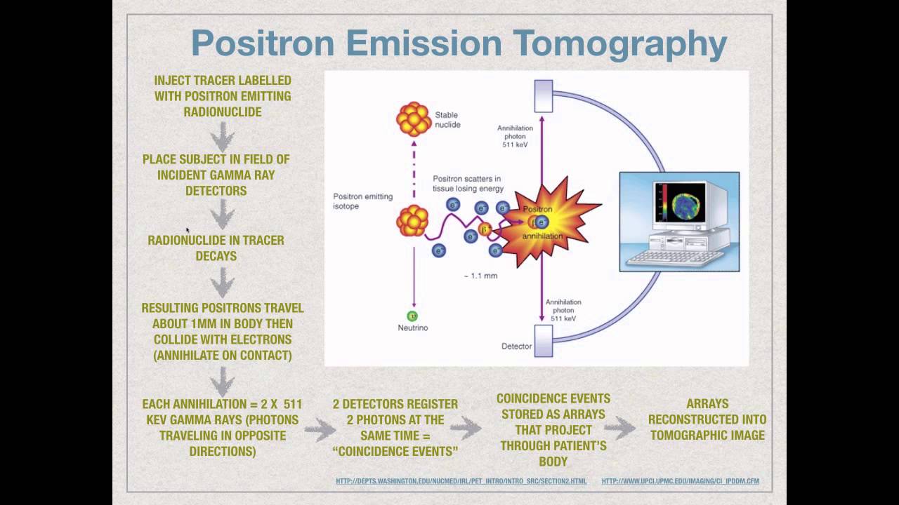
17:12
PET vs SPECT in Molecular Imaging: Principles and Nanomedicine Applications
@kimberlykelly1479
84.4K views•2015-11-02

14:39
Coagulation Cascade: Understanding Blood Clotting (Part 1)
@khanacademymedicine
1.3M views•2014-08-18

10:28
Neuroanatomy: Central and Peripheral Nervous System Divisions Explained
@AKLECTURES
136.2K views•2014-09-20

3:05
Stages of Labor and Vaginal Birth | Childbirth Animation
@nucleusmedicalmedia
52.1M views•2017-08-18
Related Study Plans & Knowledge Roadmaps
Structured learning paths in Medicine
medicine
Abnormal LFTs: Diagnostic Workup & Pathology
01Liver Anatomy and Basic Physiology
02Introduction to Liver Function Tests (LFTs)
03Hepatocellular vs. Cholestatic Patterns
+ 3 more learning modules
12h6 modules
Explore Roadmap
medicine
Analyzing Blood Smears: RBC, WBC & Platelets
01Introduction to Blood Components & Smear Preparation
02Normal Morphology of Blood Cells
03Erythrocyte Pathologies & Morphological Variants
+ 2 more learning modules
12h5 modules
Explore Roadmap
medicine
Mechanical Ventilation: Waveforms & Alarms
01Anatomy and Physiology of Breathing
02Introduction to Mechanical Ventilation & Core Modes
03Ventilator Settings and Waveform Interpretation
+ 2 more learning modules
12h5 modules
Explore Roadmap
medicine
Neuro Exam: Localization & Stroke Syndromes
01Foundations of Neuroanatomy and Vascular Supply
02The Neurological Examination: Step-by-Step
03Principles of Neurological Localization
+ 3 more learning modules
18h6 modules
Explore Roadmap