











![Endocytose/ Exocytose/ Membranfluss (Stofftransport durch die Biomembran) - [Biologie, Oberstufe]](https://i.ytimg.com/vi/8YAp558E8O0/sddefault.jpg)

















PET (Positron Emission Tomography) and SPECT (Single Photon Emission Computed Tomography) are molecular imaging techniques that measure physiological functions in vivo by detecting radioactive tracers; PET uses positron-emitting isotopes (like F-18, C-11) that annihilate to produce coincident 511 keV gamma rays detected in opposite directions, while SPECT uses gamma-emitting isotopes (like Tc-99m, In-111) detected through collimation, with PET offering higher sensitivity but requiring on-site cyclotron production and SPECT being more widely available with longer-lived isotopes suitable for nanoparticle tracking.
PET vs SPECT in Molecular Imaging: Principles and Nanomedicine Applications
Added:Basic nuclear physics, including types of radioactive decay (specifically positron emission versus single-photon emission) and isotope half-lives.
Fundamental concepts of medical imaging, distinguishing between structural imaging (such as CT and MRI) and functional or molecular imaging.
Principles of nanotechnology, including the types of nanoparticles (e.g., liposomes, polymeric nanoparticles) and surface functionalization.
General biochemistry, specifically ligand-receptor interactions, cell-surface biomarkers, and mechanisms of cellular uptake.
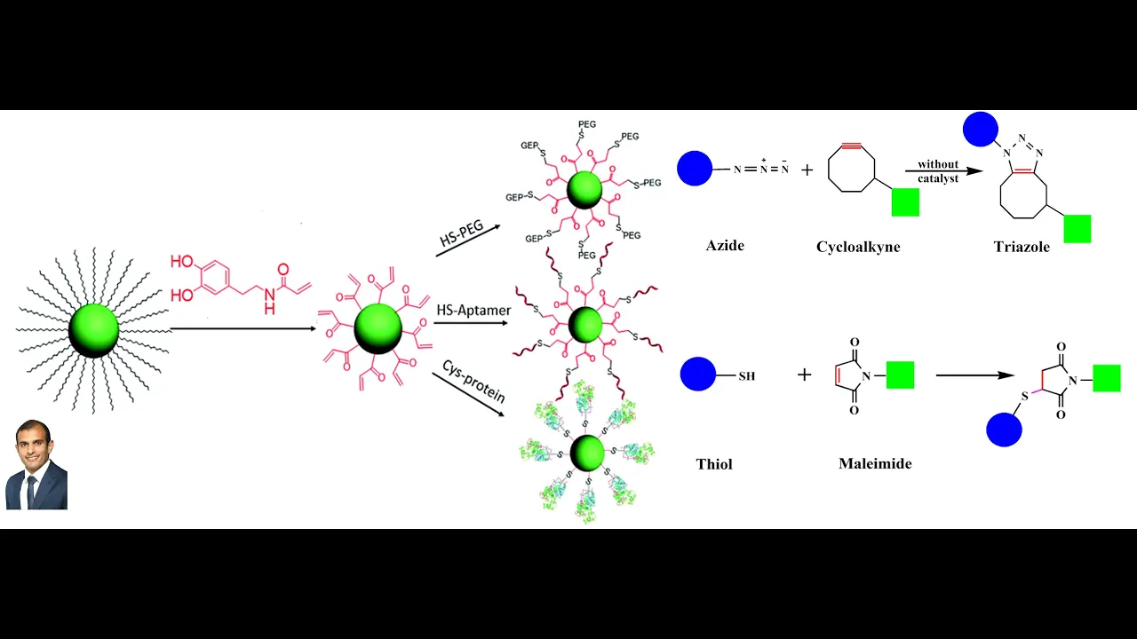
Advanced radiochemistry and bioconjugation techniques used to stablely label nanoparticles with radioisotopes.
The clinical paradigm of theranostics, which integrates diagnostic molecular imaging and targeted nanoparticle therapy (e.g., radioligand therapy).
Hybrid imaging modalities such as PET/CT, PET/MRI, and SPECT/CT, and their relative clinical strengths in oncology and neurology.
Pharmacokinetics, biodistribution modeling, and clearance pathways of nanomedicines in living systems.
Translational and regulatory hurdles in bringing novel radiolabeled nanomedicines from pre-clinical development to clinical trials.
84.4K views597likes17:12Original Release: 2015-11-02
Hi everyone and welcome to the PET spec uh lecture. So PET stands for posetron emission tomography.
Inspect a single photon emission computed tomography.
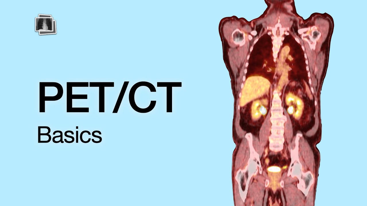
And so with the tomography in its name you know that this is a slice or a 3D uh capabilities of this. So before we get into this a little bit more nomenclature should have done this with the MRI but there are different planes to imaging.
So the axial or transverse plane slices someone in half um not across this plane. And so this is what that image would look like going up and down the body with the vertebrate always down um at the bottom of the screen. The coronal plane goes from front to back and you can see it in this image right here where you can see the heart and then the sagittal plane goes from side to side. Um and so again with the spine to the right you can see this in the very last image. So those are the three planes of imaging.
So MR and CT we've seen provide anatomic images. There are contrast agents from from them but they mostly are used in the clinic for anatomical contrast. So what information would you get from pet inspect?
Well pet inspect uh both require a contrast agent. They use gamma rays and we will get into that but there are no gamma rays in the body and so you always need a contrast agent.
This is unlike the MRI and the CT or ultrasound where anatomy will generate the contrast and so posetron emission tomography it measures regional tissue functions in vivo. So you can get blood flow which is profusion, blood volume vascularity, oxygen utilization, glucose metabolism, pre and post synaptic receptor density and affinity, neurotransmitter release, enzyme activity, drug delivery and uptake, gene expression. So you can get all of that information. It's a molecular imaging technique. However, you must have a contrast agent that will tell you all of those different things. It's very accurate and very sensitive. So, sensitive means that you can see something very small. And why is that?
Well, it's the physics of how positron emission tomography works. So, you have to make a tracer. It's labeled with the positron emitting radionucleide.
You place the subject in the field of the gammaray detectors and so this is the PET scanner.
The radionuclide in the body uh will decay the posetrons and in this case posetrons will will annihilate or the radioisotope will annihilate. it will emit a posetron and it will emit them in equal in a 180 degree linear pattern.
And so the posetron scatters in tissue, it loses energy. That's your attenuation. And so here's your posetron.
It annihilates. And when it annihilates, you have a release of a photon at 511 kileron volts. always 511 at 180 degrees at the same time. And so this is why this is very sensitive because only only photons that are coincident it's called that 180 degrees hitting the detectors in equal and opposite directions.
Uh will it be picked up by the detectors and will it be used in the algorithm?
So very important key factors coincident photons are released and um coincident photons are released and they're both at the same photon 511 kileron volts and that is a property of all posetron emitters. So F18 is the most common one or florine 18. Uh there's iodine 123 uh zirconium gallium um but all of them emit at the same time. So you cannot use two different of these uh positron emitting isotopes and and look at two different things. You can only look at one thing at a time.
So the resulting positron so getting back to our diagram the radionuclide in the tracer decays. So this is what's wherever it is in the body. The resulting posetrons will travel about 1 millter in body. Then they'll collide with the electrons. They annihilate on contact. Each annihilation um each annihilation will equal 2 * 511 ker electron volt gamma rays. The photons again are traveling in opposite directions. The two detectors register two photons at the same time. That's the coincident event. Coincident events are stored as arrays that project through the patient's body and then the arrays can be reconstructed into tomographic image.
So positron emission tomography is a type of radioactive decay where a proton in a radionuclide nucleus is converted to a neutron. It releases a posetron and an electron neutrino.
A radionuclide is an atom with an unstable nucleus. Neutrino is similar to an electron but no net charge.
These are gamma rays and they have high frequency electromagnetic radiation so they can penetrate pretty quickly.
Commonly used isotopes and their half- livives are carbon 11. This is very quick 20.3 minutes. Uh 13 nitrogen 9.97 minutes, 15 oxygen 2.03 minutes and 18 florine 109.8 minutes. It is absolutely essential that if you are going to use PET for any kind of imaging that you pair the correct radionuclide halflife of the isotope with your bio distribution. So if you choose an 11 a C11 so carbon 11 and your blood halflife is 18 hours before your particle clears your particle will be mostly in the blood when the halflife is already gone for the carbon 11. And in general after five half- livives you can no longer detect um the isotope.
And so for bigger things like an antibbody or for um for a nanoparticle you should use zirconium.
Um these things are just too short in in order to be able to see them.
So here's our electromagnetic spectrum.
Radio waves 10 the 4 10 the 2 uh microwave uh infrared 10 the minus2 uh visible light is 10 the minus5 ultraviolet 10 the minus 6 xrays are 10 the minus 8 that is um what is the CT and then the gamma ray your wavelength is 10 -10 to 10us12 so that's an atomic nuclei so this enables the highly sensitive detection of these particles. So a photon is a particle representing a quantum of electromagnetic radiation and a quantum is discrete quantity of energy with magnitude proportional to the frequency of radiation it represents.
So how can you imagine PET being used in nano medicine? So take about five minutes, write down what you're thinking in terms of how you can imagine PET being used in nanome medicine.
Now that you've thought about it, let's see how many of these that you um have been able to list. And so one is to label up your polymer nanoparticles or any kind of nanoparticles with a PET tracer. Again, you have to use the same um you have to make sure that you marry your half-life to the particle pharmacocinetics.
And so if you label this um with a PET tracer, you'd be able to image, which allows you to quantitate. And PET is quantitative. So you'd be able to image exactly where your nanop particle is going and quantitate how much is there.
And then you'd also be able to keep track of its biodistribution and get percent injected dose per gram. And if you know how much drug was loaded into these nano particles, then you would be able to calculate how much drug actually reached the tissue as opposed to just guessing based on the blood profile and the pharmaccoinetic um mathematical uh algorithms that we've talked about.
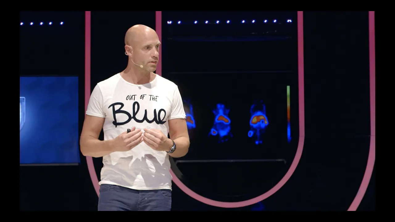
So in this particular example um we were able to not only image so doing the PET imaging in the ABCD this is a nanoparticle so the majority of it will go into the liver um and then the percent injected dose per gram that you can see that there's a lot of it in the spleen some in the kidney a lot in the liver and a lot in the blood. So again, this gives you um exact knowledge of exactly where your your radio tracer is. Now, the reason why I say that is because we're tracking the radio tracer. And so if the radio tracer is cleaved in the bloodstream or any anywhere else and is released from the nano particle, then you're no longer tracking the nano particle. You're only tracking the the radionuclide. So a lot of studies have to be done ahead of time to see what that cleavage rate is in serum or plasma or whatever so you can figure out um how well intact or how stable the nanoparticle complex is but it's a pretty decent approximation.
Um here's another example with a pet dendrimer. Um here you can put uh F18 or iodine. You can see the liver and intestines, the eskeemic muscle tissue and then also the bladder. And so here this is looking at eskeeia. Most of these things aren't really done in uh nano medicine. Nan medicine is mostly used to track where or pet imaging in nanom medicine is mostly used to track where the particles are um and to potentially image things. Uh so being able to image the tumor because you can get more taken up there. But with everything there's always a tradeoff and so if there is no if it's staying around in the bloodstream for a long time then a lot of your organs will get radiated with these um radionuclide emitters and so that's typically not a very good thing.
But these can track pH. Um they can track eskeeia. That's the oxygen sensing part. Um they can track profusion. So you can look at it and how long it stays in the bloodstream. That's that can tell you and you can see with the resolution that you can really tell where these particles are. So it's really sensitive and it allows you to track where your particles are going.
So on the other hand is spec single photon emission tomography um and some people put computed tomography so there'd be a C which makes it spec it's like PET um but gamma emitters it's a single photon it's not a posetron it's a gamma emitter it's a single photon um and that means that it's not as sensitive as PET because in order to do this you have to be able to decide position and the only way to do that is to use what's called a columator um which makes it and we'll do this in the worksheet which makes it so that way the photon or the photon can only hit the detector at a specific geometry and using that geometry you can back calculate exactly where the or roughly where the photon uh came from but that means you have to throw out quite a view of the photons and so it's not as sensitive. Um, TE 99 is one of the uh radioisotopes. It's half life is about six hours. The other isotope is indium 111. Uh, this has about a 4hour halflife no uh 2.8 day halflife. So that is a really good isotope for nano particles.
The advantages are that there there are a lot of spec cameras in hospitals.
There are about 10 times more spec cameras than there are PET detectors in hospitals. And so you can you have a bigger chance of being able to use it in the different hospitals.
And the radionuclides are readily available.
They can be shipped from different uh regions throughout the country. pet.
However, because of most of their short half- livives, you have to produce them on site in a cyclron. And so, because of those reasons, spec is typically cheaper than PET. Again, its disadvantages. It is less sensitive than PET because of the columnation.
There's a difficult attenuation correction because of the unknown path length and no single photon emitting of basic basic biological elements. Uh so labeling with tracer may change biology.
So for example in F18 there are biologic elements with a florine. So just substituting out iodine or florine doesn't change the biology.
You're just using a different isotope.
That doesn't happen with spec. And so depending on what you're doing, you would have to um you have to really make sure that you haven't changed the biology or the affinity or specificity of the tracer. Plus most of the radionuclides that are compatible for spec um are usually chelated with macrocyclic or heterosyclic chilators.
And so some of these require high heating 90° Fahrenheit for 10 minutes or or 20 minutes in order to open up that chilator and allow that radioisotope to um go in and bind. And so if you have a peptide or an antibbody, it may not tolerate being heated at such a high at such a high level. So your tracer may not be compatible um with that. And so spec is a little bit more difficult.
Also because it's not conjugated because it's chilated instead on that chilator.
um you have an equilibrium between the on and the off state of that uh that radio tracer. So it's more common for the radio tracer or the radionucli to actually get out of the chilator and so you'll have free metals that are radioactive throughout the body. But again with that being said there are advantages to spec equipment in the radionuclides are readily available and it doesn't require very difficult chemistry.
Up Next

18:58
Intermediate Risk Prostate Cancer: Treatment Options Explained (MUSC)
@muschollings
70.6K views•2023-09-11

14:39
Coagulation Cascade: Understanding Blood Clotting (Part 1)
@khanacademymedicine
1.3M views•2014-08-18

10:28
Neuroanatomy: Central and Peripheral Nervous System Divisions Explained
@AKLECTURES
136.2K views•2014-09-20

3:05
Stages of Labor and Vaginal Birth | Childbirth Animation
@nucleusmedicalmedia
52.1M views•2017-08-18
Related Study Plans & Knowledge Roadmaps
Structured learning paths in Medicine
medicine
Abnormal LFTs: Diagnostic Workup & Pathology
01Liver Anatomy and Basic Physiology
02Introduction to Liver Function Tests (LFTs)
03Hepatocellular vs. Cholestatic Patterns
+ 3 more learning modules
12h6 modules
Explore Roadmap
medicine
Analyzing Blood Smears: RBC, WBC & Platelets
01Introduction to Blood Components & Smear Preparation
02Normal Morphology of Blood Cells
03Erythrocyte Pathologies & Morphological Variants
+ 2 more learning modules
12h5 modules
Explore Roadmap
medicine
Mechanical Ventilation: Waveforms & Alarms
01Anatomy and Physiology of Breathing
02Introduction to Mechanical Ventilation & Core Modes
03Ventilator Settings and Waveform Interpretation
+ 2 more learning modules
12h5 modules
Explore Roadmap
medicine
Neuro Exam: Localization & Stroke Syndromes
01Foundations of Neuroanatomy and Vascular Supply
02The Neurological Examination: Step-by-Step
03Principles of Neurological Localization
+ 3 more learning modules
18h6 modules
Explore Roadmap