




























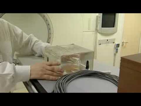
A linear accelerator (linac) generates high-energy X-rays for cancer treatment by accelerating electrons using radio frequency waves in a waveguide, directing them through a slalom path with magnets to focus the beam to approximately 1mm diameter, then striking a tungsten target to produce X-rays; the beam is shaped using collimators and a multi-leaf collimator (MLC) with computer-controlled tungsten leaves to match the tumor's exact shape, while dose is measured and controlled by ionization chambers to ensure precise delivery of radiation to cancer cells while sparing healthy tissue.
How a Linear Accelerator Works in Radiation Therapy
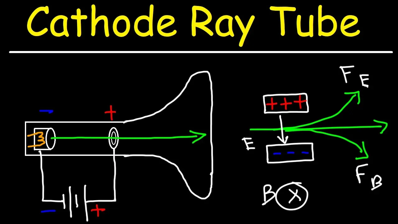
Added:Basic concepts of electromagnetism, specifically how electric and magnetic fields are used to accelerate and guide charged particles like electrons.
The nature of electromagnetic radiation, particularly the production and properties of high-energy X-rays and electron beams.
Fundamental radiobiology principles, including how ionizing radiation interacts with cellular DNA to damage and destroy cancer cells.
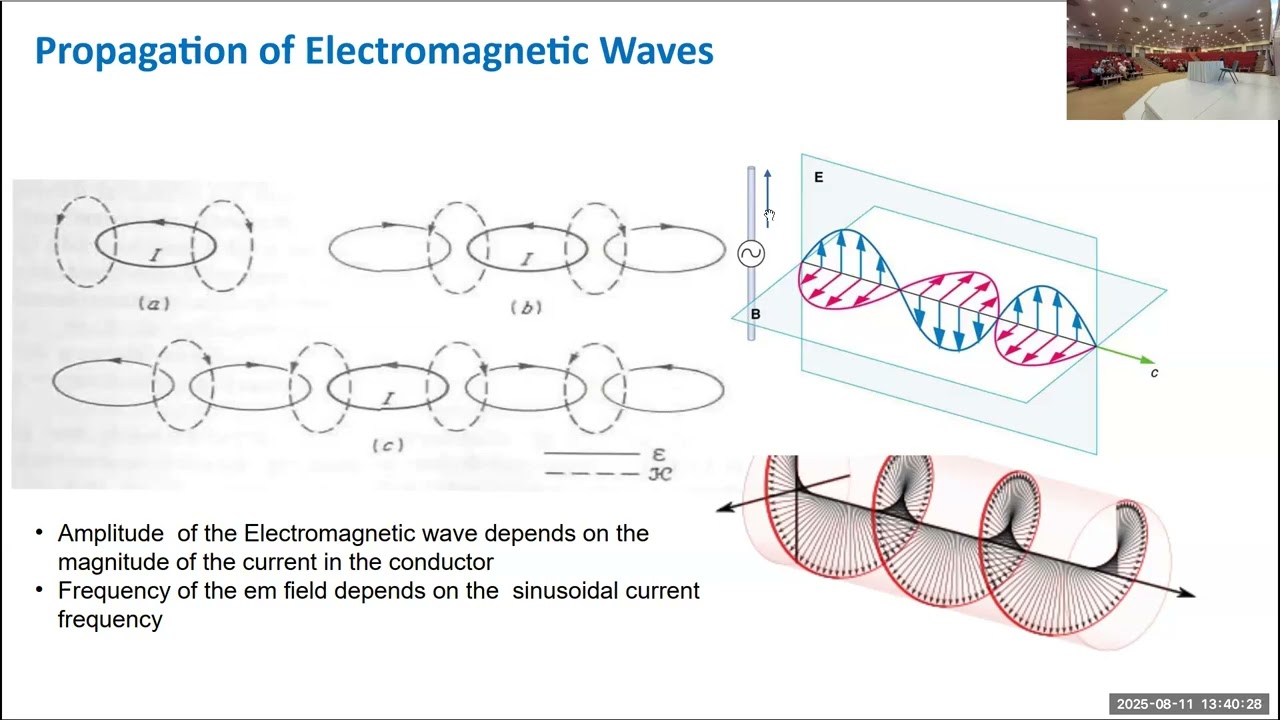
An introduction to wave physics, especially radiofrequency (RF) waves, which are crucial for understanding the operation of linear accelerator waveguides.
Advanced radiation delivery techniques, such as Intensity-Modulated Radiation Therapy (IMRT) and Volumetric Modulated Arc Therapy (VMAT).
The integration of Image-Guided Radiation Therapy (IGRT) and real-time tumor tracking systems during treatment.
Radiation dosimetry and clinical treatment planning, focusing on how medical physicists calculate and optimize 3D dose distributions in patient anatomy.
Quality assurance (QA) protocols, calibration procedures, and safety standards required to maintain linear accelerators in a clinical setting.
898.5K views9.1Klikes8:24Original Release: 2010-12-26
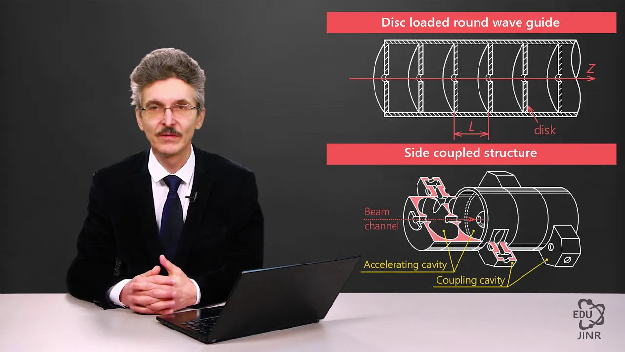
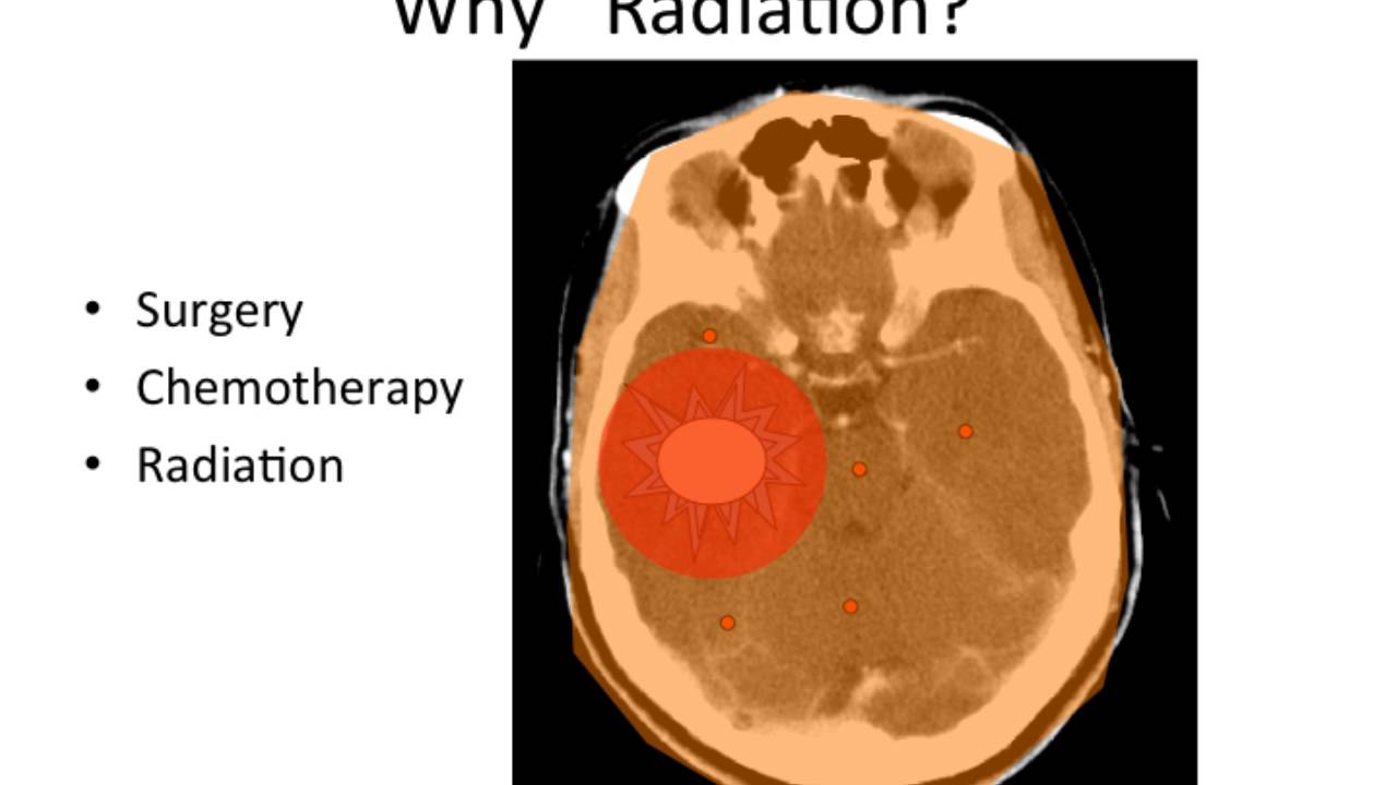
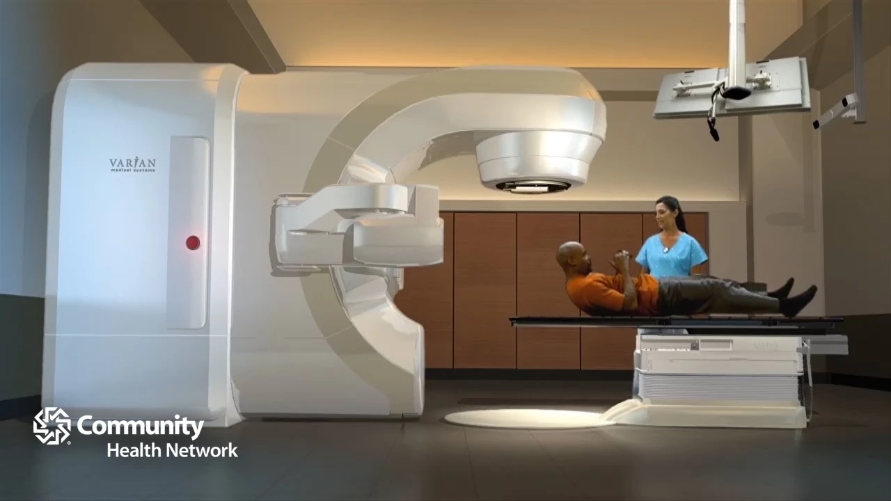
how the linear accelerator works all body cells can be damaged or killed by radiation but tumor cells are more sensitive to radiation the normal cells radiation therapy uses this principle to damage beyond repair or kill the abnormal cancer cells in a tumor successful radiation therapy depends on the ability of the linear accelerator or linic to deliver a tumor seidel dose of radiation to the tumor while ensuring minimal radiation of normal tissue the linic accurately produces monitors controls and conforms the radiation beam to the planned target beam generation radio frequency waves are pulsed into the waveguide by the magnetron this is synchronized with the injection of electrons into the waveguide by the electron gun the radio frequency waves accelerate the electrons along the waveguide to a speed approaching the speed of light the x-ray beam is created when the electrons hit and interact with a tungsten target at the opposite end the magnetron controls the power and frequency of the radio frequency waves which determine the energy of the x-rays produced the digital accelerator uses a diode type electron gun situated at the end of the waveguide the electrons are produced by heating the tungsten filament within the cathode and are then injected into the waveguide the number of electrons injected is controlled by the temperature of the filament the electrons are accelerated along the waveguide toward the target the waveguide contains a series of copper cells small holes or irises between these copper cells allow the electrons to travel along the waveguide and help to focus the beam a vacuum is created to ensure that the electron beam is not impeded by other particles the path of the negatively charged electron beam is controlled by two sets of quadrupole magnets called steering coils that surround the waveguide an additional two sets of focusing coils help to further define the electron beam so that it is very fine with a diameter similar to that of a pinhead when it hits the target the entire system is cooled by water the electrons exit the waveguide and enter the flight tube where the beam is redirected towards the target the electrons travel along a slalom path within the flight tube three pairs of magnets on either side of the flight tube cause the electron beam to bend through the turns of the slalom this process not only positions the beam to strike the target but it also further focuses the beam to a diameter of one millimeter the design of the magnets enables them to focus electrons of slightly different energies onto the same point on the target this is called a chromatic behavior this slalom bending is unique to elect the linear accelerators it helps to minimize the size of the machine and ensures that its ISO Center remains low which is important for patient setup the high-energy electrons hit a small tungsten target where the electron energy is converted into photons or x-rays the high-energy photons emerge from the target in a variety of directions the primary collimator only allows forward traveling x-rays to pass through creating a cone-shaped beam the primary collimator minimizes leakage and therefore excess total body dose by absorbing scattered x-rays traveling in the lateral direction it also defines the maximum size of the resulting clinical radiation beam at this stage the photons are not uniformly distributed across the beam and so a flattening filter is placed in the path of the beam the cone-shaped filter absorbs more photons from the center of the beam than from the sides creating a uniform photon beam dose measurement the photons now pass through the ion chamber for dose measuring and beam quality monitoring the dose delivered to the patient is measured and controlled simultaneously in two independent ionization chambers one chamber is the primary two Scimitar it measures the radiation and terminates the beam when the required dose has been delivered the secondary ion chamber acts as a backup and we'll stop the irradiation if the primary chamber fails the treatment machine must replicate the beams modeled within the planning system this is critical to the accuracy of treatment delivery a beam quality function is performed by a third ionization chamber which uses seven electrodes to monitor different sections of the radiation field the x-ray beam is almost ready to treat the patient before that further beam shaping is required to ensure that the shape of the delivered x-ray beam matches the shape of the tumor this is done using a multi-leaf collimator a number of fine tungsten leaves which move independently of one another and can create a variety of complex treatment shapes Lynnette control one computer system controls both the linic and the multi-leaf collimator this eliminates doe symmetric errors due to communication delays it also ensures synchronization between the delivered dose and the multi-leaf collimator position allowing complex deliveries such as intensity modulated radiation therapy and volumetric modulated Arc therapy all electromagnets steering and focusing coils are digitally controlled all mechanical positions of flight tube filters and foils are automatically selected from the control console and all radiation beam settings are grouped in calibration blocks for each energy these are stored digitally on the linnekar disk for flexible and easy beam adjustment calibration and servicing clearance clearance is the free space available under the linic for patient treatment and it varies with different protocols and fixation devices it is a combination of the distance between the lower surface of the radiation head and the ISO Center 45 centimeters and the head diameter 62 centimeters a wide clearance around the ISO Center means improved access for patients set up freedom to use the best possible patient positioning and immobilization accessories freedom to rotate the gantry between fields without needing to move the patient finally it means that treatment techniques using non coplanar beams are not compromised the large clearance offered by Alecto machines ensures flexibility in providing the best possible treatment for the patient
Up Next

21:14
Qwilfish and Overqwil: The Poisonous Pufferfish Mythology of Hisui
@Gnoggin
418.6K views•2022-02-19

105:00
The Internet's Own Boy: Aaron Swartz Documentary (2014) | Digital Rights & Activism
@moviemaniacs
2.4M views•2014-07-01

9:49
FastAPI vs Flask vs Django: Choosing the Right Python Web Framework
@TechWithTim
302.5K views•2024-05-26

1:42
Game of Thrones Opening Credits: A Cinematic Analysis
@gameofthrones
46.3M views•2011-04-18
Related Study Plans & Knowledge Roadmaps
Structured learning paths in General & Interdisciplinary Studies
general-studies
Elevator Engineering: History, Physics & Cities
01From Hoists to Safety Brakes: The Dawn of Vertical Lift
02Hydraulic vs. Traction: The Engineering Physics of Elevators
03Modern Speed, Safety, and Algorithms
+ 2 more learning modules
12h5 modules
Explore Roadmap
general-studies
Rubber: Material Science, History & Geopolitics
01Origins of Natural Rubber & Mesoamerican History
02The Chemistry of Natural Rubber & Vulcanization
03The Geopolitics of the Rubber Boom & Colonization
+ 2 more learning modules
12h5 modules
Explore Roadmap
general-studies
Containerization: Evolution, Logistics & Trade
01Ancient Trade & Bulk Cargo: Amphorae to Barrels
02The Industrial Revolution & Cardboard Packaging
03The Containerization Revolution: Malcom McLean
+ 2 more learning modules
12h5 modules
Explore Roadmap
general-studies
Textile Tech: Evolution, Labor & Global Impact
01Pre-Industrial Textiles: Handloom & Cottage Industry
02Mechanization: The Spinning Jenny to the Power Loom
03The Factory System & Global Labor Transformation
+ 2 more learning modules
10h5 modules
Explore Roadmap