


![ماهو تأثير DDT على الإنسان و الحشرات ؟ الوحدة 5 : الإتصال العصبي ] .](https://i.ytimg.com/vi/Hwoj9Xit4VQ/hqdefault.jpg)










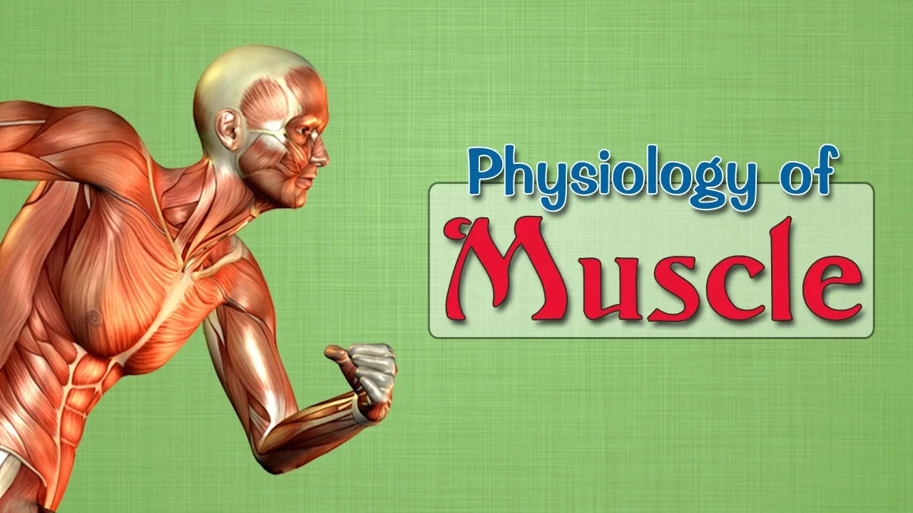


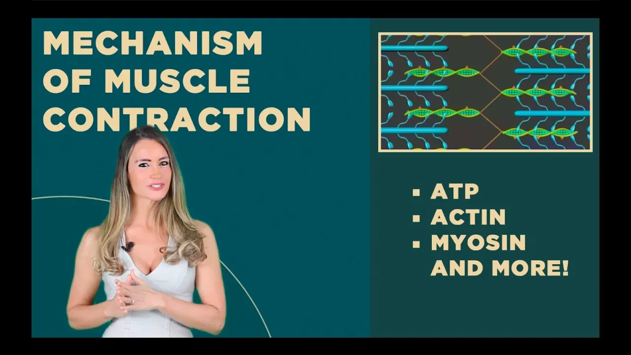



Muscle contraction occurs through the sliding filament model, where action potentials generated at the neuromuscular junction trigger calcium release from the sarcoplasmic reticulum; calcium binds to troponin, shifting tropomyosin to expose actin binding sites, allowing myosin heads to form cross bridges and pull thin filaments toward the sarcomere center, shortening the I bands and H zone while maintaining constant A band length.
Muscle Contraction Mechanism: Sarcomeres & Neuromuscular Junction
Added:Basic anatomical structure of skeletal muscle, including muscle fibers, myofibrils, and the sarcoplasmic reticulum.
The concept of cellular membrane potential, ion channels, and how action potentials are generated in neurons.
An introduction to chemical synapses, specifically how neurotransmitters like acetylcholine transmit signals across a synaptic cleft.
Fundamental knowledge of cellular proteins, particularly the structural roles of actin and myosin.
The detailed steps of the cross-bridge cycle, including the role of ATP hydrolysis and calcium binding to troponin.
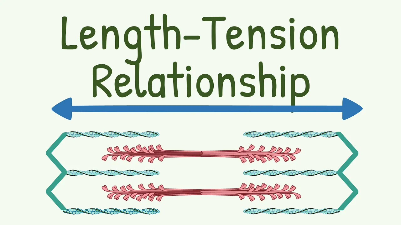
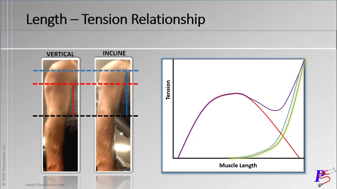
Muscle mechanics and fiber recruitment, exploring the length-tension relationship and differences between isometric and isotonic contractions.
Metabolic pathways of muscle contraction, studying how muscles generate ATP via creatine phosphate, glycolysis, and aerobic respiration.
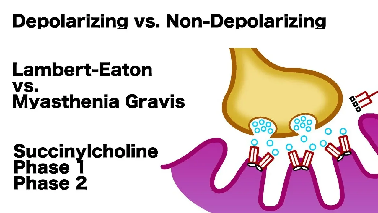
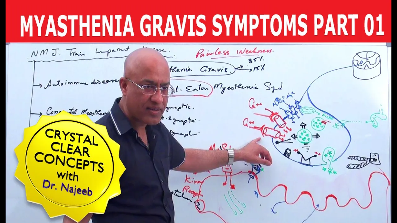
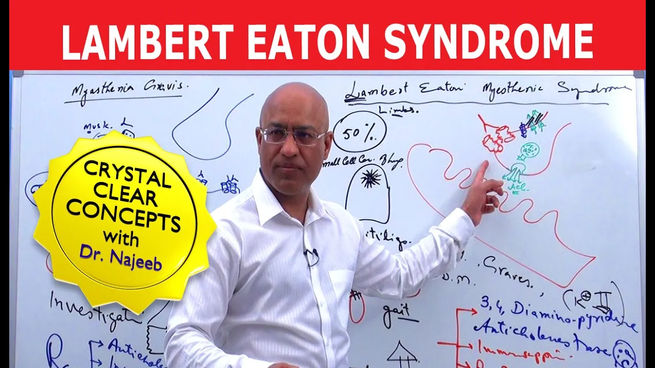
Clinical applications and pathologies related to neuromuscular transmission, such as Myasthenia Gravis, Lambert-Eaton syndrome, and the physiological cause of rigor mortis.
2M views39Klikes12:35Original Release: 2019-02-07
Professor Dave here, let’s talk about action potential.
We just learned about the structure of muscle, like skeletal muscle, which is what allows you to move your body around.
But how exactly does this work?
If you decide to lift your arms up in the air, what is happening on the molecular level that produces this motion?
It’s not magic, it’s just a staggering amount of chemistry, so let’s dive back down into those muscle fibers and get a closer look.
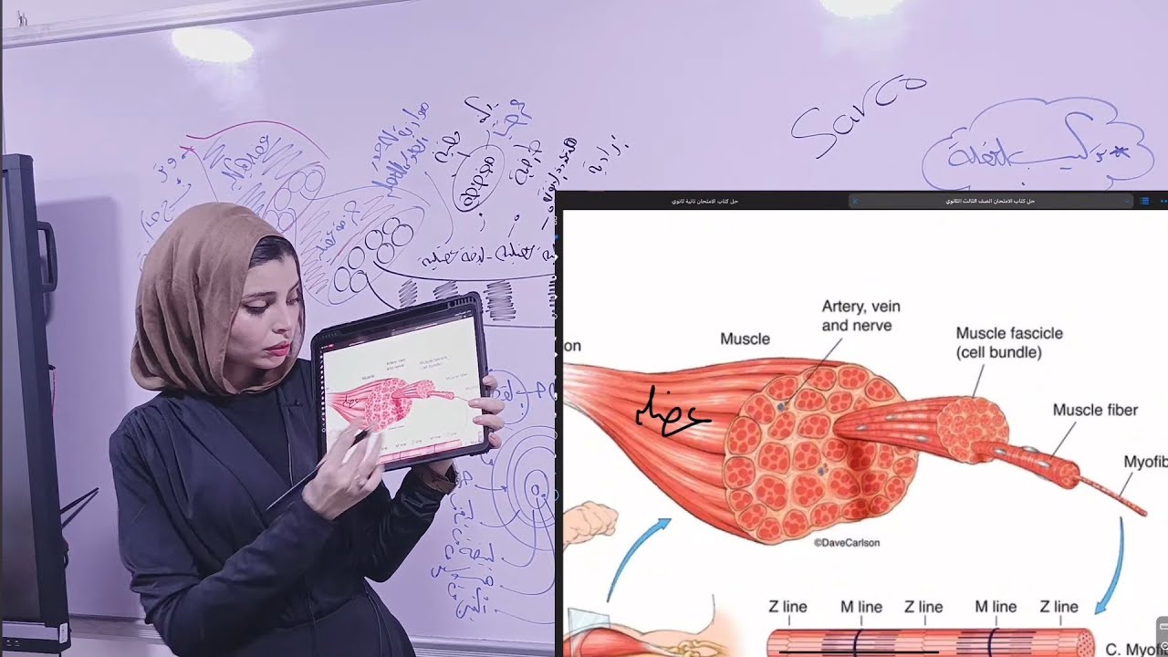
As we recall, any skeletal muscle is made of fascicles, and each fascicle is made of muscle fibers, which are the individual multinucleated muscle cells.
From there, we can zoom in farther on one of these myofibrils, which are in turn comprised of myofilaments arranged into sarcomeres, and this is the contractile unit, the one that lets muscles do what they do.
Now let’s get a closer look at these structures, because this is where all the action happens.
Looking at a sarcomere, we can label a few regions.
We can see these darker A bands and the lighter I bands, which are aligned and responsible for the striated appearance of skeletal muscle.
Within each A band we can find a lighter region called the H zone, and this H zone is split down the middle by something called the M line, made of a protein called myomesin.
The I bands are also split down the middle, by a region called the Z disc.
We can identify an individual sarcomere as being a section from one Z disc to the next, and this is the functional unit of skeletal muscle.
If we zoom in even more, we can see the myofilaments that make up these regions.
As we discussed previously, the thick filaments contain myosin, and these extend across the A band, connected at the M line.
The thin filaments contain actin, extending across the I band and into the A band.
There are also elastic filaments made of titin, spanning from the Z disc to the thick filament and then continuing on, acting as the core of the thick filament.
Now let’s zoom in even further on these two main types of filaments.
Myosin is a protein with two globular heads pointing outwards, and a long tail which contributes to the structure of the filament.
The heads are the site of all the activity, since there are ATP binding sites, as well as actin binding sites, meaning this is where the thick and thin filaments will interact, by making cross bridges.
It’s important to note the lack of myosin heads in the center of the sarcomere, just as there also are no thin filaments.
Speaking of those, as we said, they are made predominately of actin, and two actin filaments will twist together to form the backbone of the thin filament.
Each actin subunit has an active site where myosin can bind, and when a muscle fiber is relaxed, these are blocked by spiraling strands of tropomyosin.
There is also troponin, a globular complex of three polypeptides, one of which binds to actin, one of which binds to tropomyosin, and one of which binds calcium.
Beyond this, we must be aware of the sarcoplasmic reticulum, a series of tubules that surround each myofibril.
These regulate levels of calcium, which is needed for muscle contraction, through storage and release.
This structure includes the T tubules, which sit at each A band – I band junction, encircling each sarcomere and helping signals reach every region of the muscle cell.
Now that we’ve examined each structural component, let’s describe how everything works, using the sliding filament model of contraction.
This says that when the nervous system stimulates muscle fibers, the myosin heads on the thick filaments will interact with the binding sites on the actin subunits.
These attachments will form and break several times as the thick filaments pull the thin filaments in towards the center of the sarcomere, thus pulling the Z discs towards the M line.
The I bands shorten, and the H zone disappears.
The overall effect is that the A bands from adjacent sarcomeres get closer together, so the entire muscle cell will shorten.
This is the mechanism by which muscles contract.
Now the question is, how is all of this activity initiated?
As we said, this begins with a signal from the nervous system.
We will examine that system in detail a bit later, but for now we can just examine the interface between the nervous system and a skeletal muscle.
This is called the neuromuscular junction.
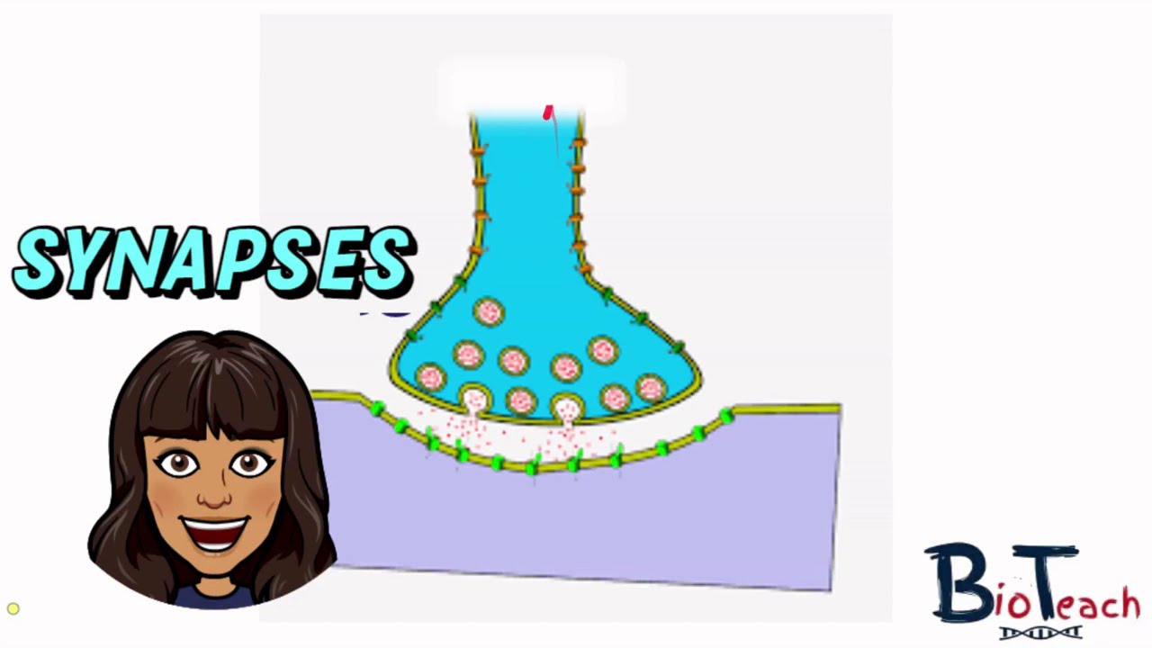
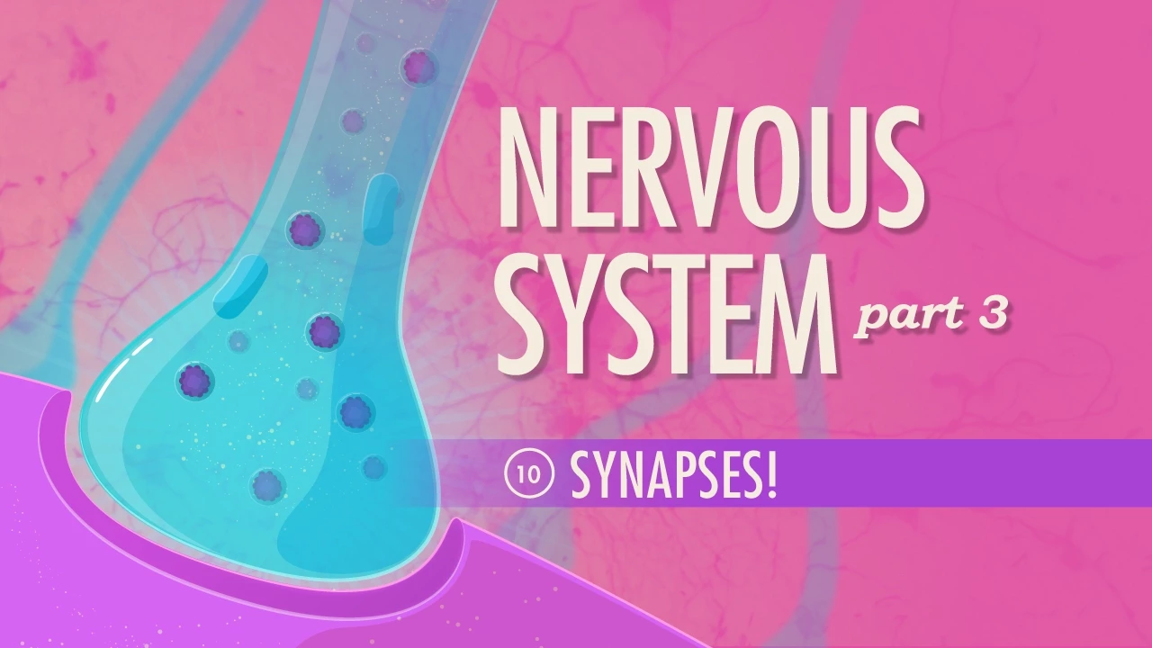
Each muscle fiber has one, and these sections, called axon terminals, are nearly touching the muscle fiber, separated only by a thin space called the synaptic cleft, where the muscle produces junctional folds within the postsynaptic membrane.
The axon terminal has lots of synaptic vesicles, which are like little bubbles, containing acetylcholine, which is a neurotransmitter.
When a nerve impulse reaches the end of an axon, by a mechanism we will discuss later, the axon terminal will release acetylcholine into the synaptic cleft.
The junctional folds contain acetylcholine receptors, so these will bind the approaching acetylcholine, which causes a conformational change.
This protein will then act as an ion channel, which allows for sodium ions to enter, and potassium ions to leave, although not in equal number.
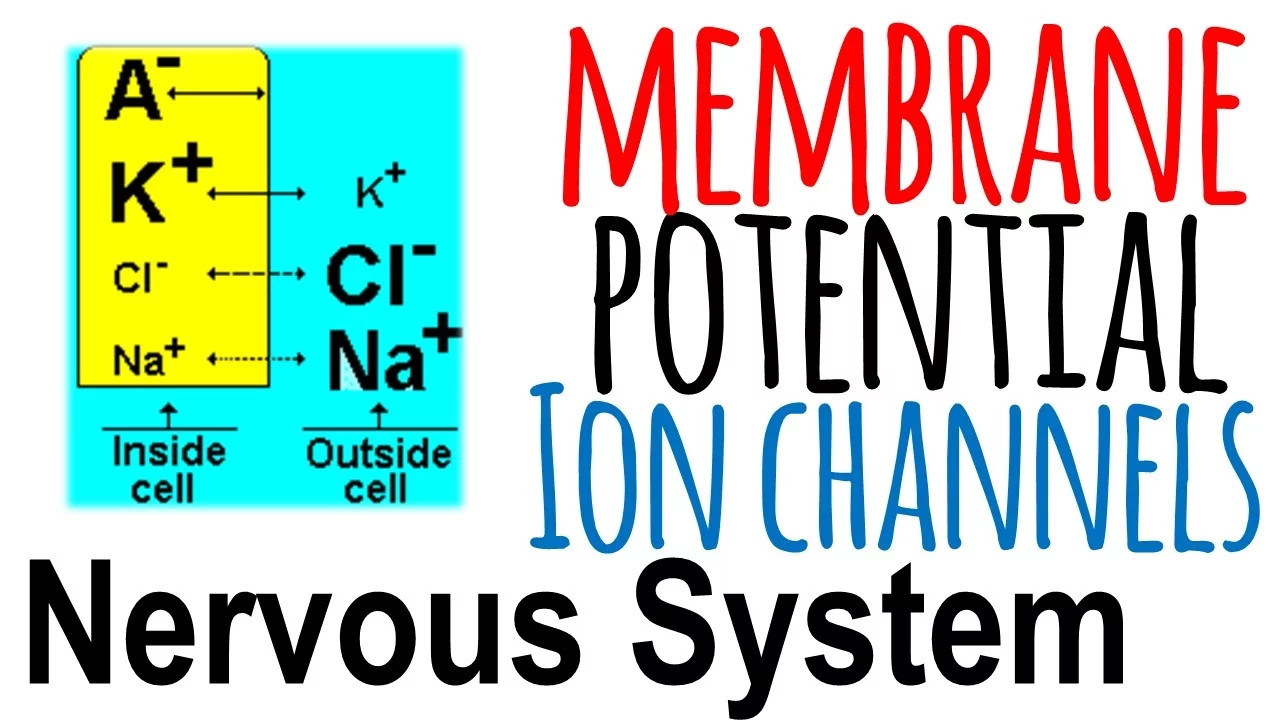
Sodium will cross the membrane in greater quantity, and this affects the membrane potential, or the potential difference across the membrane.
For more information on electric potential, check out my physics tutorial on this subject, otherwise, we just need to understand that the way that positive and negative charges are distributed within a structure can be a driving force for a particular process.
Prior to the opening of the ion channels, the inside of the cell was more negatively charged than the outside, but as sodium ions enter, depolarization will occur.
This causes other nearby sodium channels to open, and more sodium ions will enter, following the electrochemical gradient.
Once a particular threshold voltage is reached, this will generate an action potential.
Sodium can now diffuse into the cell anywhere along the membrane.
While this is happening, an enzyme in the synaptic cleft called acetylcholinesterase will break down acetylcholine, and the ion channel closes, preventing further muscle contraction until another nerve impulse arrives.
But the action potential will continue to propagate along the sarcolemma, and down the T tubules, which opens up calcium ion release channels, which we will discuss in a moment.
Finally, after the depolarization wave has completely propagated, repolarization will begin, due to the changes in charge density.
Potassium channels will now open, and potassium ions that are more highly concentrated within the cell will diffuse out of the muscle fiber, which restores negative charge inside, and the sarcolemma goes back to normal.
At this point, the muscle fiber can be stimulated again if another impulse arrives.
So we can see that charge distribution and electric potential are key concepts here, but how does that relate to muscle contraction?
Well the action potential generated will initiate excitation-contraction coupling.
As we said, the propagation of the action potential causes a rise in levels of calcium in the cytosol, and this is what causes the filaments to slide.
Let’s recall that when a muscle cell is relaxed, tropomyosin blocks the myosin binding sites on the actin subunits.
But as more and more calcium ions become present, these will bind to troponin, and once two ions bind, it will change shape, which will push tropomyosin off of the myosin binding sites, making them available for cross bridge cycling, the process we mentioned before.
In this process, the myosin heads will pivot and bend, pulling the actin filament along and using ATP in the process.
The two positions possible for the heads allow it to pull, detach, change position, bind, pull, detach, and so forth, many times, until contraction is complete.
As calcium levels deplete, troponin returns to its original shape, tropomyosin blocks actin’s binding sites once more, and the muscle fiber relaxes.
There is a lot more to discuss regarding muscle contraction.
We could talk about the time frames associated with each step in muscle contraction.
We could talk about graded muscle responses, or the differences in contraction for smooth muscle versus skeletal.
But we will have to leave that for another day, since it’s best to really understand the basics first.
Before moving forward, let’s quickly review what we discussed.
Muscle contraction in a skeletal muscle begins when a signal arrives at the neuromuscular junction.
Acetylcholine is released, which binds to receptors on the sarcolemma.
Sodium and potassium ions move through the ion channels, which results in a local change in membrane voltage, also called depolarization.
This initiates an action potential, which travels across the sarcolemma in all directions, eventually along T tubules.
This is where calcium ions are released, which interact with the myofilaments such that myosin and actin are able to bind, and contraction begins.
Now that we understand the structure of a muscle, as well as the mechanism by which muscles contract, let’s move forward and look at the entire muscular system as a whole, so that we can get the big picture regarding how we move around.
Up Next
3:32
Proteasome Function & Protein Recycling Explained | Molecular Animation
@WEHImovies
42.5K views•2024-05-24

50:12
Bacterial Communication: Quorum Sensing and Biofilm Formation Explained
@JHUAAP
12.6K views•2011-06-28

10:50
Protein Structure Explained: Primary to Quaternary | Biochemistry
@ProfessorDaveExplains
1.4M views•2016-08-27

7:08
Bacteriophages: Earth's Deadliest Killers and Future Antibiotics
@kurzgesagt
34.6M views•2018-05-13
Related Study Plans & Knowledge Roadmaps
Structured learning paths in Biology
biology
RNAi: Biogenesis, RISC & Gene Silencing
01Molecular Biology Foundations: DNA to Protein
02Introduction to RNA Interference (RNAi)
03Molecular Biogenesis of miRNA and siRNA
+ 2 more learning modules
10h5 modules
Explore Roadmap
biology
Mycorrhizae: Signaling, Biophysics & Ecology
01Foundations of Mycorrhizal Symbiosis
02Molecular Signaling and Root Colonization
03Biophysics and Bioenergetics of Nutrient Exchange
+ 1 more learning modules
11h4 modules
Explore Roadmap
biology
Circadian Clocks: TTFL, SCN & Periphery
01Foundations of Molecular Biology & Gene Expression
02Introduction to Chronobiology and Actograms
03The Core TTFL: Molecular Architecture of the Clock
+ 3 more learning modules
14h6 modules
Explore Roadmap
biology
Population Genetics: Math, Drift & Selection
01Genetic & Probability Foundations
02The Hardy-Weinberg Principle and Baseline Models
03Mathematical Modeling of Natural Selection
+ 3 more learning modules
18h6 modules
Explore Roadmap