










![[모두를 위한 모든 생명의 분자생물학] 10강. DNA로부터 단백질까지](https://i.ytimg.com/vi/VS-Ant2fCvo/maxresdefault.jpg)


![[코로나19 예수회 특강] 제 8강 면역학의 기초: 중심가설(central dogma) - 최준열 다미아노 신부](https://i.ytimg.com/vi/jCHsJ8i_Q68/maxresdefault.jpg)
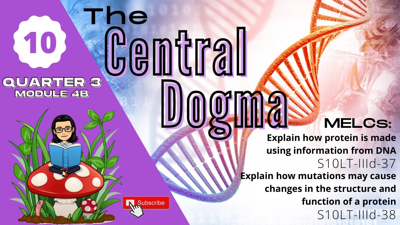

The central dogma describes how genetic information flows from DNA to RNA to proteins: DNA serves as a template for transcription (synthesis of messenger RNA), and mRNA is then translated at the ribosome to assemble amino acids into proteins; DNA is a double-stranded nucleic acid composed of deoxyribonucleotides with four nitrogenous bases (adenine, thymine, cytosine, guanine) that follow Chargaff's rules (A=T and G=C), forming a double helix structure where purine bases pair with pyrimidine bases via hydrogen bonds, while proteins are polymers of amino acids with four structural levels (primary, secondary, tertiary, quaternary) that determine their function.
Central Dogma: DNA to Proteins | Animated Molecular Biology Guide
Added:Basic structure of DNA, including its double-helix shape, sugar-phosphate backbone, and complementary nitrogenous base pairing (A-T, C-G).
The fundamental differences between DNA and RNA, such as their single vs. double-strand structures, ribose vs. deoxyribose sugars, and the replacement of thymine with uracil.
An introductory understanding of cell anatomy, specifically identifying the functions of the nucleus, ribosomes, and cytoplasm.
The basic concept of proteins as macromolecular structures composed of chains of amino acids that perform diverse functions in the body.
Post-transcriptional and post-translational modifications, including RNA splicing (introns and exons), 5' capping, poly-A tailing, and protein folding.
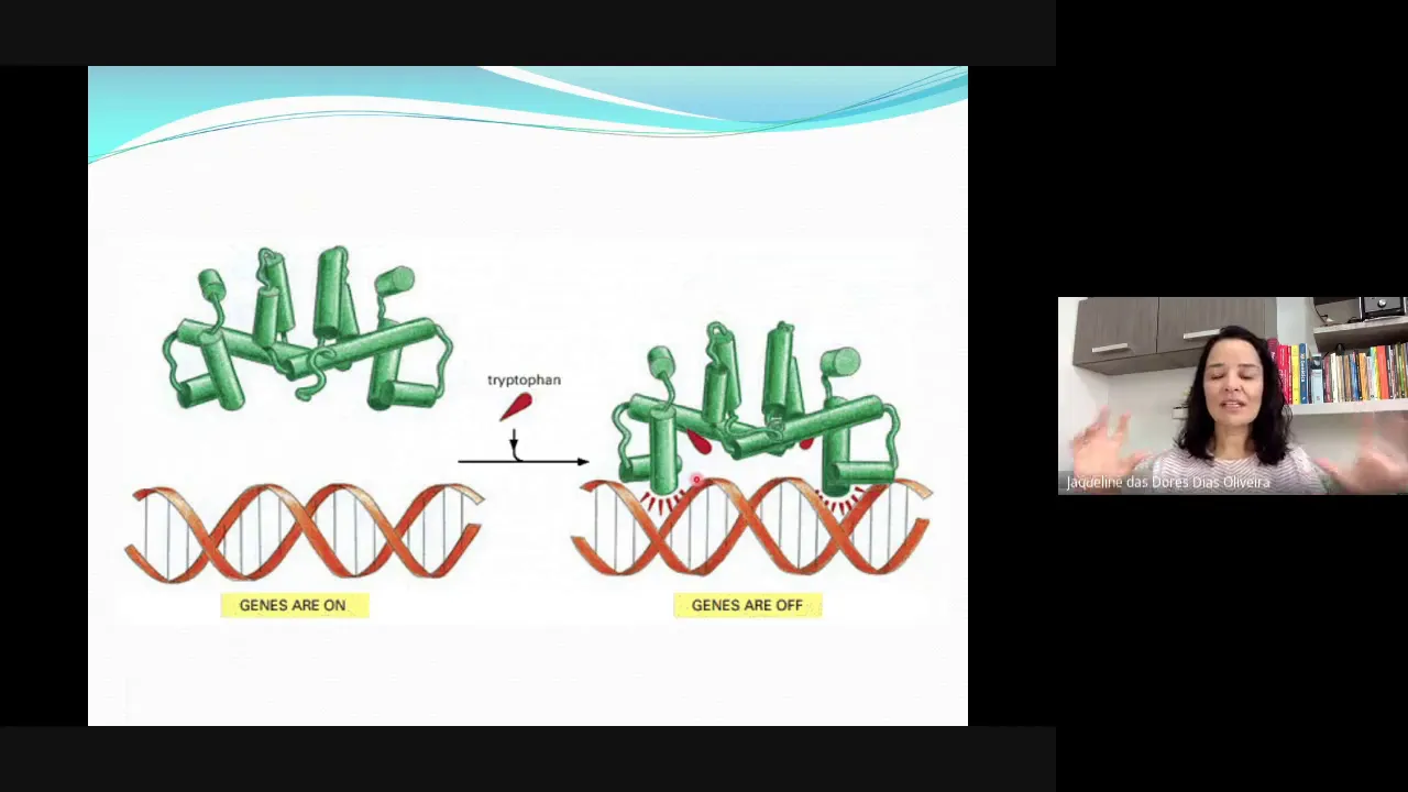
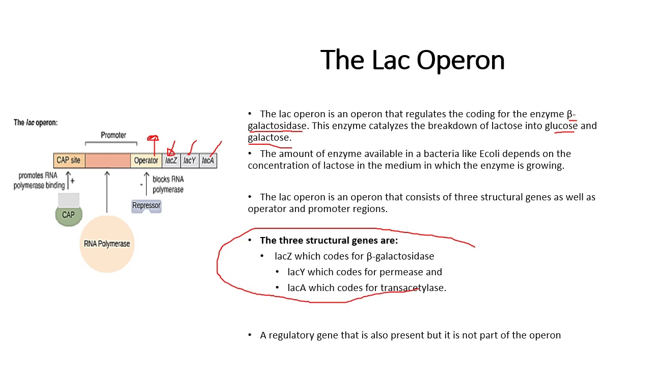
Mechanisms of gene regulation, studying how cells control which genes are expressed and when (e.g., operons in prokaryotes and transcription factors in eukaryotes).
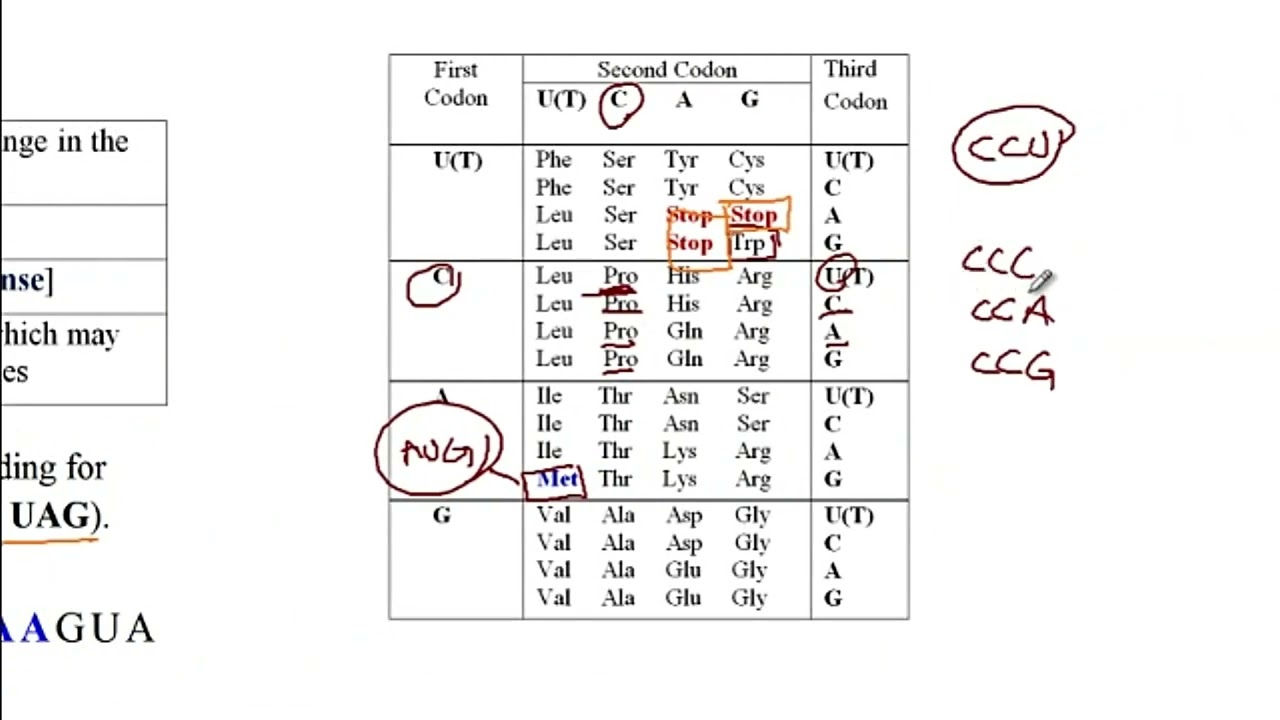
The effects of DNA mutations (such as silent, missense, nonsense, and frameshift mutations) on the final protein sequence and cellular function.
Modern biotechnological applications of the central dogma, such as mRNA vaccine technology, recombinant DNA production, and CRISPR-Cas9 gene editing.
162.4K views2Klikes27:27Original Release: 2017-04-20
[Music] genetically you are who you are because of DNA but how does DNA make you you in this lecture you will learn the basics of how DNA makes proteins and investig the molecular structure of nucleic acids and proteins as our understanding of biological molecules increased in the 20th century researchers discovered that all living organisms share a genetic code in 1956 Francis Crick proposed that DNA is an informational storage molecule capable of replicating itself further he proposed that the information that was transmitted had to be read by a manufacturing body within the cell which puts amino acids together in a specific sequence ultimately synthesizing a protein this became known as the central dogma of molecular biology specifically DNA serves as a template for the direct synthesis of a messenger RNA molecule also known as mRNA and a process known as transcription secondly mRNA is read at a ribosome by transfer rnas also known as TR rnas which work together to assemble a specific chain of amino acids which collectively assembled to generate a protein in a process known as translation proteins are the cell's internal Machinery similar to parts of a car each protein has a specific three-dimensional shape that determines its function any change in the shape potentially changes the function of the protein consider an analogy of transcription and translation to printing an essay from a computer once your essay is complete you store the document to the hard drive similar to DNA the hard drive stores information DNA is a gen genc storage molecule data in a computer is stored in a binary language when it's time to print your essay you send a command to the computer to send a message to the printer this message is akin to mRNA a genetic messaging molecule similar to the binary message sent to the printer DNA and RNA share a chemical language based on nucleotides hence why the information exchange from DNA to RNA is called transcription an exchange of information in the same language once the information is received at the printer it is translated from a binary language into a different language a language of ink analogously mRNA is read by the ribosome and translated into the language of proteins which are made up of amino acids thus the process from RNA to proteins is known as translation translating from the language of nucleotides to amino acids the ribosome is akin to the printer serving as a facility for the process of translation the molecules that actually translate the MRNA at the ribosome are a different kind of RNA Transfer RNA or TRNA in the process of translation a TRNA reads the MRNA and links a specific amino acid to a growing protein for your essay to represent your idea the ink must be physically arranged in a specific manner any malfunctioning and the positioning of the letters would not convey the same idea similarly proteins have a specific three-dimensional shape that determines their function any chains in that shape can potentially alter its function in our analogy DNA is the stored file in the hard drive mRNA is the message sent to the printer the printer is the ribosome the sa is the protein and the letters represent the amino acids DNA indirectly codes for proteins DNA directly creates all of the intermediate players of transcription and translation DNA's day-to-day function is the production of RNA molecules messenger RNA is directly generated by a specific segment of DNA that segment of DNA is known as a gene the MRNA travels to a ribosome which is made up of a protein and another type of RNA ribosomal RNA or R RNA at the ribosome the MRNA serves as a code for the synthesis of protein by linking specific amino acids in an exact sequence the overall collection of an amino acid chain is the protein DNA is also capable of self-replication necessary for the creation of new cells DNA and RNA are biological molecules known as nucleic acids nucleic acids as well as proteins are polymers or molecules made up of a linking chain of repeating molecules the repeating components are known as monomers the monomers of nucleic acid are nucleotides which are composed of three components a sugar a phosphate group and a nitrogenous base DNA or deoxy ribonucleic acid is a double stranded nucleic acid composed of monomers known as deoxy ribonucleotides a deoxy ribonucleotide is made up of three components a phosphate group the sugar deoxy ribos and one of four nitrogenous bases while the phosphate group and deoxy ribos are identical in the varying deoxy ribonucleotides DNA houses four different nitrogenous bases adenine known as a thyine t cytosine C and guanine G these four different nucleotides serve as the letters of the genetic informational storage which are transcribed into mRNA and eventually read at the ribosome to create a protein all of the biological diversity on Earth in the world world is based on the language of Life which only has four letters these nitrogenous bases can be placed into categories based on their shape thyine and cytosine are each composed of a single carbon ring skeleton and are known as perimedes whereas adenine and guanine are composed of two carbon ring skeletons connected together one six-sided and the other one five-sided and these are known as purines RI nucleic acids rnas are single stranded nucleic acid polymers made up of the monomer's ribonucleotides ribonucleotides are identical to deoxy ribonucleotides with two exceptions first ribonucleotides are made up the sugar ribos which has a hydroxide at the two prime carbon whereas deoxy ribos has a hydrogen atom at that location the carbons of ribos and deoxy ribos are notated moving clockwise from the oxygen One Prime to five Prime The One Prime carbon connects to the nitrogenous base the atoms attach to the two prime carbon differ between ribos and deoxy ribos clockwise from the two prime carbon is the three prime carbon followed by the four Prime carbon and the five Prime carbon attaches to the phosphate group so relative to RNA sugar ribos DNA sugar deoxy ribos lacks an oxygen at the two prime carbon hence deoxy ribos that single difference allows cells to differentiate between those two nucleotides second ribonucleotides differ in their Suite of nitrogenous bases three ribonucleotides have the same nitrogenous base as the deoxy ribonucleotides cytosine guanine and adenine while the fourth ribonucleotide is composed of the nitrogenous base uracil uracil is very similar to thyine except that there is a hydrogen atom at the three prime location of uracil while thyine has a methyl or ch3 group there the phosphate group is identical for both ribonucleotides and deoxy ribonucleotides nucleotides link together in long chains to form a nucleic acid individual nucleotides are connected by a calent bond that forms between the three prime carbon of the sugar molecule of one nucleotide and a phosphorus of the phosphate group of an adjacent nucleotide in this reaction a hydrogen atom is removed from the three prime carbon and hydroxy is removed from the phosphate these byproducts combine forming water in a reaction known as a condensation reaction following this reaction the two nucleotides are connected by a phoso diester bond in which a phosphate group is linked to the five Prime carbon of its original nucle nucleotide and the thre Prime carbon of an adjacent nucleotide adding a third nucleotide the nucleic acid begins to take shape in this developing nucleic acid a phosphate is attached to a sugar which is attached to a phosphate attached to a sugar and so on a phosphodiester linkage involves two of the three components of a nucleotide a phosphate and a sugar hanging off to the side of the nucleic backbone are the nitrogenous bases this repeating pattern forms the backbone of nucleic acids one end of a nucleic acid strand is bound by a phosphate group while the opposite in is bound by a sugar giving DNA and RNA directionality the phosphate group Terminus of a nucleic acid is referred to as the five Prime end of the Strand as the five Prime carbon is the closest carbon to the end of that molecule the opposite end of the nucleic backbone contains a sugar terminus called the three prime end the sequence of nucleotides in a nucleic acid is known as its primary structure scientists have standardized the notation of nucleic acid's primary structure by listing the nucleotides from the five Prime end to the three prime end five Prime to 3 Prime for example a segment of RNA adenine guanine guanine uril adenine cytosine would be notated a g g u a c RNA or ribonucleic acid is a singl stranded nucleic acid composed of ribonucleotides the nucleic backbone of RNA is bound by a phosphate group at the five Prime Terminus and ribos a sugar on the three prime Terminus each ribonucleotide has one of four nitrogenous bases adenine uracil cytosine and guanine RNA molecules are predominantly responsible for actively synthesizing proteins DNA synthesizes messenger RNA mRNA which transmits genetic information from DNA to a ribosome the primary sequence of the MRNA determines the sequence of amino acid in the resultant protein ribosomes are hybrid complexes made up of proteins and a different type of RNA ribosomal RNA R RNA at the ribosome the MRNA is red and decoded by a third RNA Transfer RNA or TR RNA in UK carots a different RNA small nuclear RNA snrna is involved in modifying the MRNA after transcription and before translation DNA is a double stranded nucleic acid composed of deoxy ribonucleotides which vary from ribonucleotides by having a different five carbon sugar called deoxy ribos in living organisms there are four deoxy ribonucleotides that vary in their nitrogenous bases adenine thyine cytosine and guanine once the primary structure of DNA was solidified the next question was how are the nucleotides arranged to create the DNA molecule or the secondary structure of DNA the discovery of DNA secondary structure was one of the most important biological discoveries of the 20th century one of the first first Clues came from analyses conducted in the early 1950s by Irwin chargo comparing the relative abundances of deoxy ribonucleotides across a variety of organisms chov discovered that the relative abundance of guanine equal cytosine and the relative abundance of adenine equals thymine and what is most interesting about this is that he found this relationship across many different species of organisms charo's Discovery was instrumental to scientists uncovering DNA secondary structure James Watson and Francis Crick suggested Charro's evidence strongly supports base pairing in DNA in which deoxy ribonucleotides of adenine attached to thyine and guanine attaches to cytosine in addition Watson and cric hypothesized that base pairing of deoxy ribonucleotides suggests that DNA was most likely double stranded to acquire evidence of the actual molecular shape of DNA rosin Franklin and Maurice Wilkins bombarded DNA with x-rays and analyzed how the radiation scattered a technique known as x-ray chromatography analysis of the Scatter Plots from this technique allowed them to measure the distance between atoms and DNA and they were able to conclude three things one DNA has a consistent width two within DNA is a repeating pattern and three the molecule must be helical in collaboration with Franklin and Wilkins Watson and cric used the measurements to define the geometry of the components of the deoxy ribonucleotides creating physical models of the nucleotides literally paper cutouts Watson and cig tinkered with the different Arrangements of the nucleotides to explain one charo's rule two a consistent width three the repeating pattern of the nucleotides and four the helical shape of the DNA by arranging base pairing nucleotides sides a with t and c with G side by side in strands running in opposite directions all of the discoveries could be explained in other words Watson and cric suggested that DNA is composed of two strands one running five Prime to three prime connected to a second strand running three prime to 5 Prime this orientation is called anti-parallel the nucleic backbone is composed of alternating phosphate and deoxy ribos sugar molecules with a phosphate on the five Prime end of of the Strand and a deoxy ribos on the three prime end the strands twist to form a double helix a spiral bounded on the outside by two nucleic back bounds running in opposite directions with the nitrogenous bases facing inward based on charos findings Watson and cric determined that the nitrogenous bases from adjacent DNA strands connect according to base pairing the discovery of a consistent width of the DNA also supported the A and CG base pairing while adenine and guanine are different molecules they're both purines and approximately the same size and shape the same is also true of the pertin cytosine and thymine however purines consist of a figure8 structure which is larger than the circular structure of the peridin for the width of DNA to be consistent with the variety of shapes found in the nitrogenous bases purines must connect with peridin a purine purine base pairing creates a larger molecule width than observed and a perimidine perimidine base pairing would be too small if a purine perimine base pairing explains the consistent width of a double stranded DNA molecule why does guanine a purine always appear to bind with cytosine a perimidine but never thyine also a perimidine why doesn't adenine a purine bind with cytosine with their physical models of the nucleotides Watson and cric deduced the nitrogenous bases of the adjacent strands were held together by hydrogen bonding due to the differential and Electro negativities the hydrogens of the nitrogenous bases are partially positive and the oxygens and nitrogens are partially negative hydrogen bonds form between the partially positive and partially negative atoms of adjacent nitrogenous bases investigating the shapes and interactions of these four nitrogenous bases they discovered that guanine and cytosine were geometrically complements of each other and held together by three hydrogen bonds while adenine and thyine are held together by two hydrogen bonds essentially the at and CG pairing are more stable than any other combination due to the complementarity of the molecular shape of the hydrogen bond orientation all right let's take a closer look at proteins proteins are biological molecules that serve as cellular machines and living organisms these large molecules are specific three-dimensional structures involved in biological processes such as cellular signaling catalyzing chemical reactions molecular transportations as well as many other functions proteins are polymers consisting of long chains of monomers amino acids of the more than 500 amino acids known only 20 appear in proteins of living organisms an amino acid is a relatively simple organic molecule attached to a central carbon by single calent bonds are one a hydrogen atom two an amine group nh3+ three a carboxilic acid group C o o minus and four an R Group also known as a side chain at around ph7 as in water the amine group of an amino acid attracts a proton becoming nh3+ and acts as a base the carboxy group is negatively charged in water due to the high electro negativity of both oxygens pulling electrons from the hydrogen and losing the proton different amino acids vary in their R group of the protein building amino acids the r groups can vary in their size shape and polarity proteins being made up of chains of amino acids vary based on the interactions of the atoms within the amino acids and water these interactions dictate the shape of the protein which in turn determines its function R groups vary in their polarity non-polar molecules have relatively equal distribution of electrons via calent bonding while polar molecules have unequal distribution of electrons the unequal distribution of electrons in polar molecules creates partially charged atoms polar R groups are hydrophilic meaning they have an affinity for water due to the hydrogen bonds between the partial charges of the R Group and water molecules non-polar R groups are repelled by water or hydrophobic therefore in a chain of amino acids ones with the polar R groups will bend towards water and non-polar R groups will bend away affecting the eventual shape of a protein proteins are polymers of amino acids chained together by calent bonds known as peptide bonds a peptide bond is a condensation reaction in which the oxygen ion of the carboxilic acid from one amino acid is removed becoming carboxy and combines with two hydrogen atoms from the amine group of an adjacent amino acid to produce water a calent bond forms between the two amino acids when the carbon of the carboxy group that lost the O during the condensation reaction combines with the adjacent nitrogen of another amino acid that lost the hydrogen atoms bonding twoj in amino acids this is a peptide bond amino acids link via peptide bonds forming long chained molecules or polypeptides proteins have four levels of structure the three-dimensional shape of a protein determines its function and the shape of proteins are ultimately dependent upon the sequence of amino acids coded for by DNA the unique amino acid sequence is a protein's primary structure in humans cell anemia is an inherited condition in which blood cells have a variant of the oxygen binding protein hemoglobin CLE cell anemia is considered a disease of the primary structure of proteins as it is caused by the variation of a single amino acid in hemoglobin a veine instead of a glutamate in the sixth position of 146 amino acid protein while normal blood cells are rounded humans with this variant produce sickled shaped red blood cells normal blood cells are elastic and flow freely through veins but sickled red blood cells are rigid and tend to get stuck where the veins Branch this blockage starves Downstream tissues of oxygen resulting in a host of medical issues including lower life expectancy the body identifies the cells when they get stuck and destroys them healthy red blood cells typically live between 90 and 120 days whereas sickled red blood cells have a 10 to 20day lifespan therefore people with CLE cell anemia must produce much more blood which is rich in iron leading to an overall iron deficiency or anemia interestingly CLE cell anemia is a evolutionary advantage in certain circumstances prior to globalization the highest rates of CLE cell anemia occur in tropical Africa the Middle East and India all malaria dominated areas malaria is a single cell eukariotic parasite transmitted by mosquitoes that some consider the most deadly human disease ever however in sickled red blood cells the malaria parasites cause the cell to rupture before they can successfully reproduce therefore people with CLE cell anemia have an evolutionary advantage over people with normal blood cells in these areas in AR's absent of malaria sickled red blood cells are highly disadvantageous due to the host of medical conditions associated with this variant all of this is caused by a single different amino acid in one protein an alteration of the protein's primary structure when amino acids are grouped into polypeptide chains neighboring amino acids can interact via hydrogen bonding these interactions can form a regular pattern which increases the molecular stability of the polypeptide chain these patterns are known as the proteins secondary structure and form either cork screw shaped structures known as alpha helices or folded ribbon shaped structures known as beta plated sheets recall oxygen has a high electro negativity while hydrogen has low electro negativity this differential in electro negativity results in hydrogen bonding between neighboring amino acids within a polypeptide backbone the hydrogen bonds can occur between amine groups and carboxy the partially negative oxygen of the carboxy combined with the hydrogen of the amine groups of other amino acids in Alpha helices and beta plated sheets hydrogen bonding occurs between amine and carboxy groups of different amino acids how hydrogen bonding occurs between amino acids determines which of the two shapes emerges within a single amino acid of an alpah Helix polypeptide chain the hydrogens of the amine groups face the opposite directions relative to the oxygens of the carboxy groups down the entire Alpha Helix the hydrogens of the amine group Face the same direction and oxygens of the carboxy group Orient in the opposite direction within the alpha Helix the hydrogen bonds forms when an oxygen of the carboxy faces the hydrogen of a different amino acid further down the polypeptide chain this accumulation of multiple hydrogen bonds stabilizes the polypeptide chain beta plated sheets are also formed by hydrogen bonding between the amine and carboxy groups of different amino acids however the orientation of these groups differs in a single amino acid of a beta plated sheet the oxygen of the carboxy and the hydrogen of the amine group face in the same direction in the adjac amino acid the oxygen and hydrogen both face the opposite direction relative to the first in beta plated sheets hydrogen bonds also occur between the carboxy oxygen and the amine hydrogen between neighboring amino acids however the orientation of these atoms causes the structures to bend in a folded ribbon shape or beta plated sheet while the secondary structure of proteins is determined by the interactions between the amine groups and the carboxy groups of neighboring acids tertiary structure is defined by how the r groups of neighboring amino acid AIDS interact these interactions result in a very specific folding patterns eventually helping to stabilize the specific three-dimensional structure of the polypeptide several types of interactions occur between neighboring R groups while the hydrogen bonding determines the secondary structure of proteins hydrogen bonding can also occur between the r groups of a polypeptide chain the 20 R groups of amino acids are either polar or non-polar polar R groups have oxygen or nitrogen atoms which characteristically have high electro negativity due to their High affinity for electrons these polar R groups tend to bond with hydrogen atoms of neighboring non-polar R groups or the hydrogen of the amine group of the peptide backbone nonpolar R groups can also form hydrogen bonds with the peptide backbone either by interacting with the oxygen of the carboxy group or the nitrogen of the amine group while hydrogen bonding is relatively weak the overarching abundance of these interactions form forms very stable polypeptide structures in living organisms proteins are surrounded by water polar R groups are hydrophilic and bend to turn towards water whereas non-polar R groups are hydrophobic and turn away from water hydrophobic R groups tend to amass in the internal section of a protein forming globular masses while hydrogen bonding is facilitated by the interactions of partial charges certain R groups have full charges and are involved in ionic bonding this happens when completely positive R groups form ionic bonds with neighboring R groups that are completely negative the overall structure of a fully functional protein is known as the quinary structure most proteins are composed of several polypeptides a polypeptide is composed of either a series of alpha helices with tertiary level interactions or a series of beta plated sheets with tertiary level interactions in the next lecture you're going to learn about the genetic code and the specifics of transcription and translation [Music] [Music]
Up Next
20:22
Plant Cell Structure and Function Explained | Basic Science Series
@biologygoal
174.1K views•2020-12-12

50:12
Bacterial Communication: Quorum Sensing and Biofilm Formation Explained
@JHUAAP
12.6K views•2011-06-28

5:35
Enteric Nervous System Explained: The Gut's Brain | Neurobiology Lecture
@alumniu6029
438 views•2018-09-12

7:08
Bacteriophages: Earth's Deadliest Killers and Future Antibiotics
@kurzgesagt
34.6M views•2018-05-13
Related Study Plans & Knowledge Roadmaps
Structured learning paths in Biology
biology
RNAi: Biogenesis, RISC & Gene Silencing
01Molecular Biology Foundations: DNA to Protein
02Introduction to RNA Interference (RNAi)
03Molecular Biogenesis of miRNA and siRNA
+ 2 more learning modules
10h5 modules
Explore Roadmap
biology
Mycorrhizae: Signaling, Biophysics & Ecology
01Foundations of Mycorrhizal Symbiosis
02Molecular Signaling and Root Colonization
03Biophysics and Bioenergetics of Nutrient Exchange
+ 1 more learning modules
11h4 modules
Explore Roadmap
biology
Circadian Clocks: TTFL, SCN & Periphery
01Foundations of Molecular Biology & Gene Expression
02Introduction to Chronobiology and Actograms
03The Core TTFL: Molecular Architecture of the Clock
+ 3 more learning modules
14h6 modules
Explore Roadmap
biology
Population Genetics: Math, Drift & Selection
01Genetic & Probability Foundations
02The Hardy-Weinberg Principle and Baseline Models
03Mathematical Modeling of Natural Selection
+ 3 more learning modules
18h6 modules
Explore Roadmap