The nervous system enables rapid communication and integration between organs and the environment through electrical nerve impulses generated by neurons; it consists of the central nervous system (brain and spinal cord as the central processing center) and peripheral nervous system (nerves and ganglia), which further divides into somatic (controlling voluntary skeletal muscles and reflexes) and visceral/autonomic (regulating internal organs, heart rate, and breathing independently of conscious control) subdivisions, with sensory fibers transmitting information to the CNS and motor fibers carrying instructions to effector organs.
Nervous System Overview: Central & Peripheral Divisions
Added:Deep Dive
Basic cellular biology, specifically the structure and function of a neuron (axon, dendrites, myelin sheath).

مراجعه احياء تانيه ثانوي الترم التاني | مراجعه ليله الامتحان احياء ثانيه ثانوي الترم الثاني 2025
42.3K
Neurons consist of a cell body containing the nucleus, dendrites for receiving signals, and an axon for transmitting signals. Neurons lack the ability to divide because they do not contain a centrosome, which is why their number decreases with age. The myelin sheath is a fatty substance produced by Schwann cells that wraps around the axon. Myelin acts as an insulator that increases the speed of nerve impulse transmission, which is essential for rapid communication between neurons and between neurons and muscles.
Click to watch →

#4 متوسط ملخص الاتصال العصبي ( الجزء 1 )#فروض وإختبارات
690.3K
Neurons (nerve cells) are the basic functional units of the nervous system. Each neuron consists of three main parts: (1) Cell body (soma) - contains the nucleus and organelles, (2) Dendrites - branched extensions that receive signals from other neurons, and (3) Axon - a long projection that transmits signals away from the cell body. The axon can extend up to a meter in length. The axon is covered by a myelin sheath (produced by Schwann cells in the PNS and oligodendrocytes in the CNS) that insulates the axon and speeds up signal transmission.
Click to watch →

10th SCIENCE BIOLOGY |Unit 15 Nervous System LONG ANSWER part-1 | Qn.1 | structure of a neuron |2020
110.9K
The axon has specific structures: the axolemma (plasma membrane) and axoplasm (cytoplasm). The myelin sheath is a protective covering formed by Schwann cells that insulates the axon. It ensures rapid and efficient transmission of nerve impulses by preventing external interference. The myelin sheath breaks at regular intervals called nodes of Ranvier, with the sections between nodes called internodes.
Click to watch →
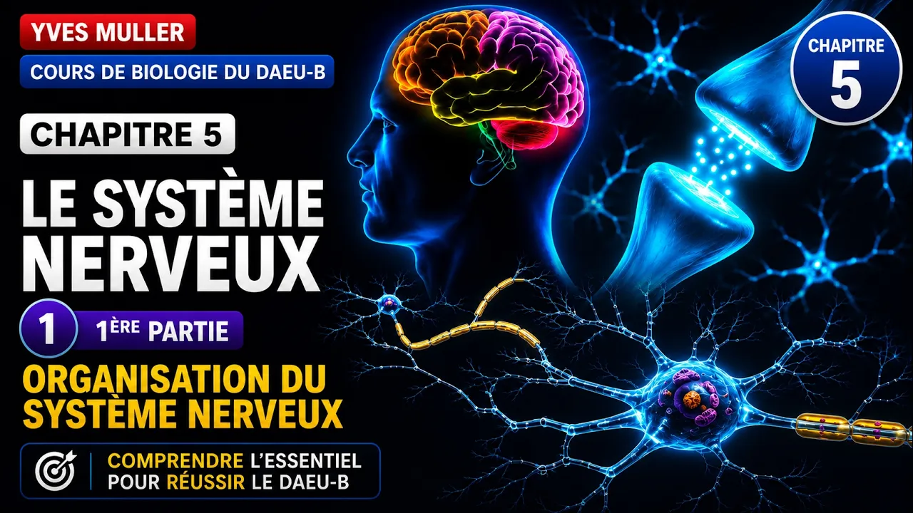
Chapitre 5 - 1ère Partie : Organisation du système nerveux et du neurone - Cours de Biologie
656K
The myelin sheath is a fatty insulation layer surrounding many axons, formed by glial cells. Functions include: (1) Electrical insulation - prevents leakage of electrical current between axons; (2) Protection - shields axons from damage; (3) Accelerated conduction - enables saltatory conduction where action potentials jump between nodes of Ranvier, increasing speed to up to 150 m/s compared to 1 m/s in unmyelinated axons. Myelination is critical for rapid neural signaling.
Click to watch →

RRB group d 2025 | Neuron structure हर शिफ्ट में आ रहे है प्रश्न By Kajal Maam #groupd
35.8K
The axon is a cylindrical tube that carries messages forward from the cell body. The myelin sheath (मलीन शीत) is a protective covering over the axon. Schwann cells (स्वन सेल्स) form the myelin sheath. The axon terminal (एग्जोन टर्मिनल) is the last part of the neuron where messages are transmitted to the next neuron.
Click to watch →
Deep Dive
The concept of electrochemical signaling, including how action potentials transmit information.

Nervous System (Part 2): Electrochemical Signaling, Action Potential & Electrophysiology in Neuron
5.3K
Neurons transmit information through electrochemical signaling, where electrical signals (action potentials) travel along axons via a series of ion channel openings and closings. The process begins with the resting membrane potential of approximately -70 mV, maintained by the sodium-potassium pump. When a stimulus reaches the threshold potential of -55 to -60 mV, voltage-gated sodium channels open, causing rapid depolarization as sodium ions rush into the cell. This is followed by potassium channels opening, initiating repolarization as potassium ions exit the cell. The membrane briefly hyperpolarizes below resting potential before returning to its baseline state. This entire action potential cycle takes about 1 millisecond and enables rapid signal transmission throughout the nervous system.
Click to watch →

Física - Sesión 10 - Bioelectricidad - Joseph Zamora - 2025 II
409
Electrochemical equilibrium occurs when chemical and electrical forces balance, resulting in no net ion movement. At equilibrium, potassium is -90 mV, sodium is +60 mV, and chloride is -70 mV. Resting membrane potential (-70 mV in neurons) results from unequal ion distribution, selective permeability (potassium has high permeability), and sodium-potassium pump activity. The Nernst equation calculates equilibrium potential: E = (61 mV/Z) × log([ion]out/[ion]in). Action potentials are rapid electrical signals with phases: resting potential, depolarization (sodium entry), repolarization (potassium exit), and hyperpolarization. The threshold (-55 mV) triggers the full action potential. Refractory periods prevent immediate re-firing: absolute refractory period (sodium channels inactivated) makes re-firing impossible; relative refractory period (potassium channels still open) requires stronger-than-normal stimulus. Action potentials propagate along axons. Larger diameter axons conduct faster. Humans use myelination (insulation by myelin sheaths) enabling saltatory conduction, where action potentials regenerate only at nodes of Ranvier, allowing rapid, efficient transmission.
Click to watch →

2) Sinir Sistemi - Nöronda İmpuls Oluşumu ve İletimi
449K
Impulse transmission involves both electrical and chemical changes. Sodium and potassium ions are critical for creating the electrical gradient. Action potentials consist of three phases: polarization (resting state with negative inside), depolarization (positive inside when stimulated), and repolarization (return to negative inside). In myelinated neurons, action potentials jump between nodes of Ranvier (saltatory conduction), enabling faster transmission. The sodium-potassium pump actively maintains ion distribution, powering these electrochemical changes through ATP production from aerobic respiration.
Click to watch →
Электрическая сигнализация. Часть 2. Биопотенциалы. Законы реагирования возбудимых тканей
5.3K
Action potentials solve the limitation of local potentials by propagating without decay over long distances. They require voltage-gated sodium channels in trigger zones (first node of Ranvier, axon hillock). Local circulating currents spread depolarization from the stimulus site to the trigger zone. When threshold is reached, all sodium channels open simultaneously in an all-or-nothing fashion, generating an action potential. This discrete, binary signal (like computer bits) allows reliable information transmission across the entire nervous system.
Click to watch →
Signal Transmission - Control and Coordination | Class 10 Biology
82K
The complete process of signal transmission between neurons is called electrochemical signal transmission. An electrical signal travels down the axon to the presynaptic terminal, triggering chemical release into the synaptic cleft. The chemicals cross the gap and bind to receptors on the next neuron, converting back to electrical signals. This cycle repeats as signals propagate through neural networks.
Click to watch →
Deep Dive
The fundamental definition of homeostasis and the general role of the nervous system in maintaining bodily balance.
The whole of AQA HOMEOSTASIS and RESPONSE. 9-1 GCSE biology or combined science revision for paper 2
179.6K
Homeostasis maintains constant internal conditions through controlled variables: blood glucose, water, and temperature. The brain serves as the control center coordinating responses via the nervous system. The nervous system comprises the central nervous system (brain and spinal cord) and peripheral components including neurons, receptors, and effectors. Reflex actions bypass the brain for rapid responses, while voluntary actions involve the brain. Neurons transmit electrical signals along long cell bodies, but chemical synapses slow signal transfer between neurons. The brain contains specialized regions: the cerebral cortex handles higher thinking, the cerebellum controls balance, and the medulla regulates automatic functions.
Click to watch →
![[#정목스님] 세상의 모든 어려움을 해결하는 만병통치약 '사무량심' #다시보는명법문](https://i.ytimg.com/vi_webp/Jp9K1pkHTXo/maxresdefault.webp)
[#정목스님] 세상의 모든 어려움을 해결하는 만병통치약 '사무량심' #다시보는명법문
92.1K
The sympathetic nervous system provides energy for survival activities, while the parasympathetic nervous system calms the body and reduces energy consumption. These two systems work in balance through homeostasis - the process of maintaining equilibrium. When energy expenditure increases, the parasympathetic system brings it back down; when conservation is needed, the sympathetic system brings it back up. This constant adjustment maintains an average value or equilibrium. The vagus nerve extends throughout the entire body, including the brain, internal organs, and lungs. When this balance is disrupted, it constitutes death. The state of balance between these two systems represents life itself.
Click to watch →
Grade 10 SCIENCE | Quarter 3 Module 3 | The Power and Control of the Nervous System
183.7K
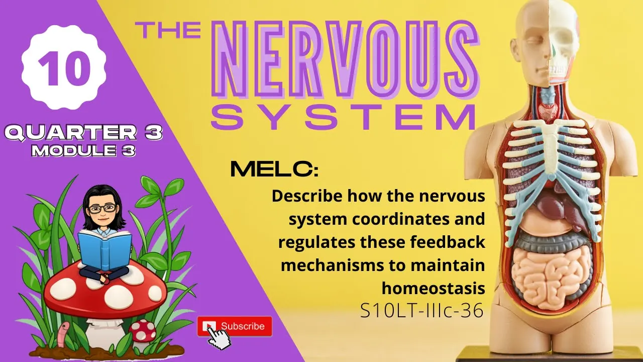
The nervous system is the major control system of homeostasis, which means it helps maintain equilibrium or balance in the body. The main learning competency is to describe how the nervous system coordinates and regulates feedback mechanisms to maintain this balance.
Click to watch →

【社会福祉士国試対策96】ホメオスタシス&人体の器官(脳、心臓、脊髄、消化器)
69.1K
Homeostasis is the body's ability to maintain internal stability and balance, working through the nervous system, endocrine system, and immune system. The nervous system is divided into central (brain and spinal cord) and peripheral (somatic and autonomic) components. The autonomic nervous system includes sympathetic (stress response) and parasympathetic (relaxation response) branches. The brain consists of four main parts: diencephalon (basic functions), cerebrum (higher functions), brainstem (vital life functions), and cerebellum (motor control). The cerebral cortex has four lobes: frontal (thinking, judgment), parietal (sensation, spatial awareness), temporal (hearing, language), and occipital (vision). The limbic system handles emotions and memory.
Click to watch →
EL ORIGEN EMOCIONAL DE MUCHAS ENFERMEDADES Y CÓMO CURARLAS
248.5K
This section explains homeostasis as the body's self-correction mechanism that maintains balance in blood sugar, hormones, oxygen levels, temperature, and other vital functions. The autonomic nervous system controls this process automatically and has two parts: the sympathetic nervous system (like a car's accelerator) that activates during emergencies and mobilizes energy, and the parasympathetic nervous system (like a car's brake) that reduces speed, conserves energy, and enables digestion and organ repair. The body is designed to be perfectly balanced, but stress disrupts this natural equilibrium.
Click to watch →
Deep Dive
Basic anatomical terminology (e.g., medial, lateral, anterior, posterior) to understand physical relationships between body parts.
Basic anatomical terminology | Medial and lateral | Proximal and distal | Anterior and posterior
3.6K
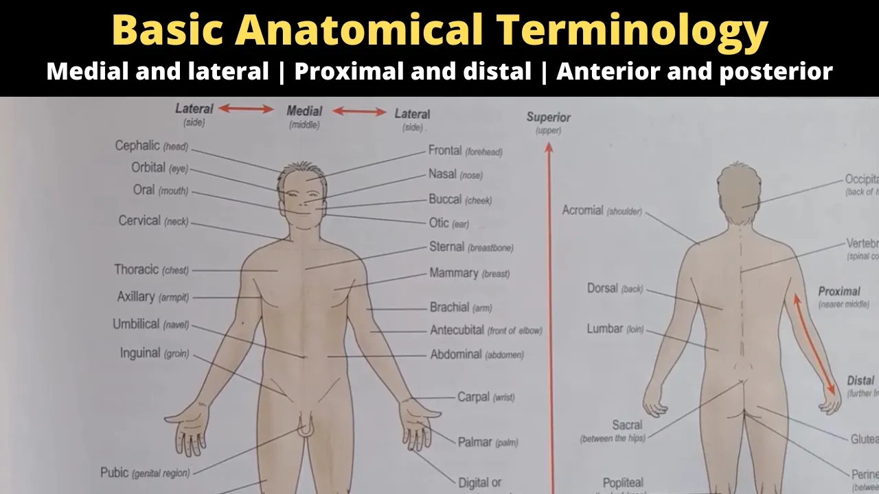
This video teaches fundamental anatomical directional terms used to describe body positions. Medial refers to structures closer to the body's midline, while lateral indicates structures farther from it. Proximal describes parts closer to the point of attachment, and distal indicates parts farther away. Superior refers to structures closer to the head, while inferior indicates structures farther from it. Anterior (ventral) describes front-facing structures, and posterior describes back-facing structures. These terms provide a standardized language for describing anatomical relationships and positions in the human body.
Click to watch →

Homeostasis | Positive , negative feedback system | basic anatomical terminology | b pharm 1st sem.
371
Anatomical terminology uses directional terms to describe body positions. Superior means 'above' (head is superior to neck), inferior means 'below' (mouth is inferior to nose). Anterior means 'front' (sternum is anterior to heart), posterior means 'back' (spine is posterior to heart). Medial means 'toward midline' (umbilicus is medial to heart), lateral means 'away from midline' (lungs are lateral to heart). Proximal means 'closer to attachment' (proximal convoluted tubule), distal means 'farther from attachment' (distal convoluted tubule). Body planes divide the body: sagittal (right-left), transverse/horizontal (superior-inferior), and coronal/frontal (anterior-posterior at 90 degrees).
Click to watch →
Introduction to Anatomy & Physiology (2026) | Anatomical Position, Body Regions & Directional Terms
313
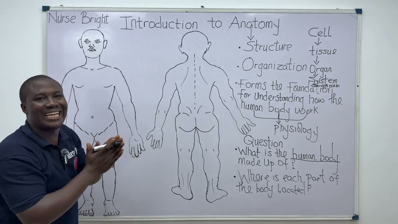
Directional terms are standard anatomical terms used to describe the position of one structure in relation to another. Medial means closer to the midline (median plane) than another structure. Lateral means farther away from the median plane than another structure. Anterior means closer to the front of the body (also called ventral). Posterior means closer to the back of the body (also called dorsal). Superior means closer to the head region (also called cranial). Inferior means farther away from the head region (also called caudal). These terms require a relationship between two structures and are used when the individual is standing in anatomical position.
Click to watch →

termos anatomicos de relação
21.5K
Anatomical terms of relation describe the positional relationship between body structures: medial refers to structures closer to the median plane, lateral to those farther away, median to structures exactly on the median plane, intermediate to structures between medial and lateral structures, and medio to structures between superior and inferior structures.
Click to watch →
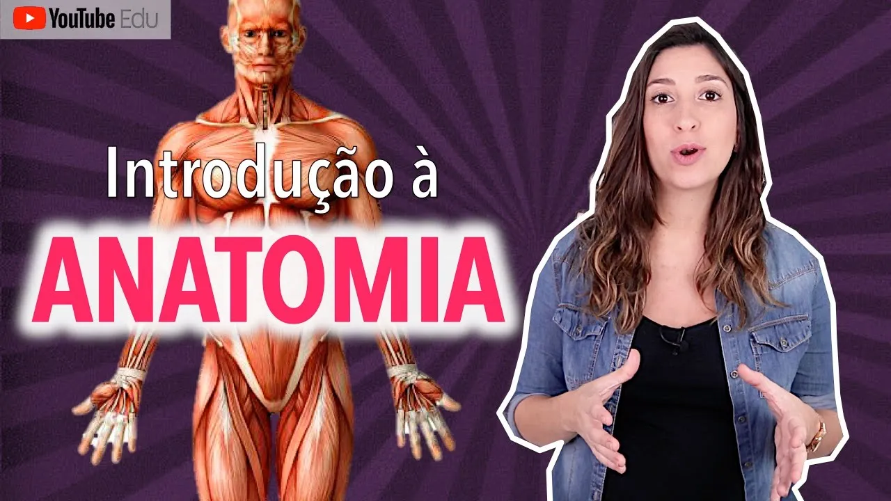
INTRODUÇÃO À ANATOMIA: Posição anatômica e termos de relação
2.8M
Medial means closer to the median plane of the body (an imaginary line dividing the body into right and left halves). Lateral means further away from the median plane. For example, the thumb is lateral to the little finger because it is further from the median plane, while the little finger is medial to the thumb.
Click to watch →
Prerequisite Knowledge
- Concept 01Basic cellular biology, specifically the structure and function of a neuron (axon, dendrites, myelin sheath).
- Concept 02The concept of electrochemical signaling, including how action potentials transmit information.
- Concept 03The fundamental definition of homeostasis and the general role of the nervous system in maintaining bodily balance.
- Concept 04Basic anatomical terminology (e.g., medial, lateral, anterior, posterior) to understand physical relationships between body parts.
Subsequent Learning
- Step 01The functional mechanics of the Autonomic Nervous System, specifically comparing the Sympathetic and Parasympathetic pathways.
- Step 02Detailed neuroanatomy of the brain (cerebrum, cerebellum, brainstem) and the structure of the spinal cord.
- Step 03The physiology of reflex arcs and sensory transduction (how the body detects and reacts to external stimuli).
- Step 04Clinical applications and neuropathologies, such as multiple sclerosis, peripheral neuropathy, and autonomic dysreflexia.
Nervous System Overview
PNS Subdivisions
Nervous System Overview
0:03Playing Section
- 1
Explains nervous system functions: fast communication via neurons and electrical impulses.
- 2
Details components: brain, spinal cord, nerves, and ganglia with their roles.
- 3
Introduces central and peripheral divisions, plus sensory and motor nerve functions.
Network Neuroscience and Functional Integration
While the traditional division of the nervous system into distinct compartments (such as central vs. peripheral, or somatic vs. autonomic) is a useful anatomical teaching tool, modern network neuroscience and systems biology challenge these rigid boundaries. Critics argue that strict compartmentalization obscures the highly integrated, bidirectional, and continuous nature of neural signaling. For instance, the enteric nervous system (the 'second brain' in the gut) operates with a high degree of autonomy, blurring the line between peripheral reflex and central control. Furthermore, the nervous system is increasingly viewed not as an isolated hierarchical controller, but as part of an inseparable, bidirectional network involving the immune and endocrine systems. Overemphasizing structural divisions can lead to a reductionist understanding that overlooks how sensory-motor loops, cognitive processes, and visceral states are dynamically coupled and distributed across the entire body rather than confined to specific divisions.
Deep Dive
The functional mechanics of the Autonomic Nervous System, specifically comparing the Sympathetic and Parasympathetic pathways.

Autonomic Nervous system
263.3K
The autonomic nervous system (ANS) is divided into the sympathetic (fight-or-flight) and parasympathetic (rest-and-digest) divisions, which work antagonistically to regulate involuntary body functions. The sympathetic system originates from thoracolumbar spinal cord segments (T1-L2/L3) and uses norepinephrine/epinephrine neurotransmitters acting on alpha-1, alpha-2, beta-1, and beta-2 receptors, causing effects like heart rate increase, bronchodilation, vasoconstriction, and glycogen breakdown. The parasympathetic system originates from cranial nerves (III, VII, IX, X) and sacral spinal cord (S2-S4), using acetylcholine acting on muscarinic and nicotinic receptors, producing effects like heart rate decrease, bronchoconstriction, GI tract stimulation, and gland secretion. Key examples include: sympathetic causes mydriasis (pupil dilation) via alpha-1 receptors in eyes, while parasympathetic causes miosis (pupil constriction); sympathetic causes ejaculation via alpha-1 receptors in prostate, while parasympathetic causes erection; and sympathetic causes renin release in kidneys via beta-1 receptors. This dual control mechanism allows the body to automatically adapt to different physiological states.
Click to watch →

Autonomic Nervous System (ANS) Overview: Sympathetic vs. Parasympathetic | Dr. Priyanka Sachdev
98.2K
The Autonomic Nervous System (ANS) is the motor division of the peripheral nervous system that controls involuntary organs through two opposing divisions: the sympathetic (thoracolumbar distribution, ganglia near CNS, post-ganglionic neurotransmitter norepinephrine) and parasympathetic (craniosacral distribution, ganglia near organs, post-ganglionic neurotransmitter acetylcholine) systems. Both divisions use two neurons (preganglionic and post-ganglionic) with acetylcholine at the preganglionic level, but differ in their post-ganglionic neurotransmitters and ganglion positions. In most organs, these systems have opposite actions (e.g., pupil constriction vs dilation, increased vs decreased heart rate), except in genital organs where they have complementary actions (erection vs ejaculation).
Click to watch →
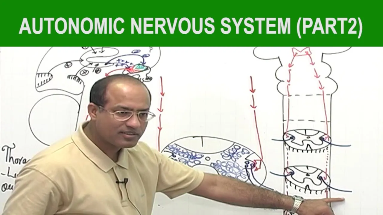
Autonomic Nervous System | Neurology | Neuroanatomy | Part 2🧠
177.1K
The autonomic nervous system regulates involuntary bodily functions through two antagonistic divisions: the sympathetic nervous system (thoracolumbar outflow) and the parasympathetic nervous system (craniosacral outflow). The sympathetic system prepares the body for 'fight or flight' by releasing norepinephrine and epinephrine, which act on beta-adrenergic receptors to increase heart rate, dilate airways, and mobilize energy stores through glycogenolysis and lipolysis. The parasympathetic system promotes 'rest and digest' functions through acetylcholine acting on muscarinic receptors, decreasing heart rate and promoting digestion. These systems antagonize each other at multiple levels including presynaptic inhibition, receptor competition at adenylyl cyclase, and opposing effects on ion channels, creating a balanced regulatory system that maintains homeostasis.
Click to watch →

Autonomic Nervous System (Sympathetic & Parasympathetic) - Overview
400.8K
The autonomic nervous system, a subdivision of the peripheral nervous system, controls unconscious bodily functions through two opposing divisions: the sympathetic nervous system (fight or flight response) and the parasympathetic nervous system (rest and digest response). Both divisions use a two-neuron chain with preganglionic neurons releasing acetylcholine, but differ in their postganglionic neurotransmitters and receptor types—sympathetic uses noradrenaline acting on adrenergic receptors (alpha and beta), while parasympathetic uses acetylcholine acting on muscarinic receptors. The sympathetic division originates from the thoracolumbar region of the spinal cord and activates organs like the heart, airways, and blood vessels to prepare the body for stress, whereas the parasympathetic division originates from cranial nerves III, VII, IX, X and the sacral region, promoting relaxation and digestion through effects on the heart, digestive system, and other organs.
Click to watch →

SISTEMA NERVOSO - FISIOLOGIA - Aula | Biologia com Samuel Cunha
1.8M
The autonomic nervous system regulates involuntary functions. The sympathetic division prepares the body for stress (fight-or-flight): increases heart rate, dilates pupils, redirects blood to muscles, and releases stored glucose. The parasympathetic division promotes relaxation and digestion (rest-and-digest): slows heart rate, stimulates digestion, and promotes urination. These divisions use different neurotransmitters - sympathetic releases noradrenaline while parasympathetic releases acetylcholine.
Click to watch →
Deep Dive
Detailed neuroanatomy of the brain (cerebrum, cerebellum, brainstem) and the structure of the spinal cord.

GENERALIDADES del SISTEMA NERVIOSO |Parte 2 de 2| Neuroanatomía | Unani 🧠🤓
113.8K
The cerebellum has two hemispheres connected by the vermis, located in the posterior fossa below the cerebrum, separated by the tentorium cerebelli. It contains gray matter in the cortex and deep nuclei (fastigial, globose, emboliform, dentate), with information transmitted via three cerebellar peduncles (superior to midbrain, middle to pons, inferior to medulla). The brainstem (midbrain, pons, medulla) serves as a connection between spinal cord, cerebellum, and cerebrum, with ten of twelve cranial nerves emerging from it. The brainstem lacks peripheral gray matter, with nuclei concentrated internally. The spinal cord begins at the foramen magnum and extends to L1-L2, tapering into the conus medullaris. It contains central gray matter in an H-shape with anterior horns (motor nuclei), posterior horns (sensory nuclei), and lateral horns (autonomic nuclei), surrounded by peripheral white matter.
Click to watch →

الحصة 03/ الدعامة البنيوية للجهاز العصبي / تحدي الحصول على 20/20!
138.4K
The nervous system consists of two main divisions: the Central Nervous System (CNS) and the Peripheral Nervous System (PNS). The CNS includes the brain and spinal cord. The brain has three main parts: the cerebrum (largest part with two hemispheres), the cerebellum (located behind the cerebrum for coordination), and the brainstem (connects brain to spinal cord). The cerebrum contains four lobes: frontal, parietal, temporal, and occipital, separated by three fissures (longitudinal, central, and lateral). The spinal cord is a cylindrical structure extending from the brainstem through the vertebral canal, composed of gray matter (central) and white matter (surrounding).
Click to watch →
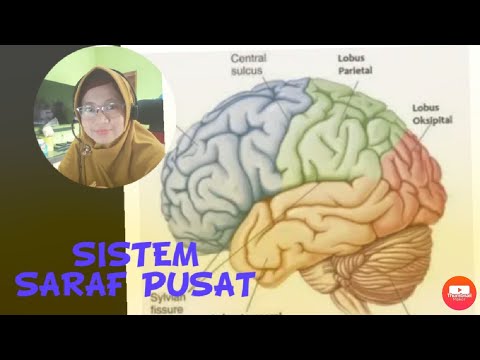
Sistem Saraf Pusat
32.7K
The brainstem combines midbrain, pons Varolii, and medulla oblongata. The pons serves as a neural bridge; the medulla oblongata connects brain to spinal cord and controls vital autonomic functions (heart rate, blood pressure, breathing, digestion, cough, sneeze). The cerebellum (cerebellum) is the second-largest brain structure and coordinates balance and voluntary movement. The spinal cord (medulla spinalis) extends from skull to L2 vertebra, serving as a reflex coordination center. Its cross-section shows white matter (outer nerve fibers), gray matter (inner with dorsal sensory root and ventral motor root), and central canal containing cerebrospinal fluid.
Click to watch →
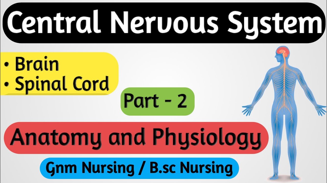
Central Nervous System (CNS) // Central Nervous System Anatomy and Physiology // Nervous System - 2
246.9K
The Central Nervous System (CNS) consists of the brain and spinal cord. The brain is divided into three parts: the cerebrum (largest part, 1000-1200g, divided into frontal, parietal, temporal, and occipital lobes), the brainstem (controls vital functions like respiration and heart rate), and the cerebellum (controls balance and coordination). The spinal cord is approximately 45cm long in adults and serves as the communication pathway between the brain and body. The brainstem connects to the spinal cord and controls involuntary body systems. The cerebellum, located behind the brainstem, coordinates voluntary movements and maintains balance.
Click to watch →
#4 متوسط ملخص الاتصال العصبي ( الجزء 1 )#فروض وإختبارات
690.3K
The brain consists of three main parts: (1) Cerebrum - the largest part with two symmetrical hemispheres, covered by the cerebral cortex (rich in blood vessels and folded to increase surface area), divided into four lobes (frontal, parietal, temporal, occipital) separated by grooves (central sulcus, lateral sulcus, Sylvian fissure), (2) Cerebellum - located at the back, involved in balance and coordination, and (3) Brainstem - connects the brain to the spinal cord, controlling vital involuntary functions like breathing and heart rate. The brain is protected by the skull and meninges.
Click to watch →
Deep Dive
The physiology of reflex arcs and sensory transduction (how the body detects and reacts to external stimuli).

الفرض الاول فى العلوم الطبيعية اولى ثانوى ادبى نموذج بالحل المفصل مع تجميعية جديدة من الفروض pdf
72.2K
The reflex arc consists of five essential components: (1) Neuromuscular spindle - sensory receptor that generates nerve impulses in response to muscle stretch, containing special muscle fibers innervated by sensory neurons; (2) Sensory neuron - transmits sensory information toward the central nervous system; (3) Spinal cord - integration center that converts sensory impulses into motor impulses; (4) Motor neuron - transmits motor commands from the spinal cord to the effector; (5) Effector organ (muscle) - contracts in response to motor impulses. The sequence is: stimulus detection → sensory signal transmission → spinal cord processing → motor signal transmission → muscle contraction.
Click to watch →

Complete SCIENCE Revision in 1 Video || Watch This Before Exam 🔴 || Class 10th Boards
182.8K
Sensory receptors detect stimuli from the environment and convert them into nerve impulses. For example, thermoreceptors detect temperature changes. The reflex arc is the pathway by which reflex actions occur: (1) Stimulus detected by receptor, (2) Sensory neuron carries impulse to spinal cord, (3) Relay neuron in spinal cord processes information, (4) Motor neuron carries impulse to effector, (5) Effector (muscle or gland) responds. Reflex actions are rapid, automatic responses that do not require conscious thought.
Click to watch →

Biología - Semana 09 - Pre San Marcos Ciclo 2024-II (Nuevo Ciclo)
308
A reflex arc is the neural pathway mediating reflex actions - involuntary, rapid responses to stimuli. The reflex arc includes: sensory receptor, sensory neuron, integration center (interneurons in spinal cord), motor neuron, and effector. Monosynaptic reflexes (knee-jerk) involve one synapse, while polysynaptic reflexes (withdrawal from pain) involve multiple synapses. Sensory receptors detect different stimuli: mechanoreceptors (pressure), photoreceptors (light), chemoreceptors (chemicals), thermoreceptors (temperature), and nociceptors (pain).
Click to watch →

Somesthetic and visceral sense receptors -part 1
382
This comprehensive section covers the complete physiology of sensory detection and reflexive responses. Sensory receptors include Merkel's disks, Meissner's corpuscles, Pacinian corpuscles, Ruffini endings, and free nerve endings for touch/pressure; free nerve endings for pain; Krause end bulbs for cold; and muscle spindles/Golgi tendon organs for proprioception. Receptors are classified by stimulus origin: exteroceptors (external: touch, pain, heat, cold), teleceptors (distant: vision, hearing), and interoceptors (internal: chemoreceptors, baroreceptors). Key properties include specificity (each receptor responds to specific stimuli), Weber-Fechner law (perceived intensity increases logarithmically), adaptation (reduced response to sustained stimuli), and generator potential (non-propagated electrical changes triggering action potentials). A reflex is an automatic, rapid, inborn response without conscious control, mediated through a reflex arc consisting of receptor, afferent nerve, center, efferent nerve, and effector organ. Reflex properties include delay (minimal in monosynaptic, longer in polysynaptic), temporal summation (subthreshold stimuli applied rapidly before refractory period ends produce response), spatial summation (subthreshold stimuli applied simultaneously at different locations produce response), and irradiation (graded responses where low-intensity stimuli cause few muscle contractions while high-intensity stimuli cause widespread contraction).
Click to watch →

Lecture10 Sensory Physiology
88.7K
Sensory physiology encompasses how the nervous system detects and responds to environmental and internal stimuli. The nervous system comprises the central division (brain, spinal cord) and peripheral division (nerves, receptors). Sensory processing enables attention, arousal, perception, memory, and emotion regulation for homeostasis. Perception is the conscious interpretation of external stimuli through brain patterns, limited by receptor types, sensitivity ranges, and central processing. Sensory receptors convert environmental stimuli into electrical signals through transduction. They are classified by location (somatic, visceral, special) and stimulus type (photoreceptors, mechanoreceptors, thermoreceptors, chemoreceptors, nociceptors). Each receptor has an adequate stimulus that triggers graded receptor potentials, which summate and trigger action potentials if threshold is reached. Stimulus intensity correlates with receptor activation frequency.
Click to watch →
Deep Dive
Clinical applications and neuropathologies, such as multiple sclerosis, peripheral neuropathy, and autonomic dysreflexia.

Multiple Sclerosis | Etiology, Pathophysiology, Types of MS, Clinical Features, Diagnosis, Treatment
463.9K
Multiple Sclerosis (MS) is a chronic, immune-mediated demyelinating disease of the central nervous system characterized by an autoimmune response where autoreactive T-cells and inflammatory cytokines attack myelin sheaths surrounding axons, leading to disrupted neural signal transmission; the disease primarily affects young adults (ages 20-40), with environmental factors like low vitamin D levels and viral infections (Epstein-Barr virus, HHV-6) combined with genetic susceptibility genes such as HLA-DR2 contributing to its development; clinically, MS presents with diverse neurological deficits including optic neuritis, bilateral internuclear ophthalmoplegia, pseudobulbar palsy, and spinal cord involvement, manifesting through four main subtypes: relapsing-remitting (90% of cases with flare-ups and partial recovery), secondary progressive (progressive deterioration after initial relapsing phase), primary progressive (continuous decline without relapses), and progressive-relapsing (continuous deterioration with non-recovering flare-ups); diagnosis relies on MRI showing characteristic white matter lesions in peri-ventricular regions, brainstem, and spinal cord, supported by visual evoked potentials and lumbar puncture findings; treatment includes acute management with high-dose corticosteroids and plasmapheresis, plus disease-modifying therapies such as interferon beta, glatiramer acetate, monoclonal antibodies, and natalizumab to suppress immune activity and prevent future relapses.
Click to watch →

Mecobalamine injection | remyline PFS | ME-12 for nerve damege | nerve pain foot pain pharma&health
59.6K
This comprehensive guide covers peripheral neuropathy and multiple sclerosis, explaining that diseases originate from five root causes: physical, psychological, direct, indirect, and vitamin deficiency. Peripheral neuropathy involves nerve damage outside the brain and spinal cord, caused by high blood sugar or Vitamin B12 deficiency, manifesting as tingling, numbness, burning pain, and weakness. Multiple sclerosis damages the myelin sheath anywhere in the body, causing disrupted electrical signals and symptoms including erectile dysfunction. Treatment involves Methylcobalamin injection (1500 mcg weekly for 5-6 weeks) for Vitamin B12 deficiency and peripheral neuropathy, with Alpha Lipoic Acid (100-300 mg daily) for nerve damage. Combined vitamin supplements with Benfotiamine, Folic Acid, and Pyridoxine provide comprehensive nerve health support.
Click to watch →

Autonomic Dysreflexia Hyperreflexia Nursing Review: Symptoms, Treatment
674.1K
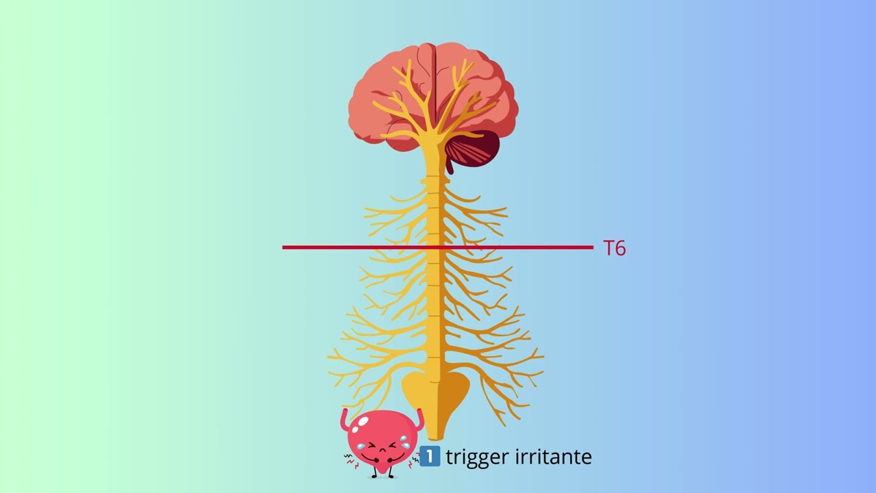
Autonomic dysreflexia (AD) is an exaggerated sympathetic reflex response occurring in patients with spinal cord injuries at T6 or higher, where the parasympathetic system cannot counteract sympathetic overactivity. The condition is triggered by irritating stimuli below the injury level, primarily bladder issues (distended bladder, UTI), bowel impaction, or skin breakdown. The pathophysiology involves vasoconstriction below the injury (causing pale, cool lower body) and vasodilation above the injury (causing flushed upper body), creating severe hypertension. Clinical manifestations include throbbing headache, hypertension with systolic BP 20-40 mmHg above baseline, bradycardia, flushing above injury, pallor below injury, sweating, goosebumps, dilated pupils, and anxiety. This represents a life-threatening medical emergency requiring immediate nursing intervention.
Click to watch →
PREVENZIONE E GESTIONE DELLA DISREFLESSIA AUTONOMICA
393
Sensory input from noxious stimuli below the lesion travels through intact peripheral nerves to the spinal cord, most commonly originating from bladder and bowel. This evokes sympathetic activation from thoracolumbar nerves, causing widespread vasoconstriction, particularly subdiaphragmatic, resulting in pale, cold lower body. Baroreceptors detect hypertension and signal the brain, which attempts parasympathetic counteraction. The brain sends descending inhibitory signals (blocked by lesion) and activates compensatory vasodilation in the upper body, causing flushing and profuse sweating. The vagus nerve attempts to slow heart rate, but compensatory bradycardia is inadequate. Clinical signs include pulsating headache (first symptom), with blood pressure elevation of at least 20 mmHg in adults or 15 mmHg in children. Baseline systolic pressure in spinal cord injury patients is typically 90-110 mmHg.
Click to watch →
Multiple sclerosis - causes, symptoms, diagnosis, treatment, pathology
2.8M
Multiple sclerosis presents with diverse symptoms depending on plaque location, typically affecting individuals aged 20-40. Charcot's triad includes dysarthria (brainstem plaques affecting speech/swallowing), nystagmus (eye movement nerve plaques), and intention tremor (spinal cord motor pathway plaques). Visual symptoms include optic neuritis (optic nerve damage causing vision loss, blurring, or central dark spots) and painful eye movements with double vision. Motor symptoms encompass weakness, spasms, tremors, ataxia, and potentially paralysis. Sensory symptoms include numbness, paresthesias, and Lhermitte's sign (electric shock sensation down spine with neck flexion). Autonomic involvement causes bowel/bladder dysfunction and sexual problems. Cognitive and psychiatric symptoms include poor concentration, depression, and anxiety. Diagnosis requires multiple neurological symptoms separated in space and time, supported by MRI showing white matter plaques, elevated CSF antibodies, and visual evoked potentials.
Click to watch →
Nervous System Overview
PNS Subdivisions
Nervous System Overview
0:03- 1
Explains nervous system functions: fast communication via neurons and electrical impulses.
- 2
Details components: brain, spinal cord, nerves, and ganglia with their roles.
- 3
Introduces central and peripheral divisions, plus sensory and motor nerve functions.
Network Neuroscience and Functional Integration
While the traditional division of the nervous system into distinct compartments (such as central vs. peripheral, or somatic vs. autonomic) is a useful anatomical teaching tool, modern network neuroscience and systems biology challenge these rigid boundaries. Critics argue that strict compartmentalization obscures the highly integrated, bidirectional, and continuous nature of neural signaling. For instance, the enteric nervous system (the 'second brain' in the gut) operates with a high degree of autonomy, blurring the line between peripheral reflex and central control. Furthermore, the nervous system is increasingly viewed not as an isolated hierarchical controller, but as part of an inseparable, bidirectional network involving the immune and endocrine systems. Overemphasizing structural divisions can lead to a reductionist understanding that overlooks how sensory-motor loops, cognitive processes, and visceral states are dynamically coupled and distributed across the entire body rather than confined to specific divisions.
Basic cellular biology, specifically the structure and function of a neuron (axon, dendrites, myelin sheath).
The concept of electrochemical signaling, including how action potentials transmit information.
The fundamental definition of homeostasis and the general role of the nervous system in maintaining bodily balance.
Basic anatomical terminology (e.g., medial, lateral, anterior, posterior) to understand physical relationships between body parts.
The functional mechanics of the Autonomic Nervous System, specifically comparing the Sympathetic and Parasympathetic pathways.
Detailed neuroanatomy of the brain (cerebrum, cerebellum, brainstem) and the structure of the spinal cord.
The physiology of reflex arcs and sensory transduction (how the body detects and reacts to external stimuli).
Clinical applications and neuropathologies, such as multiple sclerosis, peripheral neuropathy, and autonomic dysreflexia.
359.2K views4.7Klikes3:56@AlilamedicalmediaOriginal Release: 2019-09-03
The function of the nervous system is to provide rapid communication and integration between various organs, as well as with the outside environment.
It detects changes within the body and in its surroundings, and responds accordingly.
Fast communication is achieved by means of electrical signals, known as nerve impulses, which are generated and carried by specialized cells, called neurons.
The major components of the nervous system are the brain, spinal cord and nerves.
The brain, enclosed and protected in the cranium, is the central processing center.
It receives information, makes decision and coordinates the body response.
The spinal cord, enclosed in the spinal column, functions as a communication gateway between the brain and the trunk and limbs.
Nerves are cordlike structures that conduct information, similar to electricity-conducting wires.
They are composed of axons of neurons, the cell bodies of which are clustered in knot-like structures called ganglia.
Ganglia commonly serve as relay centers, where neurons synapse and transmit information to each other.
The brain and spinal cord make up the central nervous system, while nerves and ganglia constitute the peripheral nervous system.
Functionally, a nerve fiber can be sensory or motor.
Sensory nerve fibers carry sensory information from sensory receptors to the central nervous system, while motor nerves conduct motor instructions from the central nervous system to effector organs – the muscles and glands.
Nerves that contain both sensory and motor fibers are known as mixed nerves.
There are 2 major groups of nerves: cranial nerves and spinal nerves: - The 12 pairs of cranial nerves emerge from the base of the brain and relay information between the brain and the head and neck regions.
The cranial nerve X, named vagus nerve, also communicates with internal organs.
- The 31 pairs of spinal nerves arise from segments of the spinal cord and innervate the trunk and limbs.
Spinal nerves communicate with the brain via the spinal cord.
All spinal nerves are mixed nerves, they contain both sensory and motor fibers.
Typically, sensory receptors send impulses by way of sensory fibers in spinal nerves, to the spinal cord, which relays the information up to the brain.
The brain interprets the information and sends back instructions, down the spinal cord, to motor fibers in spinal nerves, to reach effector organs.
The peripheral nervous system can be divided into somatic and visceral subdivisions.
The somatic nervous system includes sensory nerves from the skin, muscles, bones and joints; and motor nerves that innervate skeletal muscles.
This system controls voluntary muscular contractions, as well as involuntary somatic reflexes.
The visceral nervous system, on the other hand, includes sensory division that detects changes in the viscera – the organs in the thoracic and abdominal cavities; and motor division that controls cardiac muscle, smooth muscle of internal organs and glands.
It produces, for example, faster heart rate and breathing rate during physical exercise, and slower cardiorespiratory rate during sleep.
The visceral motor division is also known as the autonomic nervous system because it is largely autonomous, acting independently of the body’s consciousness and voluntary control.
Up Next
50:18
Pain Pathway Part 1: Nociception, Transduction & Transmission | ACP Lecture
@nickybradley2859
880 views•2024-08-23

50:12
Bacterial Communication: Quorum Sensing and Biofilm Formation Explained
@JHUAAP
12.6K views•2011-06-28

5:35
Enteric Nervous System Explained: The Gut's Brain | Neurobiology Lecture
@alumniu6029
438 views•2018-09-12

7:08
Bacteriophages: Earth's Deadliest Killers and Future Antibiotics
@kurzgesagt
34.6M views•2018-05-13
Related Study Plans & Knowledge Roadmaps
Structured learning paths in Biology
biology
RNAi: Biogenesis, RISC & Gene Silencing
01Molecular Biology Foundations: DNA to Protein
02Introduction to RNA Interference (RNAi)
03Molecular Biogenesis of miRNA and siRNA
+ 2 more learning modules
10h5 modules
Explore Roadmap
biology
Mycorrhizae: Signaling, Biophysics & Ecology
01Foundations of Mycorrhizal Symbiosis
02Molecular Signaling and Root Colonization
03Biophysics and Bioenergetics of Nutrient Exchange
+ 1 more learning modules
11h4 modules
Explore Roadmap
biology
Circadian Clocks: TTFL, SCN & Periphery
01Foundations of Molecular Biology & Gene Expression
02Introduction to Chronobiology and Actograms
03The Core TTFL: Molecular Architecture of the Clock
+ 3 more learning modules
14h6 modules
Explore Roadmap
biology
Population Genetics: Math, Drift & Selection
01Genetic & Probability Foundations
02The Hardy-Weinberg Principle and Baseline Models
03Mathematical Modeling of Natural Selection
+ 3 more learning modules
18h6 modules
Explore Roadmap