



















SDS-PAGE is an analytical technique used to separate proteins based on their molecular weights by denaturing proteins with SDS and running them through a polyacrylamide gel under an electric field, where proteins migrate toward the anode and are separated by size due to the molecular sieving effect of the gel matrix.
SDS-PAGE: Principle, Procedure, and Applications
Added:Understanding of basic protein structure, including peptide bonds, folding levels (primary through quaternary), and overall charge properties of amino acids.
Fundamental principles of gel electrophoresis, specifically how charged molecules migrate through a porous matrix under the influence of an electric field.
The concept of protein denaturation and how physical or chemical agents (like heat or detergents) disrupt non-covalent interactions in proteins.
Basic laboratory chemistry concepts, including pH, buffer systems, and molecular weight units (Daltons/kilodaltons).
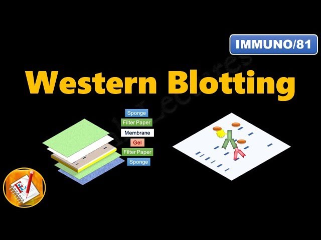
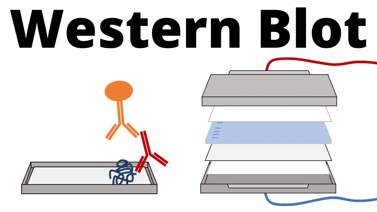
Western Blotting (Immunoblotting) techniques to transfer separated proteins to a membrane for specific antibody-based detection.
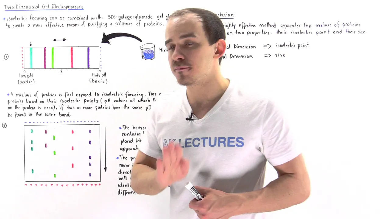
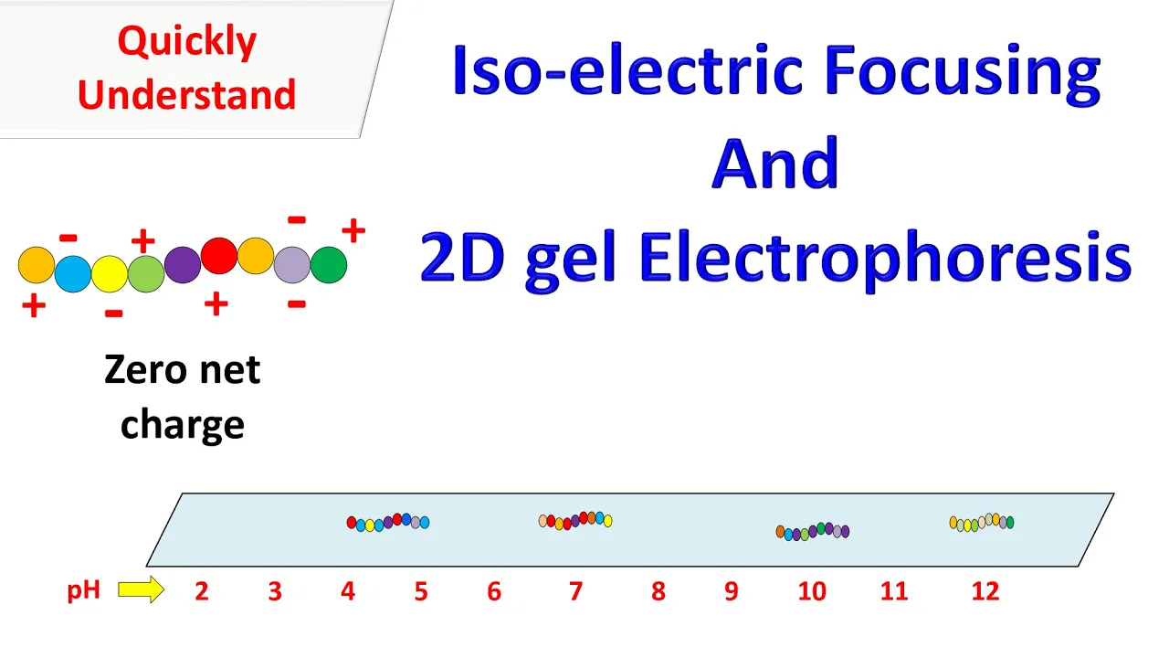
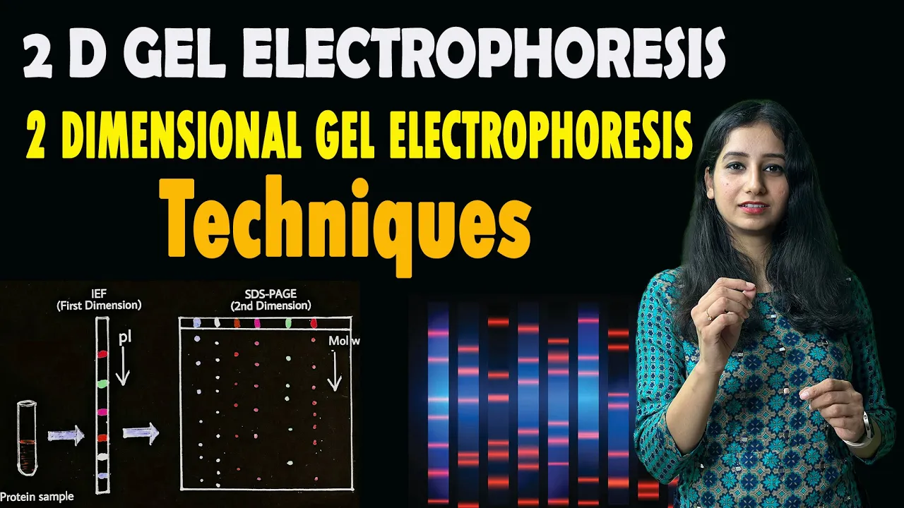
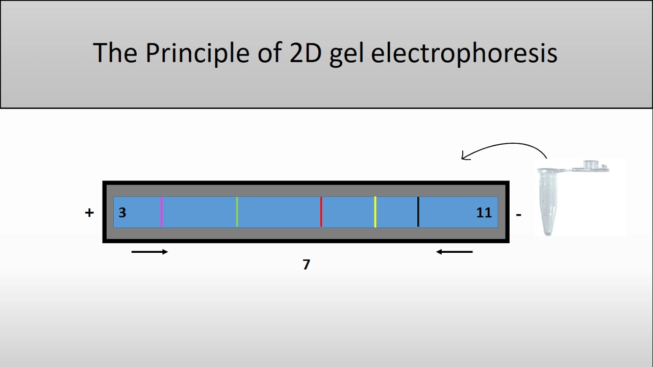
Two-Dimensional (2D) Gel Electrophoresis, which combines isoelectric focusing and SDS-PAGE to separate complex protein mixtures by both charge and size.
Mass Spectrometry (MS) sample preparation, such as in-gel digestion, to identify and characterize protein bands excised from an SDS-PAGE gel.
Quantitative methods for protein analysis, such as densitometry, to estimate the concentration and purity of separated protein bands.
997 views32likes13:31Original Release: 2024-09-08
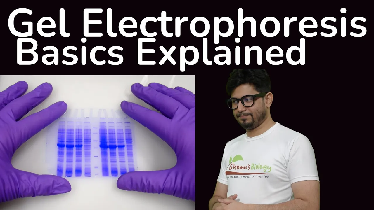
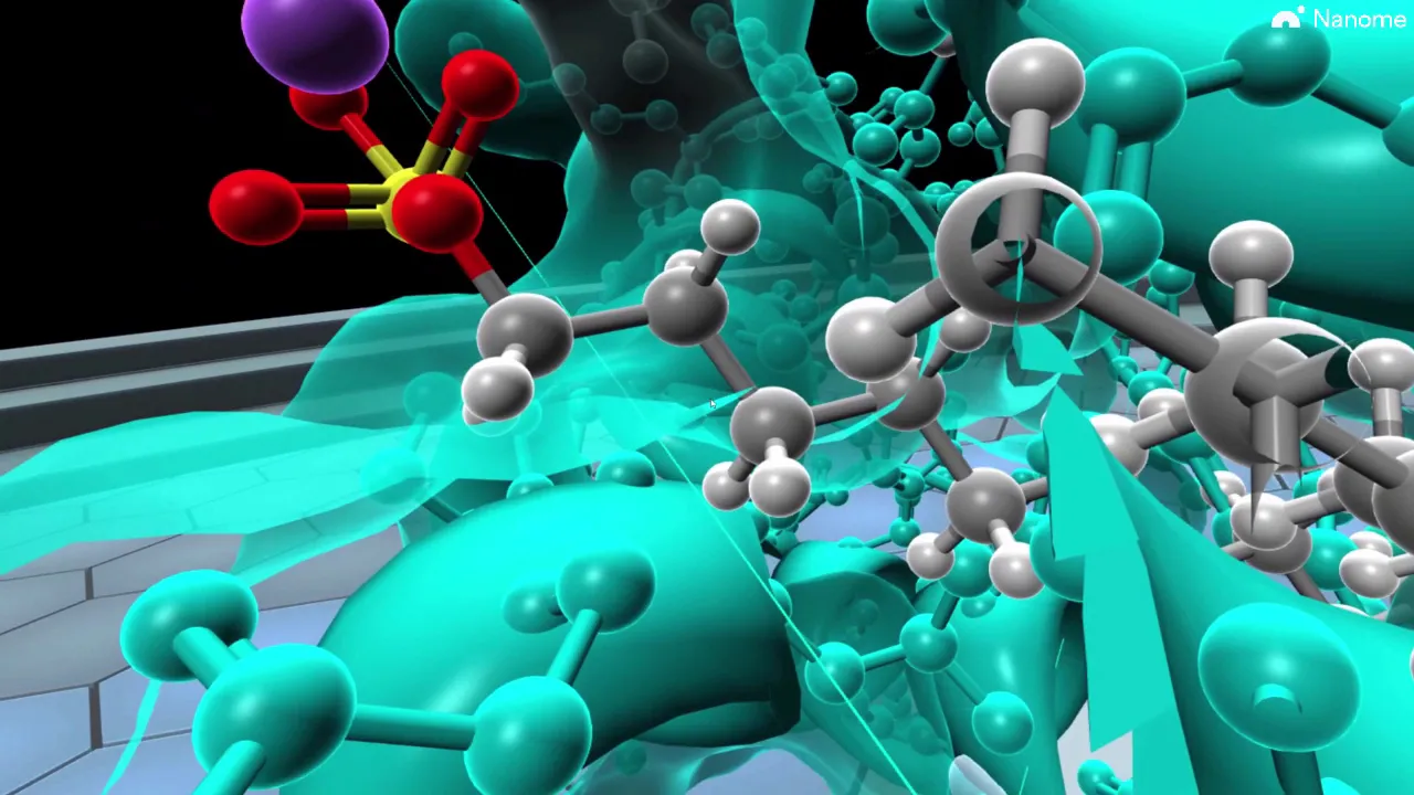
dear viewers greetings in this video we are going to see about sodium doile sulfate polyacryamide gel electroforesis in short form it is called as SDS page the contents of this video includes introduction about SDS page principle of SDS page materials used in SDS page agents used in SDS page procedure for SDS page applications of SDS page advantages of SDS pH and finally limitations of SDS pH sodium do desil sulfate polyacrylate gel electroforesis STS phage is an analytical technique used for the separation of proteins based on their molecular weights SDS is an anionic surfactant and detergent it breaks down the non-covalent links of protein molecules SDS binds to the protein backb at a constant mol ratio and has a significant protein denaturing effect poly AC gels are chemically cross-link gels formed by the polymerization of acry amide with a cross-linking agent usually y methylin bis acramite STS phase was first developed by University of Geneva Professor early lamle SDS phage is very commonly used to separate molecular Mass proteins between 5 KDA to 250 KDA and SDS phage is a widely used technique in forign six genetics biotechnology and molecular biology to separate the protein molecules based on their electrophoric Mobility principle of SDS pH electrophoric mobility in SDS phase technique depends on the molecular structure shape and charge the prot being covered by SDS or negatively charged and when loaded onto a gel and placed in an electric field it will migrate towards the anode or positively charged electrode and they are separated by a molecular seing effect based on size a smaller molecules move quickly because of their less obstruction whereas large molecules move slower after the visualization by a straining or protein space specific technique the size of a protein can be calculated by comparing its migration distance with that of a known molecular weight lader or marker sodium doel sulfate and polyacryamide help to eradicate the influence of structure and charge of proteins and the proteins are separated based on the length of the polypeptide CH materials used in SDS phage there are seven major materials used used in SDS pH they are power supply gels electroforesis chamber protein sample running buffer staining and daining buffer and finally protein lader the first material used in the SDS phase is power supply power supply is used to convert the AC current to DC current next gels gels are either self-prepared in the laboratory are purchased from the market electroforesis chamber the chamber in which sdfa gels four and protein are separated are called electroforesis chamber protein samples the protein is dissolved with an SDS Fai sample buffer and boil for 10 minutes a reducing agents such as Dio 3ol or two Mar capto ethanol is also added to minimize the D sufate linkage to prevent any treasury protein foldings running buffer the protein samples filled on the gels or run in STS pH running buffer staining and daining buffer kazis stain solution is used to stain a daining solution is used to Dain a gel protein bands can then be observed with neck I finally protein lader a ref reference protein lader is used to locate the protein of Interest based on the molecular size reagents used in SDS pH four major reagents are used in SDS pH they are beta mapto ethanol sodium doile sulfate bromoenol blue and finally polyacryamide gel the first reagent used in the SDS phases beta marap to ethanol the disulfide bridges present between the polypeptide chains are responsible for the secondary structure of protein however beta merto ethanol breaks the D disulfide bond that are present in the protein structure therefore on treating with beta merto ethanol the protein will have only a primary structure and be separated based on its size in the gel the second reagent is sodium doile sulfate or SDS SDS is used in the preparation of sample buffer to impart a negative charge on the protein after the treatment with beta merto ethanol all the proteins are in a linear structure so that SDS will import a negative charge and all the proteins will move towards the positive electrode thus separated only on the molecular size moreover SDS is also present in the gel making and it makes sure that all the proteins should stay negative throughout the gel the third reent is bromophenol blue bromophenol blue is used as a tracking D in the electroforesis as it imparts color in the protein which is being separated in the gel it is also mixed with a sample solution bromoenol blue has a slightly negative charge so that it could reach the positive electrode before for the protein molecule bromophenol plue indicates the migration of protein towards the electrode the fourth reagent is polyacrylate gel polyacryamide gel is a good choice since it is chemically inert and more importantly it can be easily prepared at a number of concentration to provide VAR poor diameter and resulting in a variety of Separation conditions that can be modified based on your demand procedure for SDS phage SDS pH contains three major Steps step one is gel preparation step two is sample preparation and the step three is electroforesis the step one of the SDS phases gel preparation the gels typically consist of 5 to 20% acry amide b acry amide or the option denaturant like SDS or Ura and a buffer with an adjusted pH the gel is prepared by the following procedure first mix all the reagents except T and once the gel is ready add tamod on it and then pour the separating gel into the casting chamber after pouring the separating gel into the casting chamber to remove unwanted air bubbles in the chamber of add butanol in it and then insert a comb in between the space of the glass plate finally allow to set the gel this polymerase gel is called as gel CD the second step in the SDS phase is sample preparation first boil water and add two mapto ethanol to the buffer sample and then add the buffer solution with the protein sample to micro centrifuge tubes and take molecular weight markers in separate tubes boil the samples for few minutes at 60° C for complete protein denaturation and then add tracking D or promol blue to the sample solution this typically has a higher electrophoric Mobility than the analytes to allow the ex to track the progress of the solution through the gel during the electrophoric Run the third step in the SDS phase is electroforesis remove the gel CD and place it in the electrode assembly fix the electrode assembly with the clam stand to fill the gel Wills pour one net electroforesis buffer in the opening of the casting frame and then add 20 to 30 ml denatured protein sample in the well using a piper after that cover the tank with a lid and connect the electroforesis unit and start the power supply after connecting the electroforesis unit allow the sample to run for one heart at 30 mamp after electroforesis process the gel is rinsed with deionized water for 3 to five times to remove SDS and buffer it may create a hinderance with the Kumasi blue strain to the proteins then the gel in the blue strain is shaken in an incubator at a room temperature the invisible bands of proteins begin to appear under UV light within a minutes this figure shows a strain polyacryamide gel used to visualize separated proteins in a sample the panel a shows a image representation of Kumasi stream STS pH data the wells and D front are both l label and large proteins are found towards the top of the gel and small proteins are found towards the bottom of the gel Lane one has a molecular weight lader and Lane two and three are samples from two separate atoms at purifying a 40 KD protein the sample in lane lane 3 is more pure than the sample in lane two since additional proteins can be seen in the sample in the lane in the panel B shows a portion of a real gel used to analyze the proteins in a purified sample of eukariotic ribosomes the eukariotic ribosomes consist of 80 proteins which is why there is such a large number of bands in the sample Lane applications of SDS phage SDS phage is used to calculate the molecular weight of molecules particularly proteins SDS phage is employed in reptite mapping it investigates post transational changes SDS phage is used in HIV test to separate the HIV proteins STDs phage is used to determine the size of protein and it is also used to calculate the proteins size SDS phage is used to compare the composition of polypeptides of various structures and it is used to calculate the protein Purity finally it is employed in Western blotting and protein ubiquination advantages of SDS phage SDS phage is a stable technique SDS phage has a greater resolving power and shows sharp bands STDs phage can accommodate large quantities of DNA without significant loss of resolution the DNA recovered from po acry gel is extremely pure the poor size of the polyacryamide gel can be altered in an easy and controllable fashion by changing the concentration of the two monomers SDS phas is good for separation of low molecular weight fragments finally limitations of SDS pH generally gel is more difficult to prepare and handle involving a longer time for preparation than the agross gel the second limitation is toxic monomers are formed during the STS phase technique the third limitation is gels are tedious to prepare and often leak and the fourth and final limitation is this SDS F needs new gel for each experiment dear viewers that's all about the STS pH technique thank you for the support thank you
Up Next

38:38
Michaelis-Menten Kinetics & Enzyme Activity Units Explained | Biochemistry
@thebumblingbiochemist
2K views•2024-09-08

39:12
Taste Matters: Why We Like the Food We Do | Professor John Prescott
@smagforlivet-video
409 views•2015-12-07

5:35
Enteric Nervous System Explained: The Gut's Brain | Neurobiology Lecture
@alumniu6029
438 views•2018-09-12

7:08
Bacteriophages: Earth's Deadliest Killers and Future Antibiotics
@kurzgesagt
34.6M views•2018-05-13
Related Study Plans & Knowledge Roadmaps
Structured learning paths in Biology
biology
Analyzing Animal Venoms: Genetics & Pharma
01Molecular Biology & Physiology Foundations
02Venom Evolution & Toxin Gene Recruitment
03Neurotoxins & Ion Channel Pharmacology
+ 2 more learning modules
18h5 modules
Explore Roadmap
biology
RNAi: Biogenesis, RISC & Gene Silencing
01Molecular Biology Foundations: DNA to Protein
02Introduction to RNA Interference (RNAi)
03Molecular Biogenesis of miRNA and siRNA
+ 2 more learning modules
10h5 modules
Explore Roadmap
biology
Mycorrhizae: Signaling, Biophysics & Ecology
01Foundations of Mycorrhizal Symbiosis
02Molecular Signaling and Root Colonization
03Biophysics and Bioenergetics of Nutrient Exchange
+ 1 more learning modules
11h4 modules
Explore Roadmap
biology
Circadian Clocks: TTFL, SCN & Periphery
01Foundations of Molecular Biology & Gene Expression
02Introduction to Chronobiology and Actograms
03The Core TTFL: Molecular Architecture of the Clock
+ 3 more learning modules
14h6 modules
Explore Roadmap