



![ONE SHOT- BREATHING AND EXCHANGE OF GASES [NEET 2024] Class 11 Comp NCERT](https://i.ytimg.com/vi/pMgwFUSkGeI/maxresdefault.jpg)

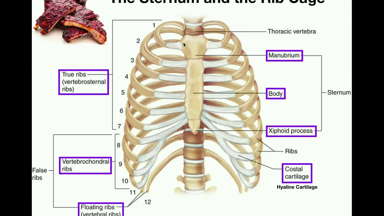







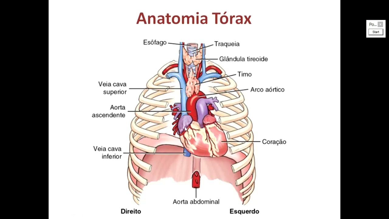


The thoracic region contains multiple interconnected systems including the skeletal framework (sternum with manubrium, body, and xiphoid process; ribs classified as true, false, or floating), respiratory system (trachea with C-shaped cartilage rings, bronchi branching into bronchioles ending in alveoli for gas exchange), cardiovascular system (heart with four chambers and associated valves, pulmonary and systemic circulation pathways), and mediastinal structures (coronary arteries supplying heart muscle, esophagus, and lymphatic system including the thoracic duct). Key muscles involved in breathing include the diaphragm (primary inspiratory muscle), external intercostals (assist inhalation), and internal intercostals/transversus thoracis (assist forced exhalation).
Thoracic Cavity Anatomy: A Complete Labeled Guide
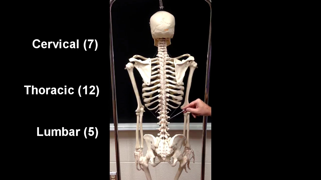
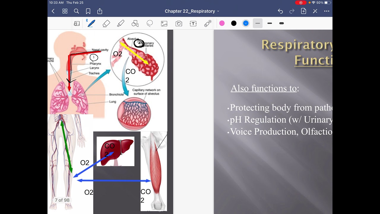
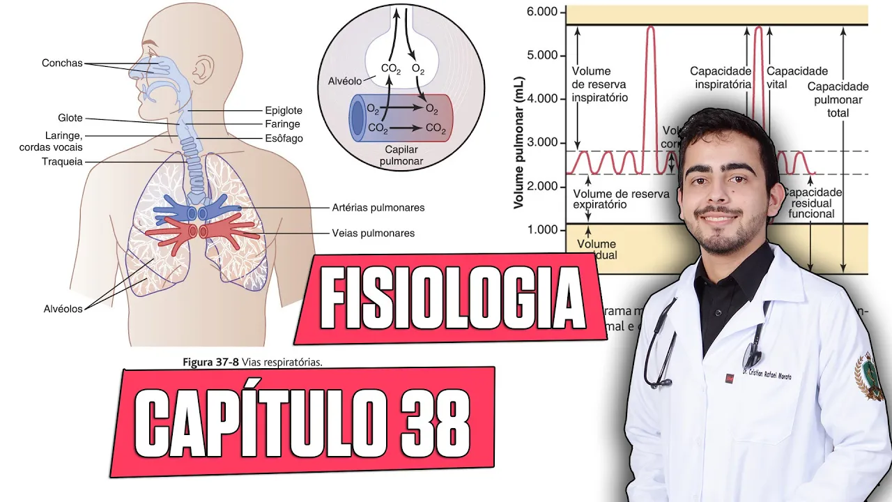
Added:hey everybody in this video you'll learn all of the structures of the thoracic region or chest region of the body we'll start from the anterior side with the most external structures then work our way posterior through the chest muscles the ribs and sternum heart and lungs and finally to the Lesser known structures in the media stum so this will be the first and a series of regional anatomy videos using my model from Anatomy Warehouse Tor the torso model who will be helping us along the way at the end of the video I'll have some blank images that you can use to help practice labeling all these structures yourself also check out the video description below for some other resources and links that you might find helpful if you're an anatomy student or teacher all right Tori you ready great let's uh jump to the model and get started so first let's take a look at some regions of course this is the thoracic region up here down here is going to be the abdominal region and really the separation between the two is going to be the diaphragm which is going to be kind of up in there but of course the ribs and intercostal muscles and costal cartilage and stuff kind of extend down below that so we'll include those a little bit in our video we said we're going to start from kind of the front and then work our way in so of course we've got skin we've got a hypodermis layer right here and then underneath that we can see all the structures over here so here we've got the sternum we're going to look a little bit more at the sternum in just a second when we look at the back side of the chest plate here we've got the pectoralis major muscle that we can see and we've got the mamry gland right here let's talk about that for a minute the mamry gland of course is going to produce milk especially after pregnancy and it's going to have different lobules throughout the mamory gland and then there's going to be lactiferous ducks and so you can see kind of a little bitty cutaway right here lactiferous ducks are going to take the milk from the lobules which are inside the mamm gland up to the nipple and then around that we've got the pigmented area that's called the Aela so we've got the mamry gland there's lobules inside and then lactiferous ducts that take the milk to the nipple and the Aela is the pigmented tissue around the nipple I'm going to remove that mammory gland so you can see some of the muscle underneath and let's turn the model a little bit so here we said we have the pectoralis major muscle it's going to have an origin on the sternum as well as the clavicle and it's going to go back here all the way to the humorous or the bone of the arm right here and what the petalis muscle is going to do is it's going to pull that humorous forward like whenever we're taking our arm and moving it like this so think of like a PEC fly if you're in the gym like this it'll also add duct the arm so think about taking the arm and bringing it back down to the body there's a pectoralis minor muscle underneath that that we can't see yet we'll take a look at that in the cross section down here we're going to have the seratus anterior muscle right there the serratus anterior is known as the boxer muscle because it's used whenever you're like throwing a punch like if you're bringing your arm forward like this it's going to actually have an insertion in the scapula so it's going to rotate the scapula forward whenever you're doing that kind of punching motion it's also going to help to lift the rib ribs like whenever you're inspiring or breathing in it'll help lift those ribs up a little bit to help you breathe in a little bit better so that's the seratus interior it's going to have an origin on ribs 1 through 8 and then an insertion on the scapula just deep to that right here we've got the external obliques those going to be used in kind of bending or twisting the trunk whenever you're twisting side to side like that or if you're if you're leaning down and then from there we can see some of the ribs right there we're going to have some false ribs down in here we've got true ribs up here kind of going up that we you can't see some of those and then we'll have a couple floating ribs further down that are obscured by the muscles right here now those ribs are going to be connected to the sternum via the costal cartilage so we have cartilage right through there which is of course going to make the rib cage a little bit more flexible so whenever we're breathing in or out those ribs can kind of expand out and contract a little bit if it was all bone it would not be as flexible now between the ribs we've got the intercostal muscles so there's really two layers of intercostal muscles right here you can see the external intercostal muscles the external intercostal muscles are going to help us to El El at um our rib cage whenever we're breathing in and then down here we've got the internal inner costal muscles and those are going to help to move the ribs inferior so I think internal inferior and they're going to help us to expire or breathe out notice the direction that those muscles run is opposite of each other so these right here are running this way the externals and then the internals are running kind of perpendicular to that almost so again we've got the pectoralis major muscle that's going to add duct to the arm or rotate the arm forward we've got the serratus anterior muscle which is going to rotate the scapula forward Ward and help us with breathing a little bit breathing in we've got the external obliques which are going to help with twisting and bending then we've got the inner costal muscles the external intercostal muscles which are going to raise the rib cage whenever we're breathing in and the internal intercostal muscles which are going to contract the rib cage or move the ribs inferior whenever we're breathing out also one note about the muscles that help with breathing whenever we breathe in that's going to be a very active process we use several muscles with that and we have to use muscles making them contract in order to breathe in whenever we breathe out we can just really relax muscles we don't have to use use any muscles to do that breathing out but if you breathe out kind of naturally and you try to force more air out than than just kind of the normal breathing Rhythm we call that forced expiration and that's when we're using some of these muscles whenever I say that they're we're using a muscle to like breathe out it's during forced expiration so like we get that extra breath coming out all right let's take this chest plate off and look at the posterior side of the chest plate so from this side of the chest plate we can see the bones a little bit easier and some other structures that we couldn't see from the front side so first of all the sternum has really three parts we've got the manubrium up here that's going to be connected to the clavicle the clavicle is going to be right here and right there that's connected on either side it's going to be connected to the first um two ribs we've got the body of the sternum and at the bottom we've got the zyo process now manubrium and the zyo process you can feel pretty easily if you take your finger and you put it in this Notch right there that's going to be the top of the manubrium this section right there and then if you take your finger and you put it right under here you'll feel a notch kind of poking down that's going to be the zyo process so the sternum has the manubrium the body and the zyo process we see costal cartilage connected to the parts of the sternum through there and then we've got the ribs coming down right there this will be rib number one 2 3 4 five 6 7 those would be your seven true ribs meaning that they're connected to one piece of costal cartilage that's connected to the sternum and then we're going to have some false ribs after that so 1 2 three false ribs false ribs are going to be connected to costal cartilage but the costal cartilage is going to come together into one piece of costal cartilage right here so these would be false ribs and then we'll have two floating ribs below that we can just see one of them right here and there's another one that's not on the chest plate floating ribs are the two ribs that don't connect to the sternum they're just sort of floating there all right coming back to this we've got a few muscles that we need to talk about and of course we got to talk about the diaphragm which is going to be probably the most important muscle whenever it comes to breathing whenever we breathe in or Inspire this diaphragm is going to contract down and that's going to expand the thoracic cavity right here whenever we expand that thoracic cavity it's going to lower the pressure and that's going to pull air into the thoracic cavity and therefore into the lungs so that diaphragm will contract down now you'll notice that you got all this muscle here there's also connective tissue that makes up a lot of the diaphragm as well and then the other muscle that helps us with inspiration or breathing in of course we already talked about is the external intercostals so diaphragm and external intercostals those are the ones that are going to help us breathe in here we can see the internal interc musles and those will help a little bit with us breathing out or expiration another muscle helping with breathing out is the transversus thoracis and you can see that that's going to have an origin in the sternum right here and then it's going to insert into the ribs and costal cartilage of ribs like three through about eight and again expiration is mostly a passive process but these are going to help with forced expiration when we breathe out more than kind of the normal breathing amount now I want to take the chest plate and turn it like this so we can see kind of a cross-section right here one tiny muscle we can see is called the subclavius right in there and that's going to stabilize the clavicle so that's the subclavius which just means below the clavicle right over here we've got the pectoralis major muscle and again we talked about that one's going to rotate the arm forward so that's that kind of external muscle right there just deep to that we've got the petalis minor which we haven't talked about because it's kind of hidden behind the petalis major so that petalis minor muscle it's going to move the scapula it's going to do rotation abduction and some other movements of the scapula it's going to have an origin in ribs 3 through 5 so it's going to connect to the third rib 3 4 five and then have an insertion in the scapula just like the serus anterior did so pectoralis minor and serus anterior are both going to help with moving the scapula all right pectoralis major pectoralis minor subclavius and then most of the rest of these muscles down there are going to be cross-sections of the seratus anterior muscles now if we look at all of the layers here of course we've got the epidermis and dermis right here that's going to be the outer skin layers we got the hypodermis which is a fat layer then we've got muscle layers beneath fat and then finally the bone which is going to be ribs in this case and in general that's kind of the order of things in the body you've got skin fat layer muscles and then bones and then other internal organs after that so skin fat muscle bone all right cool so now we got kind of this outer chest plate out of the way so next we're going to move to looking at the rest of the internal organs of the thoracic cavity after a quick uh coffee break all right let's go okay awesome so with that chest plate off you can immediately see some recognizable structures we got a right lung a left lung a heart a lot of times people think the heart is kind of off on the left side but really your heart's right in the middle the apex of the heart will point to the left and so in this left lung you see kind of a bigger Notch right in there because of that heart pointing to the left but the location of the heart is really kind of centered in the thoracic cavity all of this is going to be sitting on the diaphragm right here and of course we got the liver stomach intestines and all that down below up above we see some blood vessels we're going to talk about a little bit more here now immediately we can see that there's a different number of lobes for each lung we can see that there there's one two three loes of the right lung there's kind of a dividing line right there and right there if we switch to the left lung we're just going to have two loes so one lobe two lobes and we can see the dividing line right here we'll talk about this more in a minute but I really like that it's three lobes on the right and two lobes on the left because that matches the valves in the heart there's the tricuspid on the right and the bicuspid on the left just makes it a little bit easier for me to remember so let's take part of the lungs out so we can see a little bit more of the heart all right there's a lot going on here a lot to break down for some General locations we're going to have the right Atrium right ventricle left atrium left ventricle this is actually the Oracle I'll talk about that in a minute we can see some coronary arteries and coronary veins here and we see some bigger blood vessels coming out or entering into the heart kind of all around it let me take this off right here and talk about the chambers and then we'll go through the blood vessels and then I want to talk a little bit about the coronary arteries so here we have the right atrium and when I put this front piece of the heart back on you've got this structure right here which is called the Oracle we've got a right Oracle and a left Oracle and I've always had a hard time figuring out like what is is the Oracle is it just the atrium another name for it and in fact it used to be kind of used interchangeably but what it is the Oracle of the heart if you turn this around we can see a little bit better this is going to be the Oracle and basically it's like an extra pouch sticking off of The Atrium and over here we've got the left Oracle and again it's just a little pouch sticking off of The Atrium and so basically what the article does is it's a little extra pouch that when the Atria fills with blood there's a little bit more room for that blood to go into if we have a lot of blood in the Atria so it's kind of a separate structure but it's like a it's like a little Annex or extra room off of the Atria that more blood can fill so again right atrium we're going to have low oxygen blood here we've got the tricuspid valve right here then we've got the right ventricle we've got some muscle around it that's going to pump the blood from the right ventricle up through the pulmonary semi lunar valve and then through the pulmonary artery the pulmonary artery is going to Branch off in two directions so let me turn this around you can see that there's a branch right here on this side and there's a branch over here on this side so one's going to go to the left lung the other's going to go to the right lung if you look back over here that's where that continues on so this is the pulmonary artery or really the right branch of the pulmonary artery here's where the left branch of the pulmonary artery is going to connect to the lung so the blood's going to travel into the lung it's low oxygen blood the artery is going to Branch into smaller arterials and eventually into capillaries in the lung and we've got to get oxygen to that blood so let's talk about how oxygen gets into the body of course we're going to breathe in we're going to use that diaphragm and external intercostal muscles to breathe the air in by expanding in the thoracic cavity so air is going to travel down through the trachea which is right here the trachea you'll see has these light blue Rings those are cartilage rings and they're c-shaped so they're shaped like this and the reason we have cartilage rings there is to hold that trachea open we don't want the trachea like closing and then having to like forcefully bring it back open every time we breathe we're breathing basically all the time and so we want this held open so that we can always have air flowing in and out the one exception that of course is whenever we're swallowing food and so we have an epig gladus up there that whenever we swallow is going to close off the trachea so that food or water and things don't get down into the trachea and therefore into our lungs so the trachea is going to Branch off into the right and left bronchi the left bronchus will take air into the left lung and the right bronchus into the right lung each bronchus will split into bronchioles and so the first split will bring us into primary bronchioles and then it splits again into secondary and then into tertiary bronchioles and then eventually they'll end in little Alvis sacks which are too small to see in the model and that's where oxygen will get transferred to this blood and it'll receive carbon dioxide first to breathe out from there we're going to bring blood back into the heart so there's two pulmonary veins here on this side and two pulmonary veins here on this side now these sections of the lung that we keep talking about where all the stuff is coming in and out this is called the hilum so the hilum of the lung is where the broni are going to connect through as well as the pulmonary arteries and veins this is the hilum of the lung nothing's really coming in or out of this other part of the lung or down here or down here just the hilum it's one section of the lung where stuff is moving in and out of the lung so we've made it to the pulmonary veins now let's take the heart and take a look at the back of it again and so we've got two pulmonary veins on this side two pulmonary veins on this side bringing blood back into the left atrium so here's the left atrium it's connected to the pulmonary veins on two sides that blood is going to travel through the bicuspid valve or mital valve from the mitro valve it'll travel into the left ventricle and the left ventricle has really thick muscle around it because that blood doesn't get pumped to the lungs it gets pumped throughout the whole body so a lot of space to travel through that's going to pump the Bud from the left ventricle up through the aortic semi lunar valve which we can see right here that's going to connect up through the aorta so we've got the ascending aorta the aortic Arch and then it's going to wrap back around and descend there'll be three branches off of the aorta on the top so right there right there and right there and those are going to send blood up to the head as well as the arms this section of the aorta is going to connect back over here on our model and then this is going to be the descending a order which is going to travel downward and then it's going to pass through the diaphragm you can kind of see some of those there's there's a few holes in the diaphragm this one for the inferior vnea as well as one back there for the aorta and the esophagus all right let's put our heart back in for a moment and let's talk about some important arteries called the coronary arteries so everything we've talked about with the heart so far is about the heart pumping blood to the lungs or to the rest of the body but the heart muscle itself needs to get oxygen and so we've got the coronary arteries for that so we've got the aorta right here and then off of the aorta we've got a little Branch right here and that's going to send blood to the heart muscle itself there's also another Branch coming off this that a little bit hard to see CU it goes under the pulmonary artery but you can see a little red circle right there and that's going to be another branch of the aorta that's going to connect to a coronary artery so this first branch that we just mentioned that's going to connect to the right coronary artery the right coronary arter is going to travel down around this way and of course there's going to be little branches coming off of that there's going to be supplying blood to all these different parts of the heart muscle so this is the right coronary artery and then we've got a left coronary arter Ary so I rotate this around this is the left coronary artery right here it's going to have two main branches we've got the left anterior descending artery that's going to come down like this I think that's the one that gets blocked the most during heart attacks and stuff like that and then we have the circumflex artery which is another branch of the left coronary artery that's going to wrap around the side of the heart back over here and it's going to wrap around and Supply basically kind of this underside of the heart and the back of the heart with oxygen and then here's that right coronary artery again we can see it also wraps around and supplies some of the bottom of the heart with blood and then the blue are the coronary veins which are going to be bringing blood back to um the venne cavas and then back into the heart now why do we talk about coronary arteries so much and we talk about coronary veins very little well generally because there's clinical significance to the coronary arteries when a coronary artery gets blocked that's a heart attack and heart muscle starts to die because it's not getting blood but coronary veins are very unlikely to get blocked and General because they're converging together and the vessels are getting bigger whereas arteries are generally splitting into smaller and smaller branches making them more likely to get blocked by something like a blood clot or plaque buildup so coronary arteries again we got the right coronary artery we've got the left coronary artery the left anterior descending artery and then the circumflex artery all right let's take our heart out we already talked about the trachea bringing air in so let's take the trachea out behind the trachea we've got the esophagus and the esophagus of course is where food is going to travel through it's going to travel down the esophagus push through that esophagus muscle that's all muscle right there push down through the diaphragm and then to the stomach then from the stomach to the small intestine and so forth let's remove this aorta and esophagus now one thing I want to point out on the aorta if you look on the aorta you see all these little dots in pairs of two all down this descending aorta those are actually going to connect to these blood vessels back in there so there there there there those will be the intercostal arteries we're going to talk about those more in just a minute let's go and take out the lungs as well all right on the model there's a lot of structures that we can see back in here and they're all colorcoded which is nice so anything in yellow is going to be nerves anything in blue of course is veins anything in red arteries and anything in green will be a lymphatic vessel let's start with the nerves first so we've got the spinal cord back here we can't see it because these are really kind of part of the vertebrae right there it's got a membrane over it but that's going to be the vertebrae the spinal cord is going to be kind of inside those vertebrae but there's going to be nerves coming out and so I'm talking about those nerves right there those are the inner intercostal nerves they're going to be coming from the spinal cord and they're going to be sending signals throughout the intercostal muscle and around of this things in this chest region off of the intercostal muscles You' got two little branches off of each one those are going to be the gray and white Ramy communicantes which I'm probably mispronouncing but I had a hard time finding a consistent pronunciation online for it so so the Ramy communicantes are going to connect from the intercostal nerve down to here which is going to be the sympathetic trunk so this whole thing right there is the sympathetic trunk that's going to be part of the autonomic nervous system and there's going to be these bulges lining the sympathetic trunk those bulges are the sympathetic ganglia the ganglia are just going to be sections of nerve where there's a lot of cell bodies so those will be Sensory neurons traveling through there and the cell bodies of all the sensory neurons are going to be housed in that gangion so it makes that bulge CU cell bodies just take up a lot of room and the Ramy communicantes connect to the sympathetic ganglia which are going to connect to each other forming the sympathetic trunk coming off of the sympathetic ganglia down here it's not up here but kind of down here is going to be the greater splanchnic nerve the splanchnic nerves are going to inate with the Celiac ganglia the Celiac ganglia are going to be more ganglia with cell bodies in them they're going to connect to all of our kind of digestive organs down in the abdomen so signals from this is going to travel down and control our digestive organs so we've got the intercostal nerves the Ramy communicantes which are going to connect to the sympathetic ganglia which make up the sympathetic trunk and then we have the greater splan nerve branching off of the sympathetic trunk going down to control our abdominal organs next we have the intercostal arteries we already talked about a little bit these are going to be branches off of the aorta the exception of that is there's a couple up here which are going to Branch off some other blood vessels kind of in the neck but most of these are going to connect to the aorta and those are going to send blood around through the intercostal muscles and kind of into this uh all these muscles in the rib cage and then we've got the intercostal veins and so the intercostal veins are going to connect to this vein right here which is the azygous vein this is going to be taking blood up through here this is going to connect to the superior vnea to bring blood back into the heart and then on the left side we got the Hemi azygous vein and the accessory azygous vein that's going to be collecting blood from the left intercostal veins and that's going to bring the blood over here to the azygous vein where it's all going to be dumped into the superior vnea so intercostal veins to the ayus vein Hemi azygous vein and accessory aygos vean we going to bring blood to the aygos vean where it dumps into the superior V to bring that blood back to the heart and then finally we have the thoracic duct the thoracic duct is going to run all the way up here and connect to the subclavian vein basically it's going to take fluid lymphatic fluid that the lymphatic system has taken from the tissues of our body it's like excess fluid that drains out of the capillaries we got to get it back into our circulatory system somehow so that's what the lymphatic system does it's going to take all that fluid it's going to take it back up through here and dump it back into the subclavian vein so it's going to enter back into the circulatory system that way that thoracic duct will drain the lower half of the body as well as the left arm and then we've got another duct up here that's going to connect over here that's going to be draining kind of the upper right side of the body all right finally I'm going to give you a series of images for each one if you're trying to learn this stuff really well pause the video see if you can identify all the structures and then right after that I'll show you what those structures are so here's the first one we got the outer structures of the body and then those are the names right there now we've got the back chest plate see if you can name all of those structures all right and there's the answers if we take the chest plate off here's different parts of the lung and heart so if you can name all those structures and there's the answers to those here's a bunch of structures of the heart and lungs pause the video see if you can name those and then here's the answers to those here's some internal structures of the heart pause the video see if you can name those and there's the answer to those here's some of the structures of the media stum see if you can name those and there's the answer to those here's some of the muscles and layers that we talked about see if you can name name those and here's the answer to those and then finally here's all those structures in the posterior part of the thoracic cavity see if you can name all of those and then here's the answer to those all right hey thanks so much for watching the video and learning about the thoracic region with me if you're a teacher or a student and you're interested in getting your own torso model like this I've got a link in the description as well as a 15% off coupon code so check those out if you're interested especially thanks to my patrons on patreon for making this possible blank and label diagrams for all my videos as well as study guides for a lot of my videos are available on that patreon so check that out if that's something that you're interested in and yeah I'll catch you in the next video see you
Up Next

Sinningia Care: Propagation, Hybridizing, and Show Plants | Dale Martens
@lbhajdu
3.8K views•2021-05-31

Bacterial Communication: Quorum Sensing and Biofilm Formation Explained
@JHUAAP
12.6K views•2011-06-28

Enteric Nervous System Explained: The Gut's Brain | Neurobiology Lecture
@alumniu6029
438 views•2018-09-12

Bacteriophages: Earth's Deadliest Killers and Future Antibiotics
@kurzgesagt
34.6M views•2018-05-13
Related Study Plans & Knowledge Roadmaps
Structured learning paths in Biology