





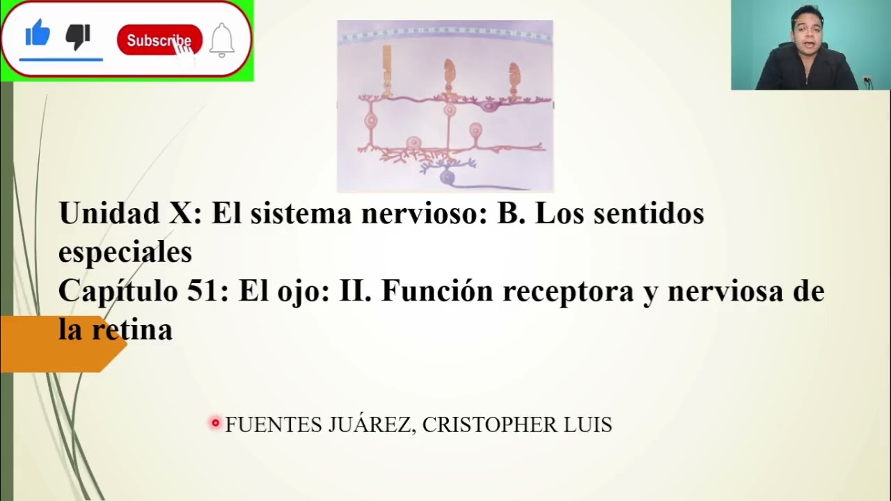






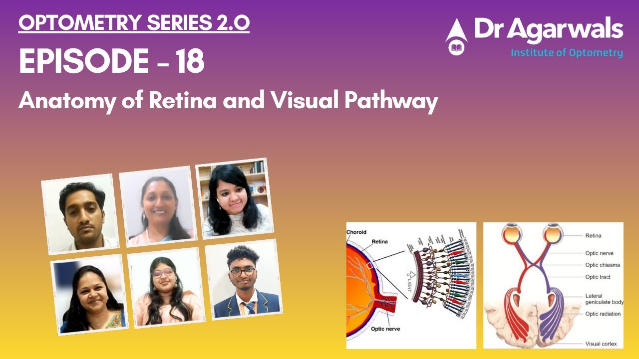

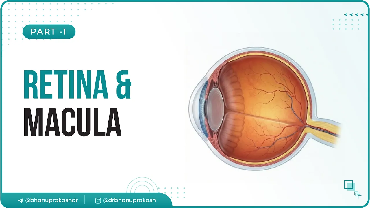











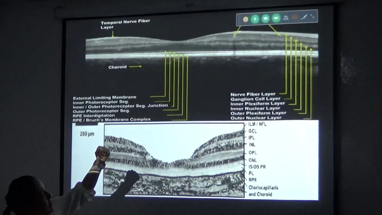

The retina consists of 10 distinct layers: pigment cell layer, outer segments of rods and cones, outer limiting membrane, outer nuclear layer, outer plexiform layer, inner nuclear layer, inner plexiform layer, ganglion cell layer, optic nerve fiber layer, and inner limiting membrane; rods and cones are first-order neurons that convert light into electrical impulses, which are transmitted through bipolar cells (first-order neurons) to ganglion cells (second-order neurons), whose axons form the optic nerve that carries visual information to the brain.
Retina Microscopic Structure: Histology Drawing Guide
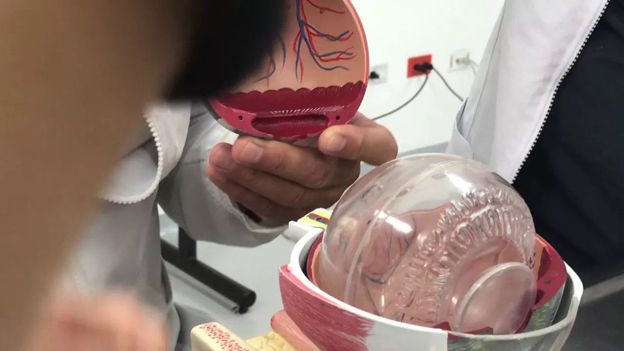
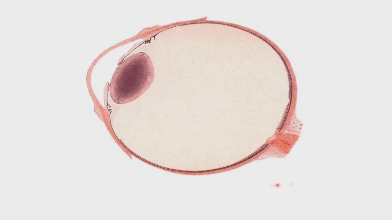
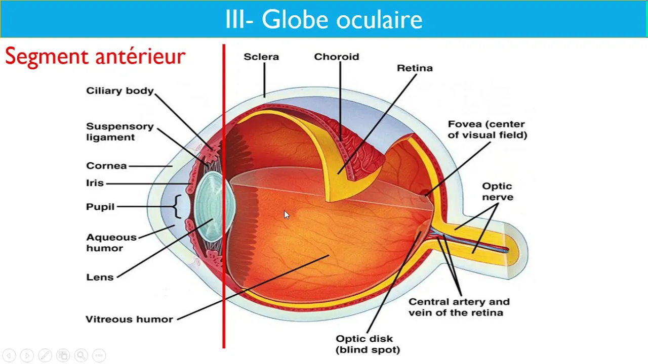
Added:Basic gross anatomy of the eye, specifically the three primary tunics (fibrous, vascular, and inner nervous tunic/retina).
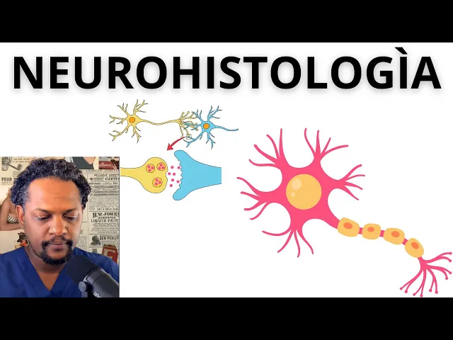
Fundamental concepts of neurohistology, including the structure of neurons (bipolar, ganglion) and glial cells (Müller cells).
The basic physiology of photoreception, specifically how rods and cones function as light receptors.
Standard histological staining techniques, particularly Hematoxylin and Eosin (H&E), to understand how cellular structures appear under a light microscope.
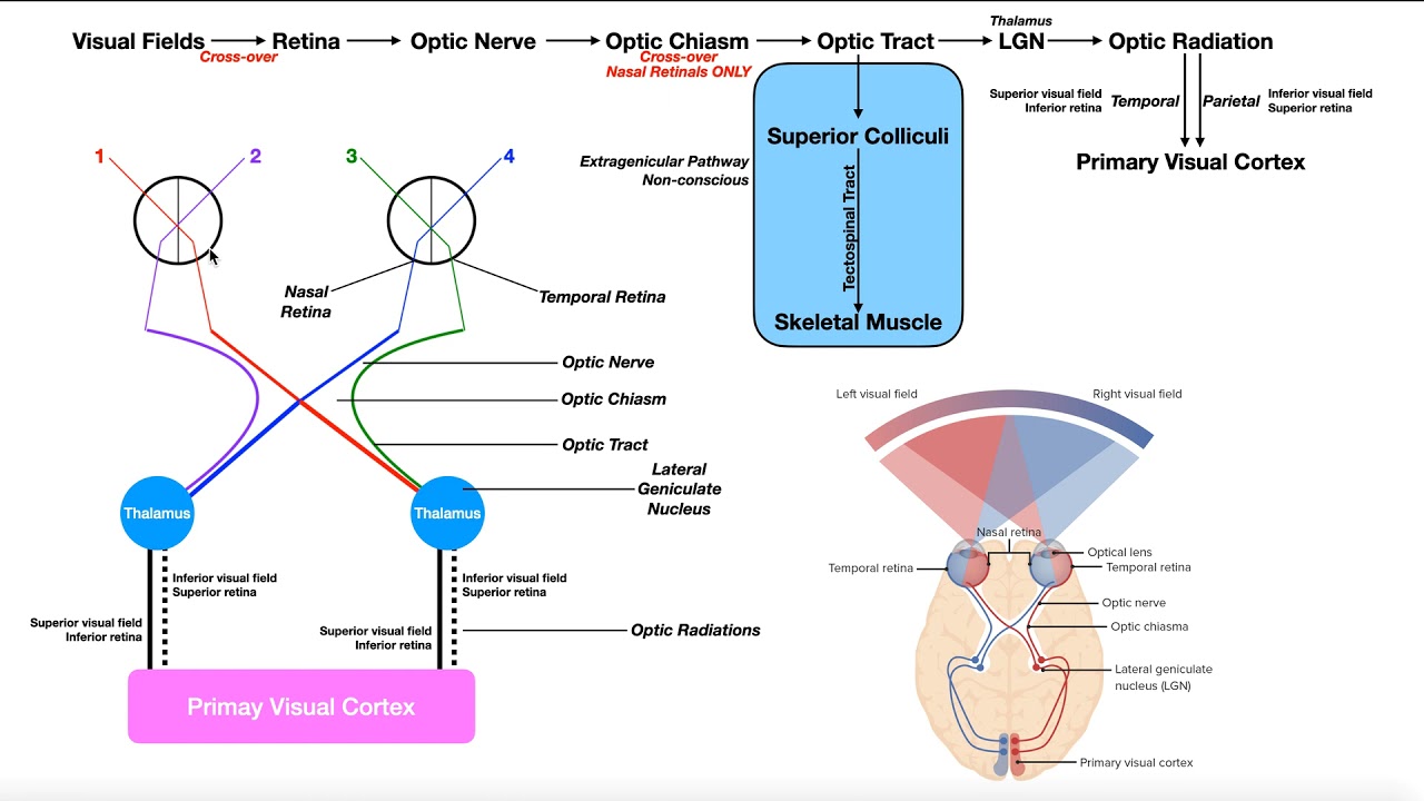
The complete visual pathway, tracing neural impulses from the optic nerve through the optic chiasm to the visual cortex.
Clinical pathology of retinal diseases, such as retinal detachment (focusing on the separation between the pigment epithelium and photoreceptor layer).
Histological variations in specialized regions of the retina, such as the fovea centralis and the optic disc.
Introduction to Optical Coherence Tomography (OCT), an advanced clinical imaging technique used to visualize these 10 layers in living patients.
212 views5likes12:18Original Release: 2025-07-25
Hello my dear students. I'm Dr. Vashali Vharbe and today I'm going to teach you how to draw the microscopic structure of retina. So let's begin by first drawing this rectangle. We going to divide this rectangle into eight parts. Now you must be wondering when retina has 10 layers why are we dividing it only into eight parts? Because two of the layers are actually just membranes. So we don't have to draw a separate section for them. Draw this division very lightly.
It should only act as a guidance for you. Okay. So let's begin by drawing the first layer of retina which is a layer of pigment cells. So draw them well.
Observe how the epical region of these cells show such microvilli like appearance. So draw those also.
The next structure we are drawing are the outer segments of rods and cones.
Outer segment of rod is thin and long.
That of cone is almost conical and triangular. So here observe the difference in their thickness.
So these are the outer segments of rods and cones. Now we have to draw the body.
So this is the body of the rod.
the body of the cone. Observe how their exonal process is going further deeper into the retina. We now focus on creating the exonal region of the rod and cone individually.
There you have your rods and cones drawn. Let's now draw the next cell which is the horizontal cell. Okay, so that's the body of horizontal cell and these are its processes which are reaching out to the rods and cones synapsing with them.
The next cell we will draw are the spindle-shaped bipolar cells. Each is having a dendritic process directed towards the rods and cones and the exonal process directed further deeper into the retina. Let's now begin to draw the dendritic processes making them sinapse with our rods and cones. Observe how the horizontal cells are part of this sinapse.
Let's now draw the exonal process of these. For that I need to draw my ganglion cell. It's a large cell and its processes are directed inwards and its exonal processes are directed further into the retina finally meeting together to form a bunch of exxons which form the optic nerve.
Now let's make the bipolar cells signapse with the ganglion cells.
Let's draw just one more ganglion cell to fill this space and let its axone go further inwards to participate in formation of the optic nerve. Now let's add one more cell which is the amocrine cell. See how the processes of amocrine cell are participating in the signapse between bipolar cells and the ganglion cells. So now we are going to add a large cell called the mueller cell which extends from the innermost portion of retina to the segments of rods and cones in this manner.
These are large cells which support all cells of the retina. On the inside these cells spread out further separating finally the retina from the vitrius body by forming the inner limiting membrane.
Let's now begin to color these cells to make us understand each cell properly.
So this is my pigment cell. The light green and the dark green are the rods and cones respectively. The blue are the bipolar cells. The pink are the horizontal cells. The orange are our emocrine cells. The yellow are my ganglion cells. And finally the peach color are the muer cells. You can color them whatever color you want because this is basically a diagrammatic representation of retina. Let's add the nuclei now to the pigment cell along with a lot of black melanin to make sure the pigment cells have ability to absorb excessive light that enters into your eyeball. Let's add the nuclei to the rods and cones to the horizontal cells to the bipolar cells. emocrine cells, ganglion cells and the muer cells.
There you have the diagrammatic representation of your retina. Let's quickly revise this. This is the pigment cell.
These are the rods and cones which act as receptors for vision. When they receive visual impulse, they convert it into a impulse that is passed on to the bipolar cells which are the first order neurons in the visual pathway. These then pass this information to the ganglion cells which are the second order neurons in the visual pathway which then transfer the information through their axons which form the optic nerve. Relaying this information finally into your visual cortex. Supporting all this activity are the horizontal cells, the emocrine cells and the muer cells.
Thus pigment cells form the first layer of retina. The outer segments of rods and cones form the layer of outer segments of rods and cones. The second layer. The innermost edge of your muer cell which is towards the pigment epithelium is called as the outer limiting membrane that is third layer of retina. The nuclear of rods and cones form the outer nuclear layer fourth layer of retina.
The sinapse between rods and cones bipolar and horizontal cells forms the outer plexififor layer the fifth layer of retina.
The nuclei of bipolar amocrine horizontal and muer cells form the inner nuclear layer which is the sixth layer of retina.
The sinapse between bipolar and ganglion cell forms the seventh layer of retina.
The inner plexififor layer.
The layer of ganglion cell forms the eighth layer. The optic nerve fiber layer forms the ninth layer. And finally the innermost membrane of the Mueller cell forms the inner limiting membrane which is the 10th layer of retina separating the retina from the vitrius body. So this was the diagrammatic representation of retina. Let's now draw the etchini diagram. Okay. So let's pick up your einy pencils and let's begin the diagram of retina.
Let's first draw the pigment cells.
Color them.
Draw their nuclei.
Add a good amount of pigment to them.
The next layer as we know will be the layer of outer segments of rods and cones. Observe how the outer segments of rods are thin whereas that of the cones is thick and almost triangular.
Make sure you add both the outer segments in this layer. Now let's draw the outer limiting membrane.
Let's now draw the layer of nuclei of rods and cones. Remember these nuclei are huge number and you have to draw a very densely packed network of nuclei here. Draw the background because every cell needs support.
This becomes your layer of nuclei of rods and cones or the outer nuclear layer. Now let's begin to draw the outer plexififor layer which is a sinapse between the exxons of rods and cones and the dendrites of bipolar cells and horizontal cells. So this is a fibrous layer. The next layer will be the layer of nuclei of bipolar emocrine horizontal and muler cells. But here make sure you draw fewer nuclei because the number of nuclei are way lesser than the number of nuclei found in the layer of rods and cones. This layer is the inner nuclear layer.
Color the nuclei.
Let's go to the next layer where the exons of the bipolar cells signapse with the dendrites of ganglion cells and imocrine cells. So again a fibrous layer. The next layer is the layer of ganglion cells. These are few cells large in size. Make sure your diagram reflects that.
Color the background.
Color the cells. Draw their nuclei. And we will now focus our attention on drawing the their exones which finally form the optic nerve layer. See how their exones are emerging out, bunching together to form the layer of optic nerve fibers.
Finally, let's draw the inner limiting membrane which is the innermost layer of the Mueller cell. This layer will separate the retina from the vitrius body. And there you have your diagram of retina. Let's understand it once more that this these are the pigment cells.
You can see the melanin pigment in them.
They form the first layer of retina.
This is the layer containing outer segments of rods and cones forming the second layer of retina.
This is the outer limiting membrane formed by the outermost layer of muer cell forming the third layer. These are the packed nuclear rods and cones forming the fourth layer.
This is the plexus of nerve fibers forming the fifth layer. These are nuclei of bipolar immocrine and horizontal and muer cells forming the sixth layer. This is again a plexus of nerve fibers. This time sinapses between bipolar and ganglion cells andrine cells forming the seventh layer. These are the ganglion cells forming the eighth layer.
These are the optic nerve fibers forming the ninth layer. And this is the inner limiting membrane forming the 10th layer. These are the 10 layers of retina.
The innermost layer separating the retina from the vitrius body. So the 10 layers one last time let's revise.
Pigment cell layer layer of outer segments of rods and cones.
Outer limiting membrane outer nuclear layer. Outer plexififor layer. Inner nuclear layer. inner plexififor layer layer of ganglion cells optic nerve layer and inner limiting membrane. Thus we complete very quickly the diagram of microscopic structure of retina. I hope you also understood the first order neuron and the second order neuron in the visual pathway. Thank you students and I'll see you in my next microscopy diagram video. Bye.
Up Next

51:36
Anatomy and Physiology of the Eye: Structure & Function
@anatomyandphysiologyvideo
167.1K views•2015-11-22

50:12
Bacterial Communication: Quorum Sensing and Biofilm Formation Explained
@JHUAAP
12.6K views•2011-06-28

5:35
Enteric Nervous System Explained: The Gut's Brain | Neurobiology Lecture
@alumniu6029
438 views•2018-09-12

7:08
Bacteriophages: Earth's Deadliest Killers and Future Antibiotics
@kurzgesagt
34.6M views•2018-05-13
Related Study Plans & Knowledge Roadmaps
Structured learning paths in Biology
biology
RNAi: Biogenesis, RISC & Gene Silencing
01Molecular Biology Foundations: DNA to Protein
02Introduction to RNA Interference (RNAi)
03Molecular Biogenesis of miRNA and siRNA
+ 2 more learning modules
10h5 modules
Explore Roadmap
biology
Mycorrhizae: Signaling, Biophysics & Ecology
01Foundations of Mycorrhizal Symbiosis
02Molecular Signaling and Root Colonization
03Biophysics and Bioenergetics of Nutrient Exchange
+ 1 more learning modules
11h4 modules
Explore Roadmap
biology
Circadian Clocks: TTFL, SCN & Periphery
01Foundations of Molecular Biology & Gene Expression
02Introduction to Chronobiology and Actograms
03The Core TTFL: Molecular Architecture of the Clock
+ 3 more learning modules
14h6 modules
Explore Roadmap
biology
Population Genetics: Math, Drift & Selection
01Genetic & Probability Foundations
02The Hardy-Weinberg Principle and Baseline Models
03Mathematical Modeling of Natural Selection
+ 3 more learning modules
18h6 modules
Explore Roadmap