






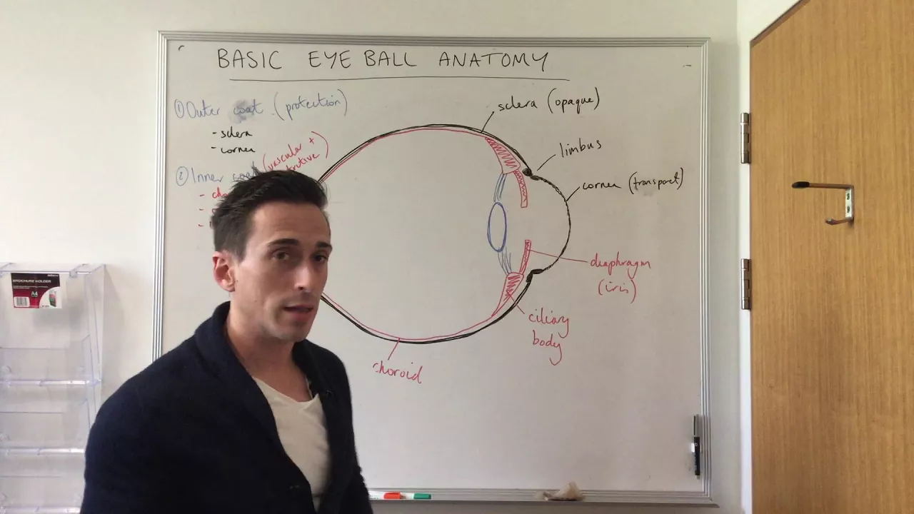









The human eye is a complex organ with three tunics: the outer fibrous tunic (sclera and cornea) that protects and refracts light, the middle vascular tunic (choroid, iris, and ciliary body) that provides blood supply and controls lens shape for accommodation, and the inner neural tunic (retina) containing photoreceptors (rods for low-light vision and cones for color vision) that convert light into neural signals. Visual information travels through the optic nerve to the occipital lobe, where it is processed. Common vision disorders include myopia (nearsightedness), hyperopia (farsightedness), cataracts (lens clouding), glaucoma (increased intraocular pressure), and color blindness, each resulting from structural or functional abnormalities in the eye's components.
Anatomy and Physiology of the Eye: Structure & Function
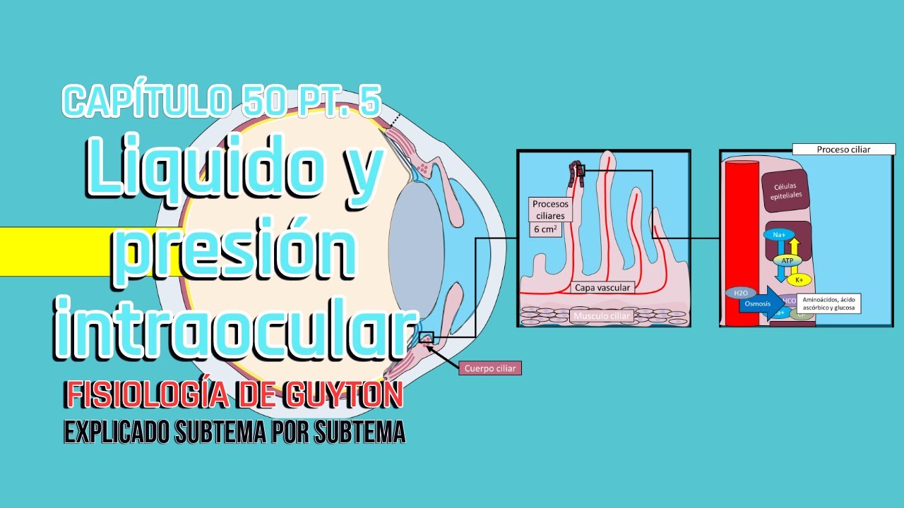
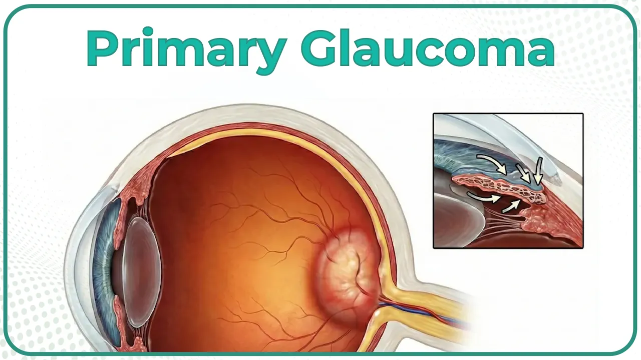
Added:hi welcome back to educator.com this is the lesson on Vision before we talk about the eyes themselves we need to look at at the accessory structures that are around the eye that have to do with the eye uh protecting them allowing them to function properly first the eyebrows uh these are one of those areas that we've retained a lot of thick terminal hairs from our Maman ancestors um of course they're really concentrated on the top of the head but if you ever wondered why do we have a lot of hair still right here part of the reason is just expression um being able to you know see someone's eyebrow movements with respect to um their their facial expressions is important for the socialization that humans have but beyond that in terms of protecting the eye when you sweat a lot above the brow the sweat will tend to get caught in these hairs and prevent a lot of it from coming into your eyes now if you sweat a lot you're not going to be able to prevent all of it but but that's a nice um function of eyebrows the eyelids the the technical term being the palpa um you know they're made up of skin muscles uh a very very very tiny bit of fat uh but you could think of those as like windshield wipers every time you blink they get a little bit of fluid over the surface of the eye helping to clean the eye lubricate the eye protect the eye and of course you know you have that automatic reaction where you blink if something comes to your face really quick uh so that's important in terms of protecting the surface of the eye eyelashes also protect the eye uh they prevent a lot of dirt microorganisms uh little bugs that we don't notice from coming into the eye itself um eyelash length is is mainly genetics unless you're um you know using some kind of product to uh accentuate them uh an interesting thing about eyelashes is the tarsal glands the tarsal glands are a modified kind of sebaceous gland now sebaceous glands are um associated with hairs in general but the modification of the tarso gland makes it so that there's even more of a lipid kind of fatty secretion that comes out with the eyelashes and uh not only um kind of protects them but it also uh makes it so that they don't Clump together it helps keep them separated so those tarsal glands associated with each little eyelash you have sometimes they can get infected sometimes a little bit of bacteria dirt can get into them uh they can get inflam fled and as a result of that inflammation you can get what's called a sty so if you ever heard somebody say that they had a sty on their eye it's because of the tarso glands getting inflamed uh skeletal muscles you have six extrinsic muscles attached to the outside of the eye on the next slide you're actually going to be able to uh to see those a little bit better you can't see them just by looking at someone's face but behind the eyelids uh attached to the white part of the eye you have all of these skeletal muscles and of course they allow you to move your eyes all all over the place the conjunctiva is a layer that's around the surface of the eye and it also curves up on the underside of the eyelids uh it's made up of a lot of epithelium that's meant to protect the eye it's just another form of protection and when it's healthy um when it's not infected you don't really notice it but when you get conjunctivitis the infection of the conjunctiva also called pink eye it's very noticeable and because of that infection you get dilation of all the blood vessels you get uh swelling in that area and it can be very irritating uh very annoying and that happens because of you know just could be um irritation of that layer of that tissue it could be a bacteria or virus that's making that happen and um generally it's something that's uh that's only going to last several days the lacrimal glands um if you remember from the skeletal lessons uh there's a lacrimal bone um and it comes from Latin word that has to do with tear like lria in Spanish um so the lacrimal glands they are those um tear producers the lacrimal gland itself is about the size and shape of an almond and um they are located in this region uh of the eye you're producing about a milliliter of Tears per day of course you can go beyond that if there's a lot of crying going on but um even when you you know Go the whole day without shedding a tear you're still producing tears they're just not exiting the eye um every time you blink you get a little bit of that fluid um going over the eyeball the nasal lacrimal duct is what leads those tears out of this area and through the lacrimal bone and part of the maxillary bone into the nasal uh con the nasal cavity you have this thing that's nicknamed aacal lake with each eye and it's kind of like uh a space for excess tears to hang out in if that lacrimal Lake gets overflowed with tears they're going to come out the eye so the act of crying you're producing so much of those tears that that Lake overflows your nasal lacrimal duct isn't able to to drain all of it out in those moments so they're going to come out the eye down the cheek Etc um the duct since it takes tears down into the nasal cone area that's why when when you are crying a lot uh you're going to get the you know like like you're you're having the sniffles at the same time because you have a lot of Tears going into that region and besides all of these you've got orbital fat uh the orbit of the eye is formed by several bones that make that you know classic looking eye socket and cushioning the eye all around the exterior attached to the white part known as the Scara you have a lot of fat and and it's meant to cushion it makes sense that you want that cushioning around the eye because you have some very hard bones that are right next to that eye so the orbital fat nice cushioning we're going to talk about the eye in terms of three tunics and we're going to start with the outer tunic this outer layer go to the middle and then to the inner so the outer fibrous tunic is the other nickname for it is incredibly fibrous it's made up of a lot of connective tissue fibers mainly collagen and eltin the white part of the eye is called the Scara very hard uh it varies in terms of the thickness and hardness depending on what part you're at if you go to the back part of the eye and this is a superior view of the right eye in the back next to where the optic nerve is uh it's actually going to be a bit harder uh it gets a little less hard when you get closer to the front or anterior part of the eye but in general the Scara is very tough uh there's a lot of science fiction movies or Tales of you know someone stepping on an eyeball and and immediately squashing as if it's like a balloon or something um it's not that soft yes with enough force on an eye you're going to collapse it but it is very tough and if you've ever dissected a cow eye or sheep eye you know that uh the Scara very tough uh you have attachment for all the muscles you can see all those extringent muscles most of them here some of the others you can't see cuz they're on the UND side the inferior side uh but besides this part of the outer tunic which the majority of which is Scara you have the cornea so the most anterior frontal portion the part that you put a contact lens on that's known as the cornea and it's made up of uh some squamous epithelium about five to seven layers thick and it is transparent at least it's supposed to be transparent and that's important because you don't want to disrupt the Rays of light that are coming through there that is the first layer that is the window of the eye in which light first passes through um so you don't want there to be anything getting in the way of light because then that would change your perception of what's outside of your head just beyond that cornea and it's actually uh hard to the touch it's it's pretty tough uh just posterior to it is a little cavity called the aquous humor and a lot of textbooks call that the anterior cavity because it's the the most frontal space just behind the cornea and there is fluid in there and that should also be clear if you look very close at someone's eye directly on the side of it you can see that little aquous humor just behind the cornea you produce fluid there and it's supposed to drain through what called the canal of schlem I I love that term schlem is spelled like this the canal of schlem uh actually drains excess fluid out of the aquous humor if that gets backed up and there's too much pressure it could actually lead to something called glaucoma which I'm going to tell you more about later uh but the interesting thing about the cornea and aquous humor is the cornea is one of those few places in the body other than the outer parts of the epidermis where there are no blood vessels directly adjacent to it so how do the cells of the cornea get nourished well it's it's through the aquous humor so you have little bits of gases and nutrients coming into that fluid cavity and since that's Direct adjacent to it that's what's nourishing the cornea um the cornea can get scratched it can get damaged with enough impact it it could be uh completely destroyed and actually one of the most common transplants done to human beings is corneal transplants and you can actually get corneas uh from cabers from dead bodies after death the majority of organs you need to take out of a Doner um while their heart is still being while the uh the the cells are still getting um oxygenated getting nourishment but one of those tissues you can take several hours after death is the corneas and so uh corneal transplants typically you get them from cadavers when we go deeper uh you have a lot going on in the middle tunic also called the vascular tunic vascular means having to do with blood transport getting blood into an area so there's a lot of circulation coming into the eye and it's thanks to the middle tunic so the choid I'm going to color this in red and it is red in a living eye if you dissect a cow eye or sheep eye the chid is going to look actually black uh because once you don't have all the oxygen and and actually you know living blood cells going into there it's no longer red it's very faint but this little layer you can see it is right in between the Scara which is here and and the retina which is actually part of that inner tunic so here's the choid in red and if you follow this all the way to the anterior portion you can see how it connects to these parts called the ciliary bodies and I'll cover those momentarily the chid is mainly uh for blood flow and it also has a lot of pigment there's also melanin in there and the purpose of that is when light enters the eye you don't want it to stick around you want light to enter the eye be noticed by the retina on the back and not be bouncing around you don't want to be uh seeing something for a second or two after you've seen it you want to get new light coming in and then going away and one of the ways that you can absorb light and get rid of it once it's been um first absorbed by the retina is by having a very dark interior that's going to absorb so having that pigment added to this Blood layer is able to like absorb light as it comes into to the uh eye and like I said it is it is providing nourishment to the parts of the eye that need it here's the iris the iris is made of smooth muscles actually two layers of smooth muscles and they're called smooth muscles not skeletal muscles because well they're not attached to your skeleton and you do not have conscious control over them they change based on how the light is outside of your face if there's a lot of light you don't need as much of the light coming into the eye so you're going to have the muscles get more relaxed and not be constricted and so that's going to actually make the iris bigger which makes the pupil smaller so the pupil is just a hole um I'm not going to draw on it because it's it's really the pupil is not uh something that contains matter it's not something that has substance it's just a hole and the hole is inside of the iris if you're wondering why the iris looks like this it's not round well this is a cross-section it's actually a transverse horizontal cross-section through the left eye and the reason why I know that is because of the angle of the optic nerve which we're going to hear more about later but the iris made of smooth muscles uh when there's not enough light you want to actually get the uh the iris expanded constrict some of those muscles to make the pupil bigger you make the hole bigger so the little bit of light that there is available you want as much of it as possible coming in uh and of course the the pupil size is connected to um the autonomic brain branch of the nervous system the sympathetic and Paras sympathic parasympathetic side um which you can review in some of those um nervous system lessons so the iris let's make it blue I have blue eyes smooth muscle unconscious control the pupil like I said that black dot it's just a hole and the reason why it looks black is because the inside of the eye is dark the lens I'm going to color in yellow now the lens should not actually be yellow uh it should be clear and the lens is very tough uh the lens is meant to last a lifetime uh it's very tough made of um crystalline fibers uh and if you actually cut it apart it almost looks like an onion like it has all these different layers of these crystalline proteins and it's meant to be clear uh it's supposed to be clear because like I said when light's coming through the cornea which which is clear through the aquous humor which is clear and through the lens which is clear that's good you don't want the light to be influenced or anything being added to it as it's coming in so you want a clear lens if your lens gets a buildup of um calcified compounds um other molecules it can make it so where it's cloudy and we're going to look at a picture that has to do with cataracts later cataract is the buildup of usually calcified products inside the lens and it makes your vision kind of Cloudy because you got this stuff in the way of the light coming through the eye and something that you can do to get rid of that is cataract surgery they take the lens out and they put a lens a new lens back in the lens is what changes in size to focus near or far so your ability to uh look at something very far away something very close up that has to do with the lens the ciliary bodies which have um little ciliary muscles in them have to do with changing the lens shape so attached to these ciliary bodies and and it's actually circular around the entire lens CU remember this is a cross-section you have suspensory ligaments uh let's do that in green so the suspensory ligaments right here and those are all along the exterior part of the lens as well if those suspensory ligaments pull on the lens it makes it flatter it actually makes it so that you're going to see far way when the suspensory ligaments kind of let go on the lens and makes it more bulging and that's what allows you to read something close look at something very close to your face uh so those are the basics of that middle or vascular tunic next up the neural tunic uh the most inner part of it and it's neural because all of this has to do with uh sensing the light that's coming in and getting those signals to the brain the most most important part of this would be the retina the retina is really what allows you to see now of course all those other parts I mentioned before very important but all those other parts could be working perfect nothing wrong with them but if the retina ain't working or the retina falls off the back of your eye it doesn't matter how well your occipital lobe is working the lens the cornea Etc the retina is kind of this drape curtain in the back of the eye that has all the photo receptors those cells that actually uh respond to those different wavelengths of light coming in um photo receptors that's the next Point these are specialized cells all along the retina you have about 130 million photo receptors per eye uh way more rods than cones and we'll discuss the differences between rods and cones in a little bit uh but those two kinds of photo receptors scattered all along the back of the eye and depending on what part of the re we're looking at the amount of rods and cones can vary the macula the macula lutetia is the full name of it is an area right back here and in the very center of this region is a point called the fobia the fobia centralis is the full name for it and the fobia is where you want light rays coming coming into the eye to be perfectly projected on you want those light rays converging to that point and if light rays are not coming on that point you're not going to see clearly so the reason why I am wearing glasses is because when I don't have my glasses on the light rays from from looking towards you they don't quite come on that point perfectly they actually come to a point converge in front of it when I put this lens in front of my eye it changes the defraction it changes the angle at which light rays come in and makes it so that they come right there and that's nice I like being able to see clearly uh so the fvia centralis is that part of the eye that you want light rays to be focused on and we'll talk more about vision and near sidess versus farsightedness in a bit so the macula or the fobia right there the optic disc is a part of the retina that's right on top of the optic nerve so notice that the optic nerve is is slightly off to the side so if we're looking down on the left eye which which we are in this case the right eye would actually have an optic nerve coming more um medial as well so we're going to look at this in a little bit how the optic nerves come kind of uh together and there's a little crisscrossing going on so with each eye there is this area right on top of the optic nerve where there are no photo receptors it's a blind spot now you don't notice the blind spot of the eyes when both eyes are open because if I consider uh light rays bouncing off of the object over there coming into my eye right on where my um optic dis would be on my right eye the reason why I don't notice that I I I don't notice it which which sounds contradictory but the the reason why I don't notice the blind spot is because my left eye is able to perceive what's there so my left eye kind of allows sort of a cover up of the blind spot of the right eye and vice versa now when you close one eye or the other eye you're able to notice that blind spot and we're going to do a test in a little bit so you can see that certain things might disappear when they're actually hitting just that spot of the retina so there are no photo receptors on that part but photo receptors all along the other parts of this uh green retina that I've colored in the vitus humor in this case they call the vitus gel is a gel-like sack that is taking up all of that space that's just deep to the retina and just deep to the lens uh it's supposed to be clear as well it's in the pathway of light rays so you want it to be clear but it's a a gel that's meant to give the eye some Integrity to give it um the right amount of pressure so that it's not collapsing uh and it's a nice medium for light rays to go through on their way to the retina uh if there's too much pressure in there it could result in something like glaucoma and if you've ever wondered um what are those little floaters that you sometimes see um you know you'll be looking at something and then it looks like there's almost like little white dot that's kind of drifting along and you blink and it's still there what is that thing well floaters can be little bits of cells that come off from parts of the eye tissues you know from parts of the inner tunic and they end up floating along in the vitus humor this this gel sack and eventually they go away eventually um you know you have white blood cells in your eye that eat up stuff that doesn't belong there uh but it's kind of funny those floaters so that's how they occur the optic nerve you have one with each eye all those different neurons Associated uh with the retina with those photo receptors come together they converge in the optic nerve and come out the back of the eye move medially and then end up going through the thalamus and eventually to the occipital loes at the back of the brain so here's that blind spot demonstration uh here's how you do it and you could switch you don't have to close your left eye you could close your right eye but if you close your left eye and stare at the star with your right eye make sure that the star is right in your center of vision so you don't want to have uh your right eye looking straight at this black dot because the black dot is what's going to be disappearing so make sure that your right eye is right in front of that star and start with your head close to the screen and with that left eye close gradually move your head back keeping your right eye looking at that star gradually slowly move your head back and then at a certain point that black dot will vanish now the reason why some people it doesn't work for them right away is sometimes they're moving their head too fast so they move their head so fast that they don't notice that disappearance because keep in mind once you move past just barely past the point where that black dot the light rays from it are are not hitting that blind spot anymore well then you can see it again so try this a few times if if it doesn't work but eventually um it should work for you if if after trying this 10 times it doesn't work U there's a lot of um optical illusion books out there um there's other things you can look up online but it's quite fascinating so when the light rays bouncing off this black disc hit your optic disc that's the blind spot but having both eyes open this demonstration would not work like I explained before so the lens function um the ility of the lens to change shape so that you can focus near versus far is called accommodation um some people's accommodations just aren't enough to cut it uh mine as I got older like once I got to high school My Lens wasn't cutting it and actually um My Lens was kind of since it's not doing enough to focus the light rays at the back of the eye uh an eye doctor would say that my eye is too long and if that doesn't make sense I'll explain something in a little a little bit to help with that but the reason why they say my eye is too long is cuz no matter how much my lens gets flat to look far far away it just can't get those light rays all the way back to my retina so if my eye was a little bit shorter then I would have Clear Vision and it's the opposite with being farsighted uh those people's eyes are in a sense uh Too Short uh for for what the lens is doing so as the lens gets more concave uh meaning the opposite of bulging so a concave um lens a by concave lens would look like this okay that's what what concave is now your lens can't literally flip itself like that but what this means is uh if this is convex getting more bulging the more flat it gets closer to this kind of inverse lens uh you can call that getting concave so getting concave means more flat and that's what happens to focus on images from far away the lens getting more convex as in this particular image right here is for images close by close to your face so for an image to appear clearly to the viewer the image must be precisely projected on the fobia which is right there and right there all right accommodation problems this is why people need glasses or contact lenses the term emat means uh proper Vision so if you have 2020 vision you don't you don't have an accommodation problem uh if you ever wondered about the 2020 thing and and why some people are 2015 or 2010 don't think that 2020 vision is perfect that's that's normal vision that's that's what you want to have that's expected here's how it works this is a good way to explain it um 2020 Vision means that uh somebody with normal vision from 20 ft away can read a sign read all the words on it properly and a person next to them also from 20 ft away can read that same image uh those same words perfect so 2015 means this that a person with normal vision actually has to be 15t away they have to move 5T closer to see that image but the person with the 2015 Vision they can still be 20 ft away and see it great see it perfectly Crystal Clear 2010 is even more exaggerated that person with normal vision uh great vision they have to be 10 feet away from from those words to read it and that other person with 2011 Vision they can be 20 feet away um so yeah you could think of 2015 or 2010 as better than perfect um uh but the the term perfect vision when we look at other species uh there are birds out there predatory birds that have Vision better than any human on earth so the term perfect vision is relative um the opposite is true uh with a relative of mine um I have a relative who had 2200 Vision 2200 Vision means that a person with normal vision could read a sign from 200 feet away uh but she had to be 20 feet away from it to read it uh after getting lasic surgery that was corrected and lasic surgery involved actually um lasering off parts of the cornea making the cornea more flat to kind of assist the lens with the lens's incapability to get even more flatter um so the lasx made it so that now she actually has close to 2020 Vision the only problem with that is now she needs reading glasses so when you make the cornea a little bit flatter to assist with seeing in the distance that ends up being a problem when you're trying to read something really close to your face but at least you have that farsightedness so back to this myopia this demonstration here shows how to to fix myopia that's being nearsighted so I am myopic I'm able to read things right in front of my face but without my glasses seeing things in the distance is quite a problem so like I said before uh with the previous slides that means that my lens just can't get flat enough to look at something far away and as a result the images are converging right here in front of or anterior to the fobia when you put a lens like this in front of your face a uh convex lens or sorry my bad concave lens uh the concave lens makes it so that uh you're assisting the lens with making those um light rays hit the fobia now you could kind of imagine how a lens inside of eyeglasses could be shaped like this but I've often heard the question well what about contact lenses when you put a contact lens on the surface of your cornea it's not shaped like that you couldn't even close your eyelid over that it would hurt that's true the the contact lens is shaped like this from the side but the way they get the lens to behave like this is by playing with the densities within the material inside the contact lens so if you change the densities uh with respect to the middle part versus the outer part that's going to to give it this property uh and the opposite is true of hyperopia hyperopia uh means that you are farsighted you actually see stuff in the distance quite well but you have problems with up close and that's not as common as myopia but hyperopia is the exact opposite uh imagine having the inability um to make your lens bulging enough to focus in on on the light rays coming from right here that makes it so that when you're hyperopic the light rays actually want to go past the retina which is not physically possible they're not going to be able to go past the retina but when you put a lens in front of somebody who's hyperopic a lens that actually happens to be uh convex then you're making it so that it hits right here and and the intensity of how convex or how concave it is has to do with how bad your vision is so somebody who's even more myopic than me like the relative I told you about maybe uh that stuff was converging even even closer to the lens um so yeah all right when we look at the red Rea all these photo receptors in the retina that are actually sensing the light and reacting to it and then sending the signal to the brain we have two main varieties rods and cones uh so the rods and cones have similar structures but very different roles here's how I remember it rods have to do with with light and dark but cones is color so imagine color with a c cones with a c c for color cones color rods is just the ability to see light versus dark in general so the rod rods these these modified neurons we call rods not very good at distinguishing a wide range of colors and when the lights dim you've probably noticed that when when it's darker in a room it is really hard to distinguish colors more so than when it's light out um you know looking at at objects in the dark you can tell that certain things are there but in terms of you know red versus orange very hard to distinguish or or blue versus green uh so the cones are meant to allow us to distinguish Ro G Biv red orange yellow green blue indigo violet the colors of the rainbow so this is in the visual Spectrum um there are three main kinds of cones um it's actually very similar to those old projection TVs if you've ever looked at those old projection TVs that have those three different colors uh projecting up you've got the red uh the blue and the green light and coincidentally uh there are red cones blue cones and green cones um and and actually the majority are most of the cones are are red uh but all three are there and um depending on what combinations of those are stimulated that actually gives you a sense of um all the different colors of the rainbow uh when we look at the retina they actually are no rods on the macula on on the fobia section it's just a bunch of cones concentrated there and so that's why um when we look at something straight on when we're focused on something um it's easy to see uh the detail uh the rich colors there because um cones are enabling you to see uh kind of that that clear um um very uh bright image whereas on the periphery where you have more rods you can tell things are there you can tell the colors are there uh but wherever we want to observe something's shape texture color we're looking straight on um when we look at the different rods and cones uh this is actually a rod the main difference structurally between a rod and a cone is actually this if we look at this part here you could see that it's kind of like a rod kind of like a pole the outer segment of a cone is shaped like this and you still would have these little lines these little discs which I'm going to talk about momentarily but imagine having another one of these right next door but instead of having the outer segment beong it's kind of triang angular but structurally other than that they're very similar bipolar cells is a kind of neuron that looks like this so here are dendrites axon cell body axon and here are the axon terminal so receiving end the sending out end uh bipolar cells are what are going to be um right next to the rods and cones so if you think about the retina as having a few different layers of cells the light first hits uh some other cells before actually entering the rods and cones portion uh so the photons stimulate bipolar cells which then uh send the signal uh to the rods and cones and depending on how the photons hit the rods and cones that's how you see all the different colors uh and different images the inner segment uh just a little bit more superficial it's like we're getting um farther back in the retina closer to uh the different tunics closer to the middle tunic closer to the outer tunic so you got a lot of mitochondria in here and then the area that's really doing most of the work in terms of actually giving you sight is the outer segment so in the outer segment are all these little discs these little discs just stacked on top of one another uh and these discs are made of uh a couple different parts we we tend to concentrate on the ropson so ropson is made up of two parts opsin is a protein and then there's a pigment called retinol and retinol is made of vitamin A or it's made from vitamin A so that's why they say like eat your carrots um carrots uh compared to other veggies and fruits have a lot of vitamin A and um it's not necessarily true to say like oh your vision's bad eat carrots and you won't e glasses no um eating carrots is allowing you to maintain your eye health uh if you get a deficiency in vitamin A it can lead to some problems over time um but yeah without vitam you're not going to be able to make that pigment retinol and opson is a protein depending on the structure of opson that determines how it's responding to Roy G Biv the the red versus The Violet versus the Green Etc so you're going to see different kinds of opsin within rhodopsin uh in the cones and the rods uh the pigment epithelium is uh just outside of that you're getting a little bit closer to um those optic tracks that are going to be taking the signal through the optic nerve U the pigment epithelium it does have pigment in it there's a little bit of melanin in those regions and there there's the ability in those cells to actually um eat up sort of swallow up uh the older discs so you can actually make new uh um discs these optic discs within uh the outer segments uh throughout your whole life now you can make these new uh discs but if you damage the cell as a whole if you destroy this cell um it's going bye-by that's why if you stare at the sun if you're looking at extremely bright lights that's that's way too much um light rays that's way too much um energy coming into the eye and it's going to fry these cells so yeah staring at the sun uh could definitely make you blind and I I have read some research suggesting that in the near future we will be able to uh regenerate retinas but as it stands now um once you lose your retina uh you're not getting one back but in the future I I'm confident we will be able to um make retinas uh from stem cells within your body when we look at the visual Pathways to the occipital lobe it's important to realize that we have stereoscopic Vision it's like having Vision in Stereo so if this is uh the visual field of my right eye this is the visual field of my left eye and they overlap and there's a few reasons why that's important number one if I close my left eye and I look around with my right eye I can see that my nose is blocking a lot of stuff over there the orbit of my eye is doing a lot to block what's what's over on the left side here but when both My Eyes Are Open I don't really notice my nose as much I I could see it's still there but um it's not as obvious to me because the parts that were being blocked by by my nose and part of the orbit of my eye uh when I had my right eye open when my left eye is also open I can see those parts clearly what was over here uh along with that having stereoscopic Vision or Vision in Stereo is good for depth perception so because the eyes on humans and primates in general are in the front we are able to see ever so slightly around the sides of objects and we can tell how far something is in front of our face unlike other animals that have eyes on the sides having eyes in the front as primates is important because if you consider our ancestors that were all arboreal all in the trees from millions of years ago swinging through the trees having depth perception is important so that you don't run into a tree or miss that branch and fall and and get eaten by something uh so it's it's important in terms of evolution and us being successful and even though now we're not in trees anymore any more as with most apes are not hanging out in trees having depth perception has a lot of importance um just think about driving a car I mean without death death perception there'd be a lot more accidents so stereoscopic Vision having Vision in Stereo uh you can see that the visual Fields do overlap here's a little depiction of those visual Fields overlapping when light rays come in uh here's the cornea the cornea um light rays I'm going to depict them in yellow the light that's coming from the right side of your face into the right eye is actually going to end up hitting the left side of the retina it's going to end up coming in on hitting the medial side of the retina and vice versa here now the funny thing about that is let's say we went back back back back into the head where these neurons actually end up crisscrossing and and we'll get to those terms in a sec but if for some reason there was uh trauma or an infection that harmed these inner parts let's say that this got completely severed where these Blue Tracks overlap think about how this is is laid out the blue parts are sensing light from the outer part so even though if this part of this little km this little um space Cas in here if that got damaged in the middle it would actually cut out everything around the outside of your visual field so it' be like just Blackness you wouldn't be able to see anything out here and you you'd have a much uh thinner or or less wide field of vision and you could see that there is some crisscrossing going on um the the red parts of the retina that are it's being depicted here is red uh don't think that this corresponds to to cones or something um but they they're depicting this part is red you can see that the Parts kind of stick on the outside the more lateral parts and the optic nerves contain these little tracks so you can see that this little black tube looking thing that's the nerve and as I mentioned before the optic nerves come out from the posterior part of the Scara of the uh the outer tunic and they come immediately they come in towards the center of your head and there's some crisscrossing going on before it ends up at the occipital loes which allow you to see so those are the optic nerves in green uh the optic Chasm and this is actually plural chasma um the the chasms right here that's where you have the little crisscrossing of some of those optic tracks so the optic tracks as you can see right here optic track the red and blue lines those are all the tracks of neurons within the optic uh nerves that are end up uh that end up going back to the occipital lobe so the occipital lobe is all of this here and you can see that that's the final destination for these tracks and remember if if you saw the uh the nervous system lessons uh the one on the brain specifically um there's the uh the visual cortex that allows you just to be able to see and then there's that Association part that allows you to make sense of what you're seeing but this is how the visual Pathways get from the eye to the occipital lobe when we look at uh Vision disorders or conditions that affect the eye I already mentioned um myopia and hyperopia remember myopia is nearsightedness and hyperopia is farsightedness so I am my opic I've got this uh concave lens that's allowing me to actually see in the distance and it's vice versa with hyperopia cataracts that's what this picture is depicting here if you shine a light in this person's eye you can see that through the pupil it's quite cloudy back here because remember just posterior to the pupil to that opening is the lens the lens should be clear uh buildup of of calcified compounds is going to make it cloudy and cataract surgeries are very common especially in older people um you can have a cataract problem as a younger individual um that would be not not that common but an injury y could actually uh cause cataracts to occur um but yes scile cataracts in in more older people um are more are more common and like I said before they will typically remove the lens and put in a new one very simple surgery these days glaucoma is an increase in the um interocular pressure sorry intraocular is a better term um uh inside of the eye where the uh the vitous humor is if you have too much pressure in there uh it's going to be pressing on a lot of the delicate Parts within the eye that allow you to see so not only is it painful but it can make you blind uh one of the causes is I think I mentioned this earlier if the aquous humor in that anterior cavity gets um too too much you have too much of that fluid production there it's not draining through the canal of schlim that buildup there can affect uh the pressure within the entire eye and uh glaucoma painful can result in blindness um glaucoma you've probably heard that um one of the treatments that you could use for it is um use of cannabis marijuana my good friend from college who's an eye doctor now has told me before that the amount of marijuana a person would have to smoke to help with glaucoma is absolutely ridiculous um so people who are given uh marijuana as a prescription for glaucoma there's actually so many uh medications out there that do a much better better job of treating that pressure within the eye and reducing that pressure than something like cannabis um speaking of which um glaucoma uh within the eye can also occur because of high blood pressure so somebody who has just high blood pressure in general um they have um high blood pressure because of genetic factors poor diet what have you that can actually affect the blood pressure of the eye as well uh so that's another source of glaucoma a stigmatism is something that affects your vision what's supposed to happen is the cornea is supposed to be a nice basically perfect uh Circle perfect sphere and then the lens should also be you know equal in terms of its shape around the outside it should be more like a baseball not like a football so if you imagine this ever so slightly not being a nice Circle not not an actual football shape but if the cornea or lens is slightly off in terms of its circular that can cause a stigmatism because the way that light is coming into the eye it's not going to have the refraction of light as it should be so some parts of light in your visual field the angle at which they come in the back of the eye is going to be a little bit off because of that misshapen so stigmatism can be corrected by um having the lenses that you wear adjust for that color blind uh it's more common in males and if you've taken Biology you might remember that because of um the sex chromosomes that's one of the ways that red green color blindness affects males more males have One X chromosome rather than two X chromosomes like a female does and that can affect um how the genotypes are inherited and being a carrier uh versus expressing it Etc so color blindness especially red green color blindness much more common in males um the inability to see color at all is extremely rare that's one in thousands and thousands of people um it's very rare for your cones not to be uh not not having the ability to distinguish between all the colors of the spectrum and if you've heard that dog see in black and white that's not true um dogs probably don't see quite as much color as us uh but think of it as more like um closer to kind of like U you know that sepia setting on a on a camera that kind of um gets to more Browns and like different Browns I'm not saying that dogs see exactly like that but um if dogs don't see Roy G Biv um they still have the different kinds of cones that allow them to see colors maybe it's not quite to the point that humans can see it uh speaking of which like I said before humans have three different uh cone varieties that allow us to see Roi Biv there are some primates out there that have four cone varieties so some of those uh monkeys could actually see more color than you and I can which is amazing when you think about it we we might be looking at two forms of purple and everyone agrees oh those are the same purple same purple but that monkey can actually distinguish between them which is pretty cool to think about uh night blindness one of the causes of it can actually be um uh vitamin A um remember vitamin A is used to make that that retinol that's inside of ropson which is inside of the rods and cones um it is recommended that you get vitamin A in your diet but somebody who doesn't get enough vitamin A in their diet you do have vitamin A reserves in your body but once those reserves run out you're going to notice that especially at night when there's less light coming into your eye it's going to be hard to see certain things especially while you're driving uh so night blindness one of the ways that you can actually um cure it or or fix it is get more vitamin A in that person's body so you can get those uh ropson molecules to what they should be how they should be functioning a scotoma is a blind spot that's not normal uh so if if other than the one that's on the optic disc someone else is having blind spots in their visual field even with both eyes are open um that could be due to an injury it could be due to um lack of nourishment um then there could be genetic factors affecting a scotoma coming into play retinitis Pigmentosa has to do with um uh a dying off of those pigment discs um in inside of the the ropson and that's something that uh could eventually um make rods and cones just not function at all um so this is something that could lead to uh blindness over time and a detached retina this is something you don't want to have happening a detached retina is usually from an injury uh some kind of injury to the Head makes it so that the retina comes off of the back of the eye and and that means you're not going to be able to see if that curtain against the back of the eye comes off that's where all your rods and cones are uh one of the ways that they can fix it if they if they do it soon enough after the accident is you can actually with a laser go in through the cornea and actually kind of solder on the sides of the retina on the back of the eye you can you can get it back on but the problem is when you laser on the sides of the retina you are damaging those parts of the visual f so that person's going to lose some of what the light that's coming in in terms of perceiving it but if it were me I'd rather have some Vision than no vision at all um detached retinas in other cases the retina comes off and it's not fixed in time and it's it's a permanent thing that that causes permanent blindness and like I mentioned before one day we'll be able to um you know remake a retina from stem cells if you've ever dissected a cow IR sheep eye uh you know this and if you haven't I suggest that you try the amazing thing is that when you cut into an eye and you look at the back where the retina is it kind of looks like wet tissue paper it looks very thin it looks very fragile and hundreds of years ago when they first were cutting into the eyes and looking at them and trying to figure out what's what they thought that the retina was just useless they kind of tossed it like Ah that's that's useless tissue because they looked at this structure and thought it's not important it's it's so tiny and so fragile but AR the retina is one of the most important tissues in the entire eye and without it you don't see thanks for watching educator.com
Up Next

Neural Coding in the Cat Visual Cortex: Orientation Detectors
@navegadorinclemente6897
21.4K views•2012-11-04

Bacterial Communication: Quorum Sensing and Biofilm Formation Explained
@JHUAAP
12.6K views•2011-06-28

Enteric Nervous System Explained: The Gut's Brain | Neurobiology Lecture
@alumniu6029
438 views•2018-09-12

Bacteriophages: Earth's Deadliest Killers and Future Antibiotics
@kurzgesagt
34.6M views•2018-05-13
Related Study Plans & Knowledge Roadmaps
Structured learning paths in Biology