












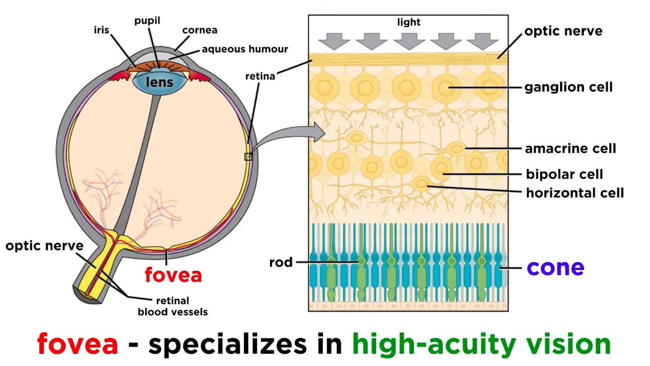
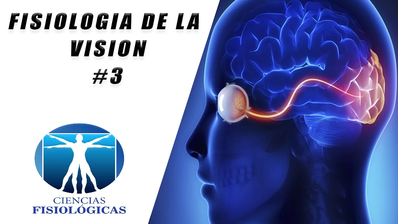
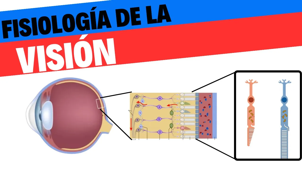






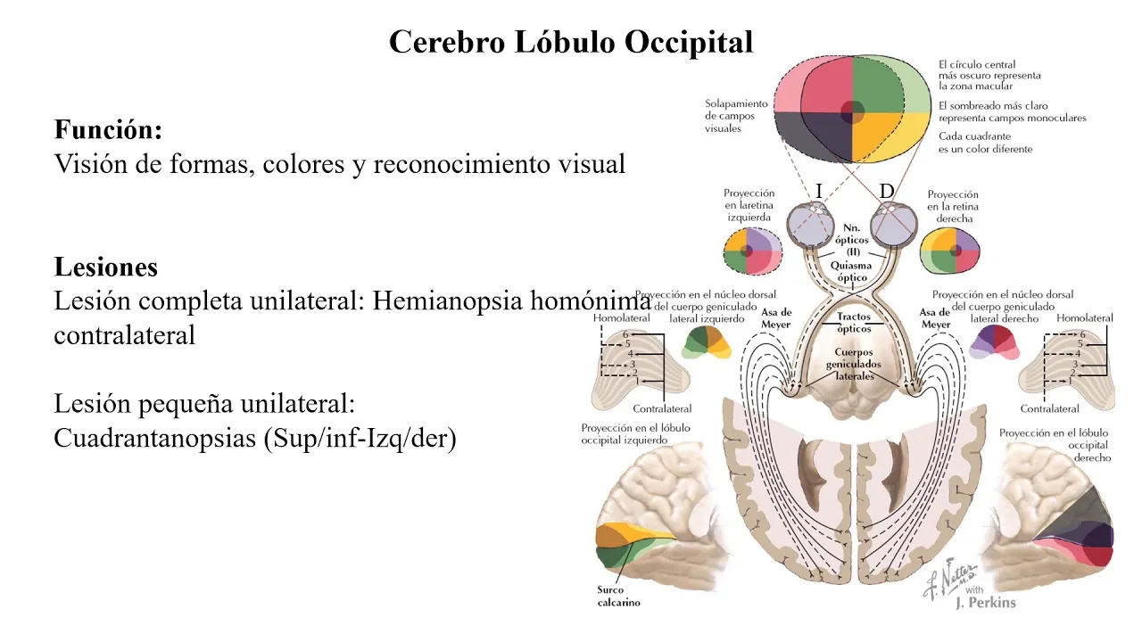



The visual cortex contains three types of orientation-selective neurons—simple cells that respond to flashed bars or edges with elongated receptive fields, complex cells that respond to moving bars and are often direction-selective, and hypercomplex cells that respond to bars of specific length and width—organized into columns where all neurons within a column share the same preferred orientation, forming a hierarchical processing system from retinal input through thalamus to cortical layers.
Neural Coding in the Cat Visual Cortex: Orientation Detectors
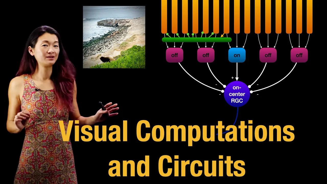
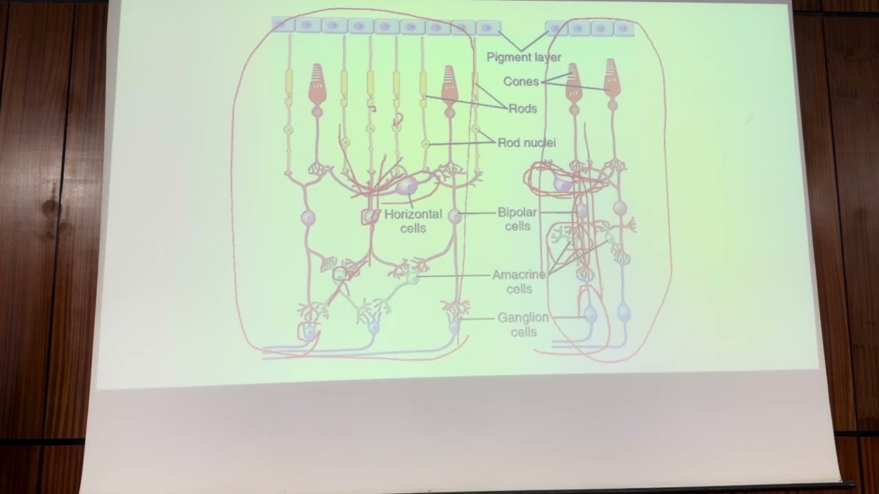
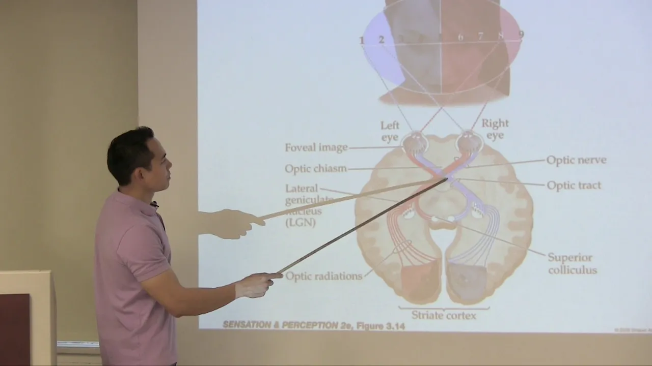
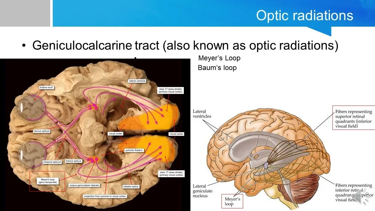
Added:Structure and pathway of the visual system, from the retina through the lateral geniculate nucleus (LGN) to the primary visual cortex (V1).
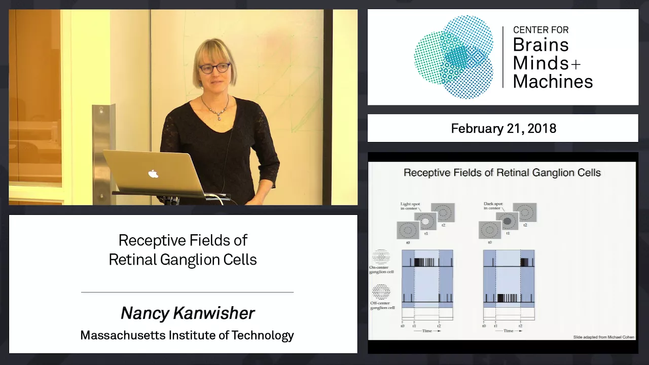
The concept of neuronal receptive fields, particularly the center-surround organization of retinal ganglion cells and LGN neurons.
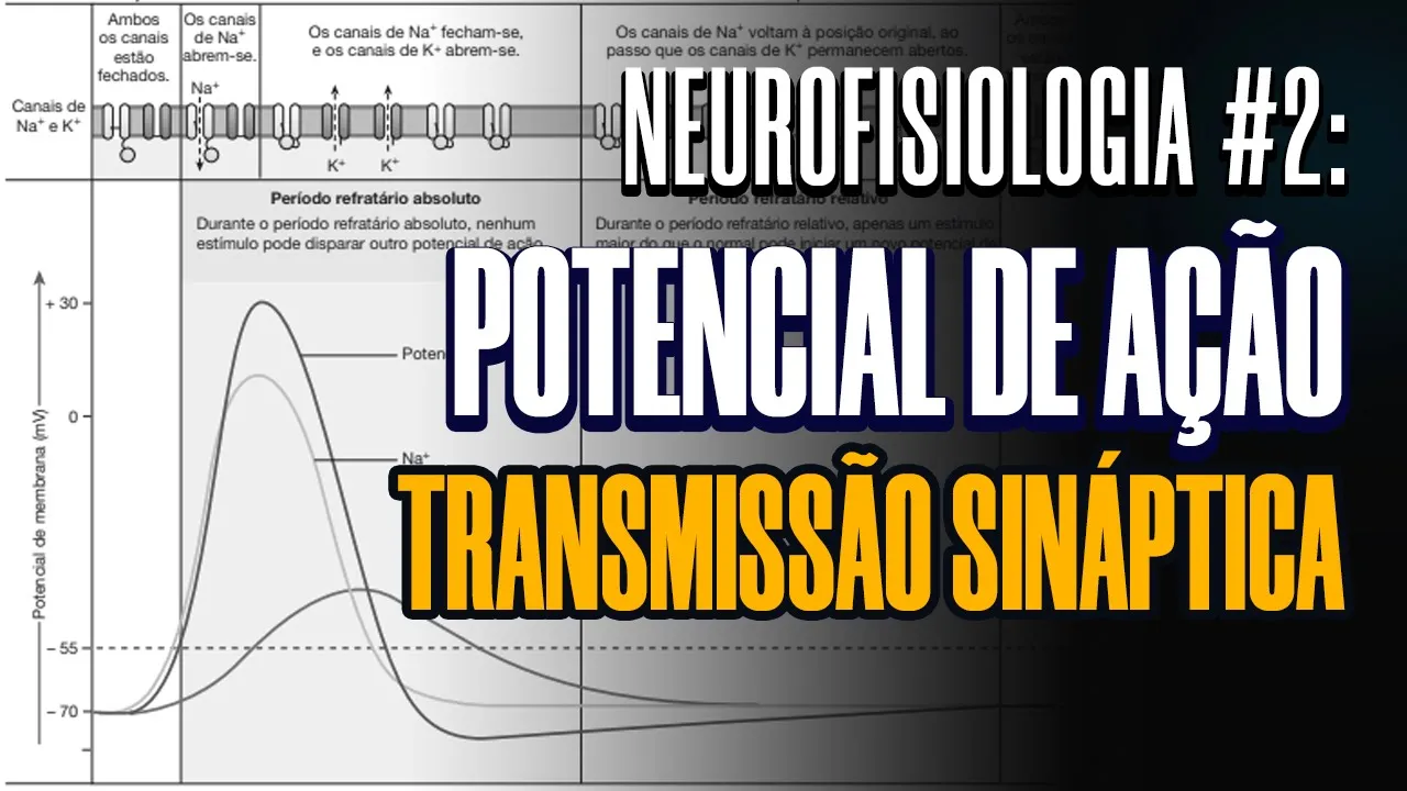
Basic neurophysiology principles, including action potentials, synaptic excitation/inhibition, and sensory transduction.
The pioneering methodology of Hubel and Wiesel's electrophysiological recordings in animal models.
Functional architecture of the primary visual cortex, including orientation columns, ocular dominance columns, and hypercolumns.
Higher-order visual processing areas (V2, V4, and IT cortex) and the divergence into the dorsal ('where') and ventral ('what') streams.
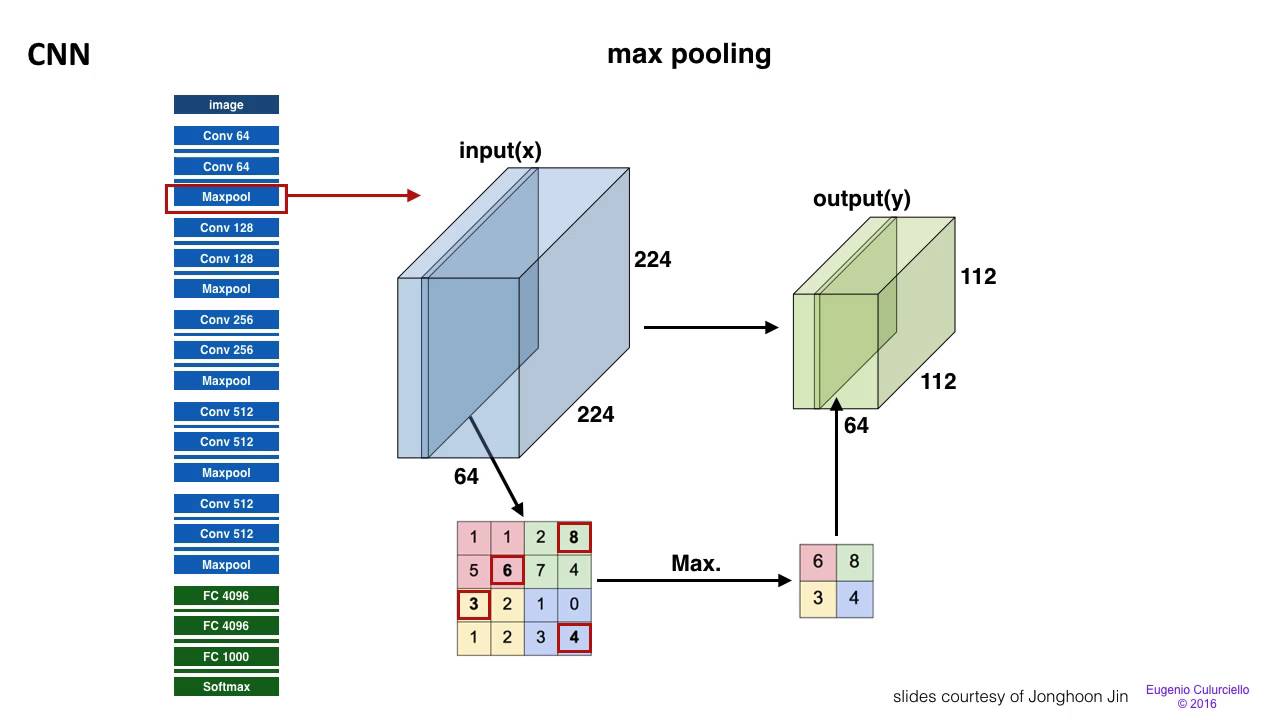
Computational models of vision, specifically how Convolutional Neural Networks (CNNs) emulate simple and complex cell hierarchies for feature extraction.
Visual deficits resulting from cortical damage, such as agnosia or cortical blindness, and cortical plasticity during critical development periods.
21.4K views212likes12:52Original Release: 2012-11-04
[Applause] [Music] nerve fibers from the retina contact cells in the lateral geniculate nucleus of the thalamus which in turn send their up to the visual cortex in the occipital lobe we you can pick up impulses from this area with a micro [Applause] electrode this anesthetized cat is fixed in a stereotaxic instrument and I'm advancing a micro electrode into the visual cortex of the cat's left hemisphere you can hear the background activity being picked up by the electrode the cat has a contact lens on each eye to protect the cornea and an extra spectacle lens in front to focus images from a screen onto the cat's retina the cat's left eye is covered [Applause] up as I move the electrode in I'm waving a pattern of lines in front of the cat's eye to help me find visually responsive NE neurons compared with retinal ganglion cells cortical cells have rather little spontaneous [Applause] activity what you heard was some action potentials growing out of the background noise as the electrode approached a cortical cell this neuron clearly responds to some thing on the patent card unlike retinal gangan cells and the cells of the lateral geniculate nucleus cortical neurons almost never respond to changes in general illumination off on you can hear that there's no response so now let's use patterns of light on a television screen in front of the cat to try to define the cell's receptive field this is a so-called simple cell it's thought that these neurons receive direct signals from a fairly small number of incoming fibers even simple cells don't respond vigorously for small flashing spots but they will always fire for a Long Bar of light now let's look at the response for a flashing bar in different parts of the receptive field on off in the middle of its receptive field this cell gives on responses it produces a burst every time the light is switched on WE indicate that on response with plus signs now let's try the areas flanking the onzone here it produces a burst of impulses when the light goes off we plot the off Zone with minus signs and on the other flank there is another area of off responses so this receptive field is rather like that of a gang Gan cell or a cell in the lateral geniculate nucleus except that the on and off areas are not round but are elongated now I'm going to try changing the orientation of the bar on off there's no response at all because the light falls simultaneously on excited Tre and inhibitory zones on off so this sort of cell will only respond if it's shown an edge or bar of the correct orientation these cortical cells are called orientation detectors the response is usually even better if the bar is moving first at the correct orientation now at slightly different angles and now perpendicular to the best orientation so this cell responds best to an almost vertical object other neurons prefer other orientations some horizontal some diagonal and so on let's search for another cell this one is a complex cell these neurons are thought to be much more complicated in their connections their receptive fields are usually larger than those of simple cells and they are often spontaneously active again this cell refuses to respond to overall changes in illumination and like many complex cells it does not even respond reliably to a flashing bar of light however complex cells always respond well to a moving Target of the correct orientation you can see that this one responds for a roughly horizontal [Applause] bar but only if it's moving up upwards not downwards so this particular cell like many others is Direction selective as well as being sensitive to the orientation of the [Applause] [Applause] target there is a third class of cortical cells we can find and because their properties are so complicated they're called hyper complex they again respond to a moving bar but not only must it be of the correct orientation it must also be of exactly the right width and length this hypercomplex cell responds to a diagonal bar moving downwards [Applause] but look what happens if I increase the length of the bar the response is inhibited if the pattern is too long and now let's try again with the optimum stimulus so so the receptive field must have strong inhibitory areas at each end that make it respond only to an edge or bar of a particular length sometimes there's an inhibitory Zone only at one end of the receptive field so the cell responds best to a 90° Corner this specificity for the length of the target is the defining characteristic of hypercomplex cells so cortical cells are orientation detectors simple cells respond to flashed bars or edges and have straightforward receptive Fields complex cells respond to moving bars and Hyper complex cells to bars of a particular length experiments with microelectrodes also tell us something about the microscopic structure of the brain they show that the visual cortex is organized into blocks or Columns of interconnected cells the incoming fibers enter the cortex at the base of each column and here in layer four are many simple cells complex and Hyper complex cells are usually found in the surrounding layers if a micro electrode is driven down perpendicular to the surface through a single column every neuron is found to respond to the same orientation if the micro electrode penetrates diagonally across many columns there are sudden changes in preferred orientation from the cells in one column to those in the next one The Columns are about half a millimeter across at the surface David hubel and toron Visa at Harvard who first described the properties of these orientation detectors have suggested that within each column the simple cells send fibers to complex cells which in turn transmit excitatory and inhibitory messages to hyper complex cells in this way all the neurons in a single column respond to the same orientation remember that only one eye was uncovered throughout this experiment although in fact most cortical cells receive similar signals from both eyes [Music] [Laughter] [Music] [Applause] [Music] [Music] [Applause] [Music]
Up Next

23:05
Grade 10 Science: Evidences of Evolution | Fossil Records & DNA
@DeahElmundo
277K views•2021-04-25

50:12
Bacterial Communication: Quorum Sensing and Biofilm Formation Explained
@JHUAAP
12.6K views•2011-06-28

5:35
Enteric Nervous System Explained: The Gut's Brain | Neurobiology Lecture
@alumniu6029
438 views•2018-09-12

7:08
Bacteriophages: Earth's Deadliest Killers and Future Antibiotics
@kurzgesagt
34.6M views•2018-05-13
Related Study Plans & Knowledge Roadmaps
Structured learning paths in Biology
biology
RNAi: Biogenesis, RISC & Gene Silencing
01Molecular Biology Foundations: DNA to Protein
02Introduction to RNA Interference (RNAi)
03Molecular Biogenesis of miRNA and siRNA
+ 2 more learning modules
10h5 modules
Explore Roadmap
biology
Mycorrhizae: Signaling, Biophysics & Ecology
01Foundations of Mycorrhizal Symbiosis
02Molecular Signaling and Root Colonization
03Biophysics and Bioenergetics of Nutrient Exchange
+ 1 more learning modules
11h4 modules
Explore Roadmap
biology
Circadian Clocks: TTFL, SCN & Periphery
01Foundations of Molecular Biology & Gene Expression
02Introduction to Chronobiology and Actograms
03The Core TTFL: Molecular Architecture of the Clock
+ 3 more learning modules
14h6 modules
Explore Roadmap
biology
Population Genetics: Math, Drift & Selection
01Genetic & Probability Foundations
02The Hardy-Weinberg Principle and Baseline Models
03Mathematical Modeling of Natural Selection
+ 3 more learning modules
18h6 modules
Explore Roadmap