







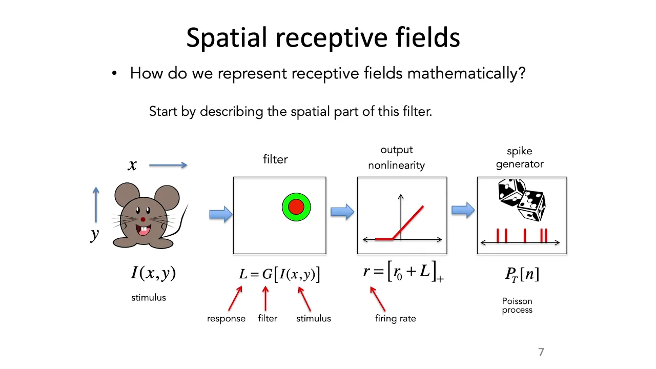


















Retinal ganglion cells (RGCs) possess receptive fields organized as concentric circles—a small center surrounded by a broader ring—falling into two categories: ON-center/OFF-surround and OFF-center/ON-surround. These patterns emerge from retinal circuitry where ON bipolar cells reverse cone signals (depolarized by light) while OFF bipolar cells preserve them (hyperpolarized by light). Horizontal cells provide lateral inhibition by receiving excitatory cone input and sending inhibitory feedback to surrounding cones. When light illuminates only the ON-center, the center cone releases minimal neurotransmitter due to hyperpolarization, while surround cones remain active, exciting horizontal cells that further inhibit the center cone, maximally exciting the ON bipolar cell. As light expands into the OFF-surround, surround cones hyperpolarize, reducing horizontal cell excitation and decreasing inhibition on the center cone, thereby diminishing RGC firing. Conversely, when light strikes only the OFF-surround, surround cones release minimal neurotransmitter, horizontal cell inhibition is minimal, the center cone depolarizes, and neurotransmitter release increases, leading to minimal excitation of the ON bipolar cell.
Center-Surround Receptive Fields of Retinal Ganglion Cells
Added:light hyperpolarizes photo receptors the signal propagates through bipolar cells to retinal gangan cells or rgc's which carry visual information from the retina to the brain rgc's have a receptive field that can be described as two concentric circles a small circular Center and a broader ring around the center called the surround these receptive Fields fall into two categories the off center on surround and the on center off surround in rgc's with an on Center offs surround receptive field when there is no light stimulus the rgc fires at a baseline rate when a light spot coincides with the on center the firing rate is at a maximum during the stimulus as the light circle expands to cover the off surround the firing pattern diminishes closer to Baseline firing when there is light only on the off surround and not on the on center then the Baseline firing is suppressed during the stimulus period the receptive fields of rgc's are established by signaling in Upstream retinal circuits there are two types of bipolar cells that signal to rgc's on and off off bipolar cells preserve the sign of the cone and are therefore hyperpolarized by light on bipolar cells reverse the sign of the cone and are deep polarized by light in addition to sending signals to the bipolar cells cones also send signals to horizontal cells which connect to surrounding cones horizontal cells receive excitatory input from the cones and send inhibitory feedback in return in this schematized on Center offs surround circuit a center cone is flanked by surround cones and connected to an on bipolar cell Downstream when only the on center is illuminated by light the center cone is hyperpolarized and therefore releases very little neurotransmitter however the surround cones are depolarized because they didn't receive light and therefore still release neurotransmitter the horizontal cell is excited by this neurotransmitter and continues to send high levels of inhibitory feedback to all the cones this inhibitory feedback causes the center cone to release even less neurotransmitter the on bipolar cell post synaptic to the center cone is maximally excited as it reverses the sign of the cone when the light expands into the inhibitory surround the surrounding cones become hyperpolarized and the horizontal cell is less excited inhibition to the center cone is reduced and the on bipolar cell is is consequently less excited compared to the previous state when Light reaches only the off surround and not the on center the surround cones release little neurotransmitter since they are more numerous than the center cone the horizontal cell is minimally excited and its inhibitory signals are low the center cone becomes depolarized due to the absence of light at the center of the receptive field as well as reduced inhibition from the horizontal cell its neurotransmitter release increases to high levels which leads to the least excitation of the on bipolar cell rgc's inherit this on Center off surround pattern from the on bipolar cell an off center on surround rgc simply receives input from an off bipolar cell that is connected to the center cone e
Up Next

Horizontal Cells and Retinal Ganglion Receptive Fields (Vision Series Part 3)
@harpinmartin
81K views•2011-08-01

Deciphering Neural Circuits Controlling Anorexia in Mice | Richard Palmiter
@AllenInstitute
2.3K views•2011-10-10

Vagus Nerve (CN X): Anatomy, Nuclei & Functions Explained
@Alilamedicalmedia
305.2K views•2022-10-31

How Exercise Benefits Your Brain: Science Explained
@TED
11.4M views•2018-03-21
Related Study Plans & Knowledge Roadmaps
Structured learning paths in Neuroscience