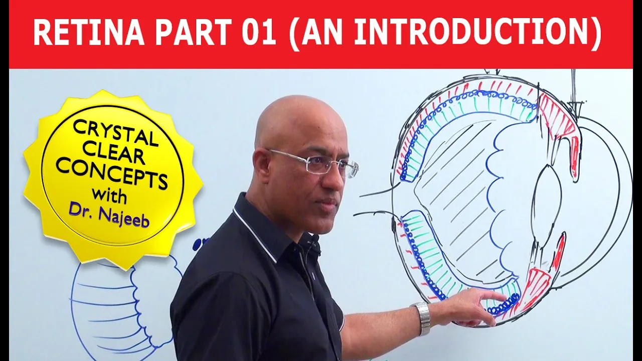






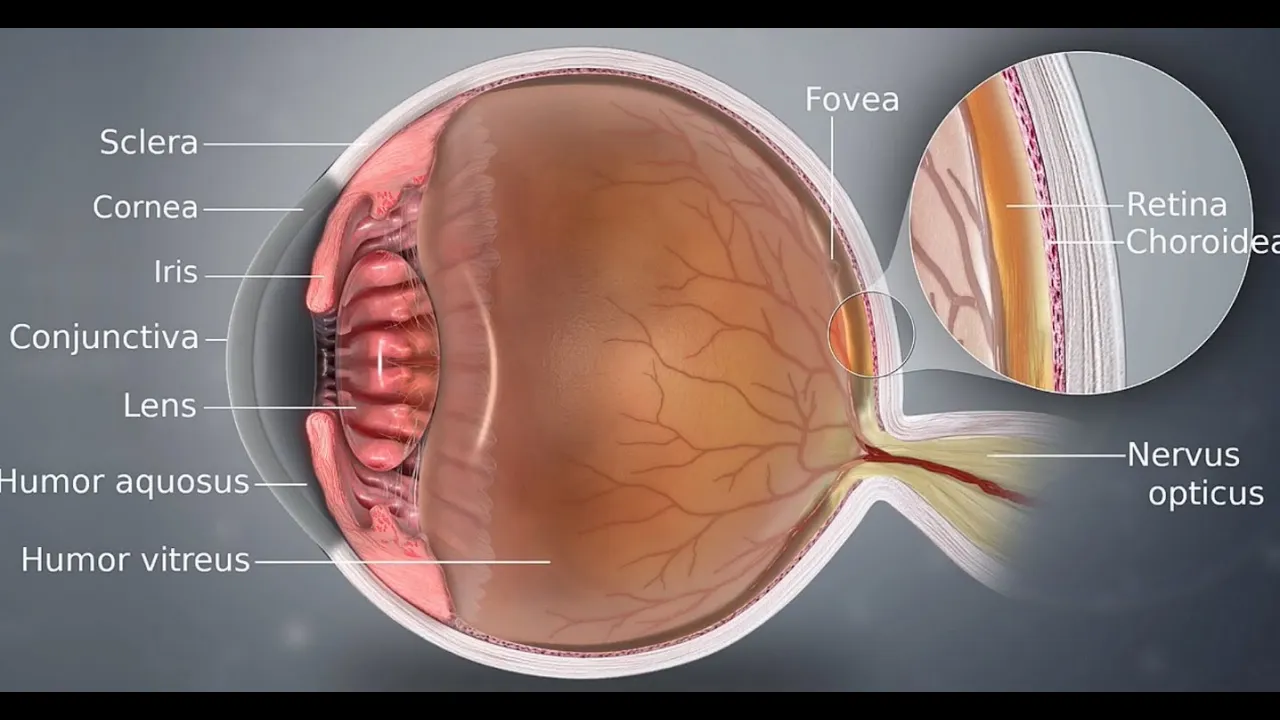








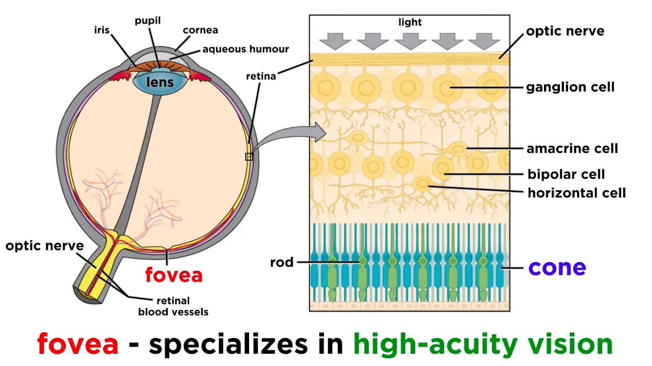
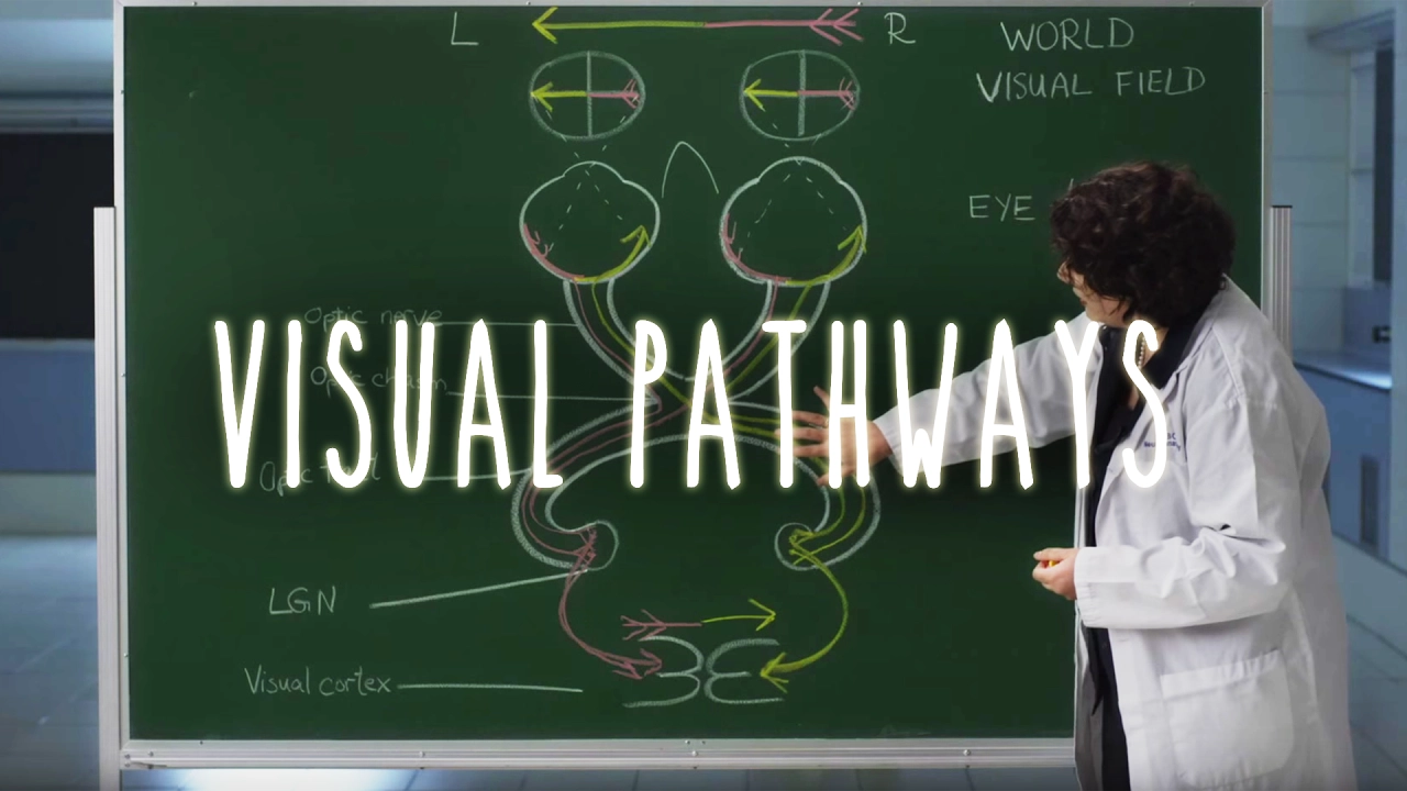
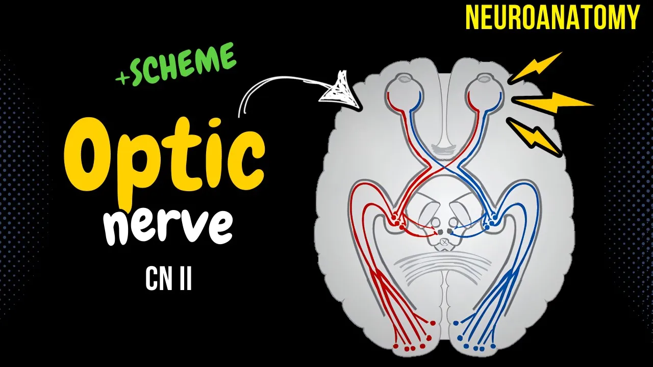
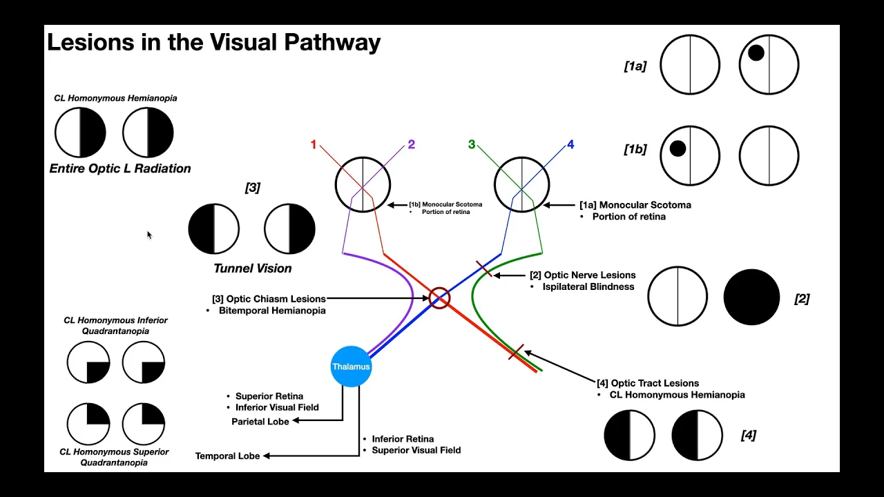
The visual processing pathway from the retina to the lateral geniculate nucleus (LGN) involves retinotopic organization, lateral inhibition, and two parallel processing streams. The retina projects to the LGN through the optic chiasm, with nasal fibers crossing to the opposite hemisphere and temporal fibers remaining ipsilateral. The LGN contains magnocellular layers (layers 1 and 2) responsive to movement and low-light conditions, and parvocellular layers (layers 3-6) responsive to color and fine detail. Receptive fields in both the retina and LGN exhibit center-surround antagonism, where neurons respond to light in their receptive field center while being inhibited by light in the surrounding area, enabling contrast enhancement and border detection. This lateral inhibition mechanism, first demonstrated in horseshoe crab photoreceptors, creates phenomena like Mach bands and enhances our ability to perceive edges and differences in light intensity.
Vision: Retina to LGN Visual Pathway Explained
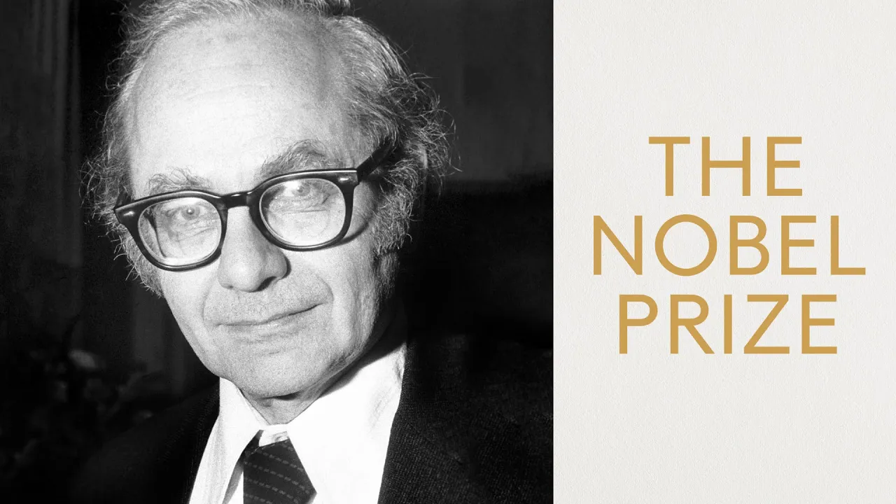
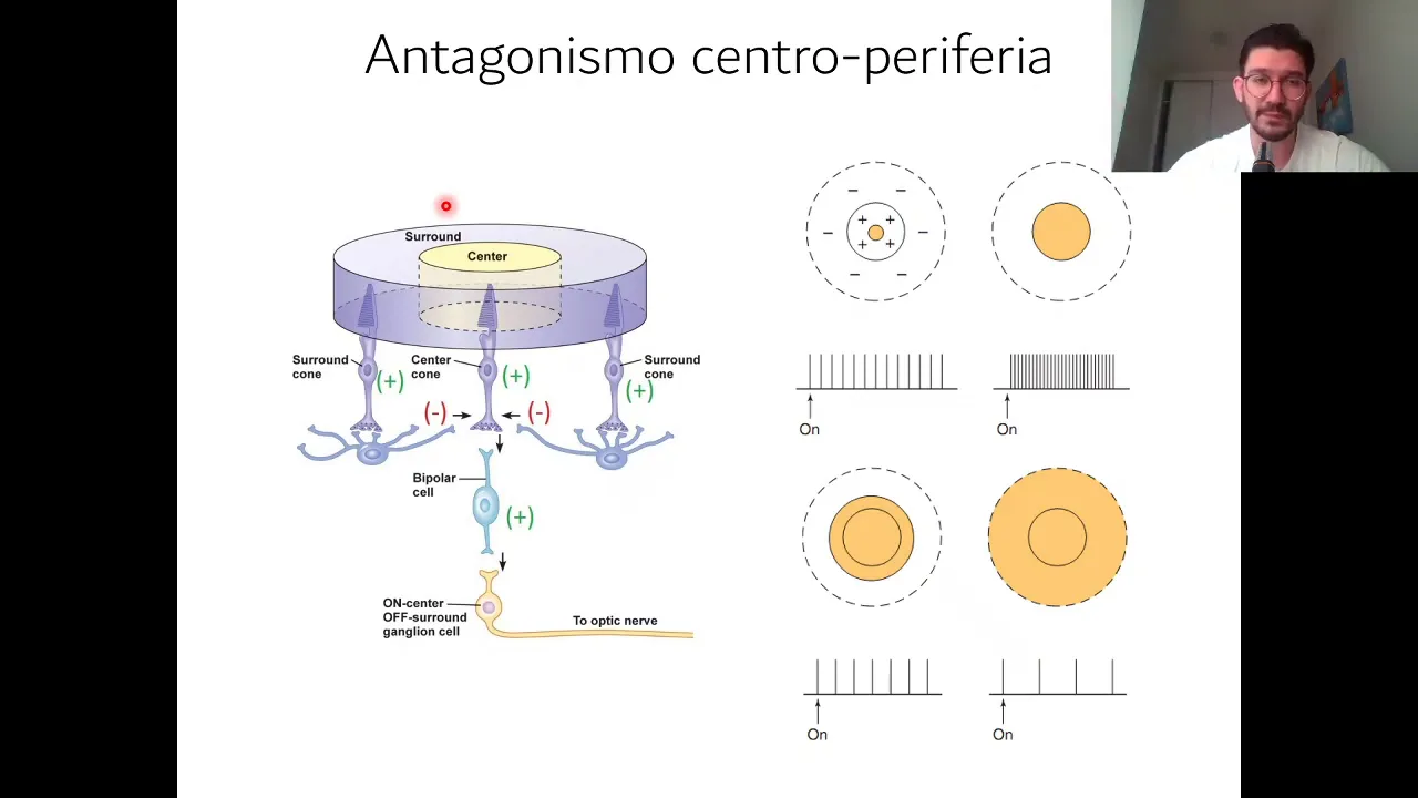
Added:hello and welcome to our fourth part of the series on a vision of visual processing be talking today about neural processing in the retina and the lateral geniculate nucleus this is the starting of our visual processing abilities and our ability to detect patterns and features in the environment so no processing starts very early on in the visual processing pathway and we'll talk about this early part of that processing and then in the fifth part we will talk about cortical processing and how the visual cortex processes some visual features so to start we're going to start with an overview of the retina geniculate striate pathway so that's the pathway that goes from the retina to the lateral geniculate nucleus to the striate cortex or the primary visual cortex in the occipital lobe we'll talk a little bit about how along this pathway this exhibits a retinotopic organization talk about the concept of lateral inhibition an important part of our ability to discern things like contrast we'll talk about the MMP layers of the lateral geniculate nucleus which are the magnocellular and parvocellular layers of the LGN and then finally we'll talk a little bit about receptive fields and center-surround antagonism so first off for an overview of this pathway you can see here that we have information projecting onto the retina the nasal part of the retina or the nasal hemorrhage as we call it these fibers travel over to the opposite hemisphere so these are contralateral so this is the right visual field they're traveling to the left hemisphere similarly the left visual field travels to the right hemisphere so here the left visual field and the right eye stays in the right hemisphere so these are itsel a t'rul fibers these cross over at an area called the optic chiasm so the object nerve exits the eye we have the optic chiasm now we have all the right visual field information in the left hemisphere and the left visual field in the right hemisphere so the nasal fibers then are contralateral the temporal fibers are EPSA lateral they then synapse at the lateral geniculate nucleus the ipsilateral fibers synapse at layers 2 3 & 5 the contralateral layers 1 4 & 6 as we discussed in earlier lectures the cortex has 6 layers as does the the LGN so we start up setting up those 6 cortical layers here in the synapses at the LGN so we start to get that layered processing that's going to become very important 2 columns and lamina in the primary visual cortex so then from the LGN they go on to layer 4 of the primary visual cortex and we'll talk more about that in our next segment so for now we'll talk about the retina lgn portion of this pathway at each level of the pathway information is organized like a map of the retina so essentially cells respond to information that are nearby those cells in the retina which are of course also representative of our visual field so our what we're looking at in the environment is actually mapped out in the LGN much like it is in the retina and similarly in the cortex important to know that the fovea is over-represented in this map because of course this is where all of our focal vision is this is where most of the cones are and this is of course where cones primarily have a one to one one cone per ganglion cell spatial summation so no spatial summation and so as a result that part of the retina is over-represented in this map which makes perfect perfect sense so this is a again at this idea of cortical magnification an important property of that starts at the retina and continued continues on in creating this some of our perceptual abilities is this idea of lateral inhibition so this is the capacity of an excited neuron to reduce the activity of its neighbors and so what we're going to do first is talk about how information from the horseshoe crab helped to create a lot of the knowledge we have in this lateral inhibition and horseshoe crabs are used to study this idea and so you can get an idea of how this function so this is the horseshoe crab the horseshoe crab has very large photoreceptors that each have a single axon and these are connected by a lateral neural network and they're easily accessible with their kind of compound eye so each of these is called a Noma titia and each of these photoreceptors then has a large single axon and so we can actually measure the activity in these axons from the horseshoe crab eye so if we have these ommatidia receptors they're connected by this lateral plexus system and a lot of a model we can think about in terms of how parts of our retina actually function so we have a single receptor fires that are rate proportional to the intensity of its stimulus so again we have an intense light here and a dim light here so these photoreceptors are firing at a much higher rate than the dim light photoreceptors but one of the things that happens is when this single photo receptor is activated it actually inhibits its neighbour so it sends out an inhibitory signal to its neighbors so within this intense light period while they're being activated by the intense light they're being inhibited by their neighbors now the dim light they're also being activated but less than the intense light but they're also sending out less inhibition so what this means is at the border between the intense light and the dim light there's a perceived intensity what happens is these neurons that are getting this intense light are spreading more lateral inhibition to this next neuron that has less activity and is getting less inhibition from its neighbor so it actually ends up with more inhibition than its dim light neighbors and so it actually appears to be dimmer at this border similarly this neuron is receiving less inhibition from this dim light neighbor and as a result this neuron fires at a higher rate as a result we get an increase in perception of intensity in the intense light side and a decrease in the perception of intensity on the dim light side so this is one of the things that highlights borders in our perceptual ability it also results in a number of important phenomenon so this is the neural basis of contrast enhancement is basically understood in terms of this idea of lateral inhibition so at a border between different intensities of Lights we actually get this sort of magnification of this intensity difference so we get a decrease in the neural firing on this side and an increase on this side which highlights this kind of border now to show you what this looks like in our visual system I'm going to show you a few examples of how this functions so these are what we call MOC bands so what you can see is when you look at the junction between these two different areas you see this differing intensity so for example it looks brighter and darker on each side of these mock bands you can see it looks brighter here here here it looks a little bit darker on the other side well again what's happening here is the brightness changes when we get these changes in intensity we get this change in lateral inhibition so this neurons are aspiring at a higher rate because it's receiving less lateral inhibition and these neurons are firing at a lower rate because they're receiving more lateral inhibition relative to their neighbors then again when we get to this next change we get an increase in firing rate because of reduction in lateral inhibition and then we get a reduction in firing rate due to an increase in lateral inhibition so we can see that here as well we get these kind of atmospheric effects even in when we look at landscapes you can see these kind of changes in intensity are highlighted by this idea of lateral inhibition other areas where we get illusions based on this lateral inhibition and we're talk about the Hermann grid and how Center trying to antagonism is part of this responsibility but when you look at any of these junctions you don't see a shadow but at the other junctions you do is actually a different version of that it makes my eyes a little crazy we're going to talk about how lateral inhibition and particular center surround antagonism has a role to play in creating these periods of reduce or these areas of reduced intensity and therefore creating this illusion of a shadow we'll talk more about this in our separate lecture on color vision but when we have two different colors that are right next to one another we get a change in color perception due to this idea of simultaneous contrast this happens with black and white as well and so this again has to do with this lateral inhibition based on its nearby perceptual asserts its nearby color so whatever colors it's presented along with actually has an effect on the color that we perceive and we'll talk more about that concept later but it is a part of this idea of lateral inhibition so this starts in the retina we then move on to the lateral geniculate nucleus now this is where we start to get a continued division of the visual system so remember in the retina we had the scope topic info topic systems now as we get into the LGN the top four layers of the LGM compromised the parvocellular system this is the system that's responsive to color find patterns or details stationary or slowly moving objects and of course its primary input comes from the photopic system which is the cones all of our detailed color vision is occurring primarily in the fovea and from the cones and so the parvocellular system then is receiving most of its input from those cones and so you can see this is actually a slice of the LGN looks a little bit like a fingerprint so these top three layers layers three four five and six are the parvocellular system again each layer at this point is monocular remember that coming from either the ipsilateral or contralateral retina so these are only coming from one eye these top four layers are the parvocellular system the bottom two layers are the magnocellular system these are layers one and two this is primarily responsive to movement and it receives its primary input from the schoo topic system so at this point we still have a monocular field or it's all layers I should the cells and layers Arman ocular so each cells on the responses responding to one I so we have no depth perception at this point again the parvocellular system is primarily getting its input from the photopic system so from the cones so this is color and detailed vision and the magnocellular system is then primarily receiving its input from the schoo topic system so it's primarily responsive to movement low levels of illumination and no fine detail we start moving into the cortex we'll start talking about how the parvocellular system primarily feeds into what's called the what pathway and then we'll talk about the magnocellular system feeding primarily into the where pathway but not entirely independent from one another but we see this sort of separating visual pathways from retina all the way up to court to cortex so one of the important components of understanding both the retina and the LGN are the receptive fields of specific ganglion cells and cells in the LGN Keeble and basal won the Nobel Prize for the discovery of receptive fields primarily the research was in cats but we do have good evidence of how this these receptive fields hold up in all species everybody's each species visual system is a little bit different but this idea of receptive fields was primarily discovered about Hebel and basil excuse me so a single neuron will respond only to light presented in its receptive field so the idea is that each neuron and the visual cortex particularly in the LGN on the retina will only respond to light that's presented within its receptive fields that's receiving input from cones in that area and then cones and rods in that area and so it responds only the light presented in its receptive field so we use the firing rate of a neuron to measure this idea neurons fire or have slow firing rates on their own when they're not being stimulated they when they're being inhibited their firing rate of course decreases when they're being excited their firing rate increases so we look at the firing rate of neurons in terms of what happens when we present light in their receptive fields so at this point we're talking about shining light into the retina and seeing what happens to neurons particularly either the optic nerve or the LGN and at this point we're really talking about the LGN so what people in Basel find is that at each level retinal ganglion lgn and the lower layer four of the permanent visual cortex the receptive fields in the fovea are smaller than in the periphery again not surprising they're a given that there's a great deal of spatial summation in the peripheral retina and there is more closely a one-to-one mapping of cones to ganglion cells in the fovea they also found that the receptive fields in this area are circular and monocular that is coming from a single eye and included both inhibitory and excitatory areas in a center surround field so essentially this looks like a doughnut and we're going to take a look at what these look like here in a moment it's called an annulus where you have a center area and the surrounding area just like a doughnut or a bagel but what happens is when you shine a light in one area you get inhibition and when you shine a light in the other area you get excitation so they're inhibitory and excitatory areas in this center surround so we can have on Center cells and off Center cells which also means on surround and off surround so here we have an on center cell so this is that annulus I was talking about and if a Lights presented in the center it will increase the firing rate of that neuron so for root present a light in the center of the receptive field of this neuron neuron could be in the LGN of the primary visual cortex it's going to increase the firing rate of that neuron now if we present a light only in the surround it's actually going to decrease the firing rate of that neuron so this is an off surround cell similarly there are off-center cells we present a light in the center of an off-center cell that will reduce the firing rate of a neuron when we present light into the on part of this receptive field we get an increase in firing rate from the neuron now why is this important well this is one of the ways in which we're able to discern contrast okay so if we shine a light over the entire receptive field of this neuron it's farm rate will not change because we've all be both exciting and inhibiting it at about the same rate but if we have a neuron right next to it and it has a differential firing rate then we know that that's where a stimulus sort of begins and ends so this is how we're able to do things like see text on a page is through this center-surround antagonism mechanism because it codes for where things start and stop in our visual world so this is basically what we're going to be looking at in our next lecture is how these center-surround antagonism cells line up to create things like perception of a line that is upright or a horizontal line or a vertical line etc and we'll talk about how these receptive fields are mapped or mapped out we'll talk a little bit more about the perceptual attributes of these this center-surround antagonism as well but this is similar to the idea of lateral inhibition where we're starting to see more complex patterns of responding and as we move up in the cortex we'll start to see more complex patterns of responding as well so this is a good place to pause and think about this idea of center-surround antagonism and how it affects processing of visual information by increasing or decreasing firing rates of neurons and then we'll take a look at how this works in the visual cortex in our next lecture
Up Next

Visual Cortex Layers: Simple, Complex & Hypercomplex Cells Explained
@Dr.Ericug
493 views•2025-07-20

Deciphering Neural Circuits Controlling Anorexia in Mice | Richard Palmiter
@AllenInstitute
2.3K views•2011-10-10

Vagus Nerve (CN X): Anatomy, Nuclei & Functions Explained
@Alilamedicalmedia
305.2K views•2022-10-31

How Exercise Benefits Your Brain: Science Explained
@TED
11.4M views•2018-03-21
Related Study Plans & Knowledge Roadmaps
Structured learning paths in Neuroscience