


![[Neuroanatomia] 6 - Diencéfalo I](https://i.ytimg.com/vi/nfCWTzwzPPw/hqdefault.jpg)





















![[Classic] ImageNet Classification with Deep Convolutional Neural Networks (Paper Explained)](https://i.ytimg.com/vi_webp/Nq3auVtvd9Q/maxresdefault.webp)
The primary visual cortex (Brodmann area 17) has six layers, with optic radiations terminating primarily in layer 4, which is divided into 4A, 4B, 4C alpha, and 4C beta subdivisions. M cells (motion-sensitive) terminate in 4C alpha, P cells (detail and color vision) terminate in 4C beta, and K cells (blue-yellow color vision) terminate in layers 1-2. The visual cortex contains three types of cells: simple cells detect lines and edges, complex cells determine line orientation, and hypercomplex cells (end-stopped cells) detect line length through excitatory and inhibitory signals. The dorsal stream processes motion and spatial information, while the ventral stream handles object perception and recognition.
Visual Cortex Layers: Simple, Complex & Hypercomplex Cells Explained
Added:Basic anatomy of the visual pathway, specifically the transition of signals from the retina through the optic nerve to the Lateral Geniculate Nucleus (LGN).
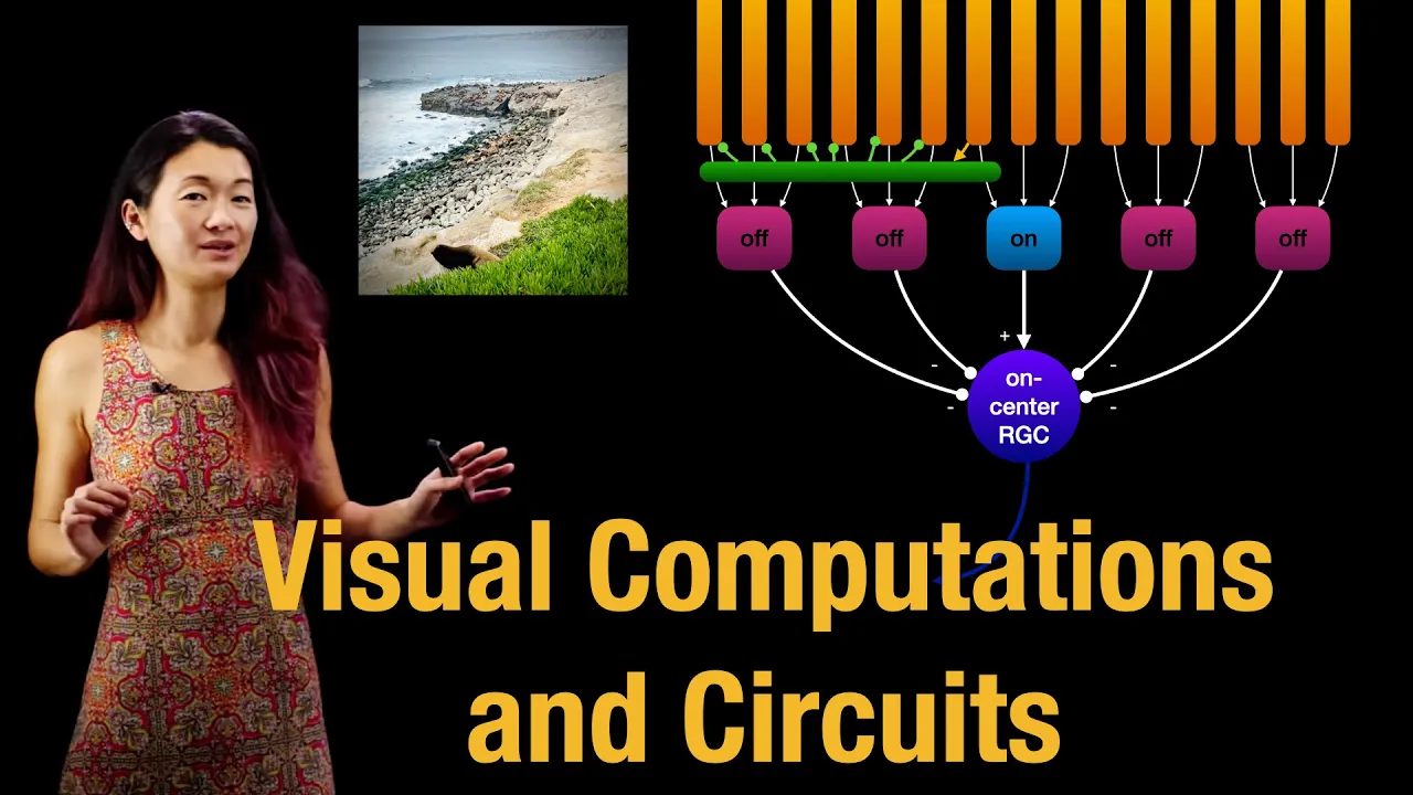
The concept of receptive fields and center-surround antagonism in retinal ganglion cells.
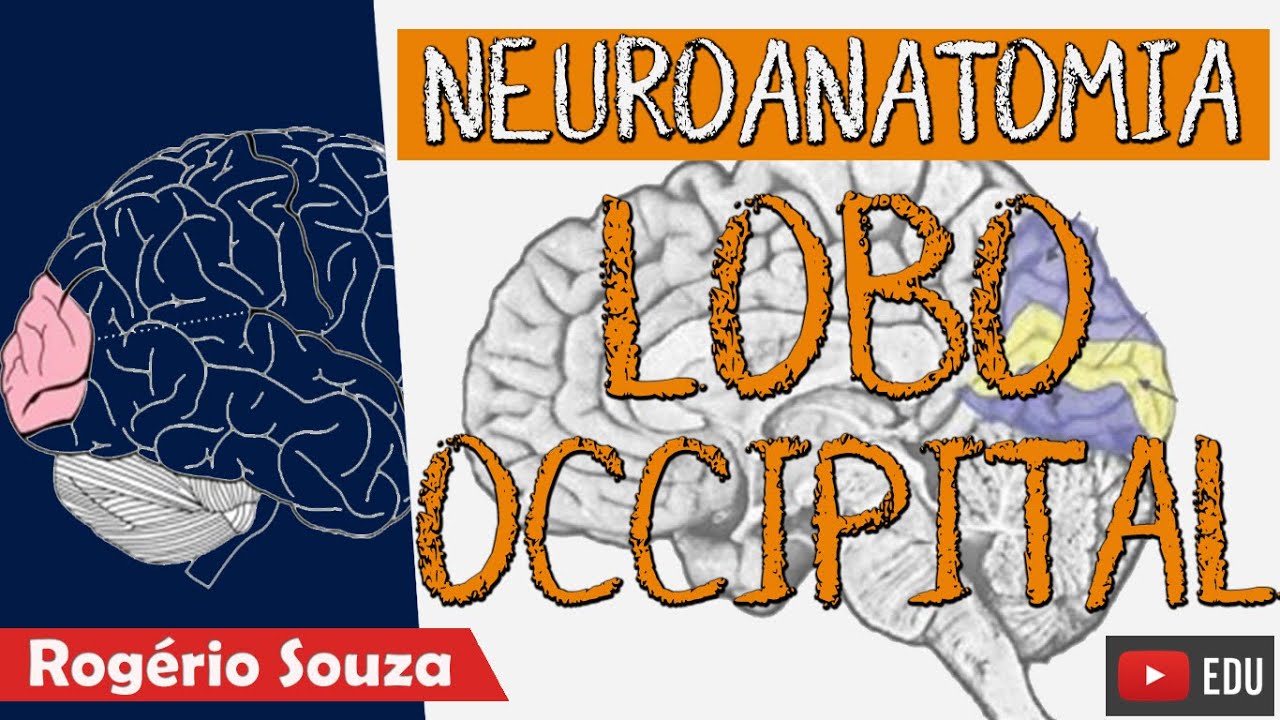
Fundamental neuroanatomy of the cerebral cortex, including the location and basic function of the occipital lobe (V1/striate cortex).
Basic principles of neuronal signaling, including action potentials, synaptic transmission, and excitatory/inhibitory post-synaptic potentials.
The Two-Stream Hypothesis of visual processing, exploring the dorsal ('where/how') and ventral ('what') pathways for spatial awareness and object identification.
Advanced processing in extra-striate visual areas (V2, V4, and Inferior Temporal cortex) and specialized regions like the Fusiform Face Area (FFA).
The clinical implications of visual cortex damage, such as cortical blindness, visual agnosia, and prosopagnosia.
How biological visual pathways inspire artificial intelligence, specifically the architecture of Convolutional Neural Networks (CNNs) in computer vision.
493 views9likes9:56Original Release: 2025-07-20
Let's look at the layers of the visual cortex now.
So layers of the visual cortex.
Layers of the visual cortex.
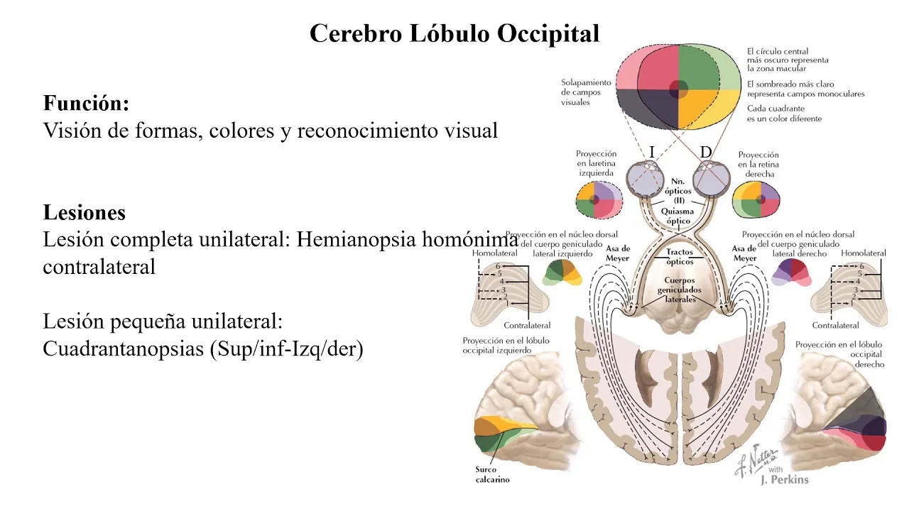
So when we looking at layers of the visual cortex uh you need to take note that from our previous discussion uh we say that we have uh optic radiations which come from the lateral nucle nucleus to the primary visual cortex which is found within the calcine fissure.
So this primary visual cortex don't forget that it can also be called the stride cortex can also be called the broadman area 17 get. So it has a total of six layers just like the lateral nucleus which we've just looked at.
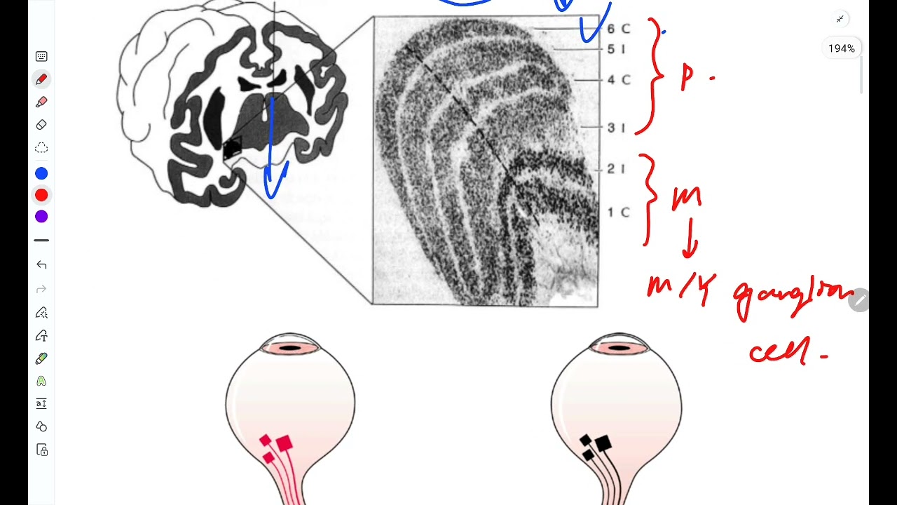
So it also has six layers. However, most of these optic radiations which we also call the genoalkkaline uh tracks will terminate in layer 4. So if you look at this where do we have our layer four? L layer four is here.
But uh you can easily tell that I've drawn drawn these uh layers. You can see that layer 4 is a bit unique in that for it has division which are in black. So I want to tell you that layer 4 has about four division. It's divided into 4 A, 4 B, 4 C and then this layer 4C is further divided into two that is alpha and beta you get. So these are the four divisions of layer four and you find that all the optic radiations will terminate into layer four. get. So it's it will be the same thing this side. So what you need is to follow. So uh we remember that we had P cells uh we had M cells and uh we had K cells.
So mainly we shall have these two.
So this side I'll put the M cells.
Then the other side I'll put the P cells. So where are they going to project or where where are they going to stop? So uh you need to see that or remember that all the M gloaline tracks will move up to layer 4 C alpha you get. So this is where they terminate the point. So all these uh tracks will terminate in layer 4 C alpha. You can draw it and draw it better in your book or or pate whatever you're using to follow. Then from layer 4 C alpha they can then spread into other layers you get. They can then spread into other layers by their termination is layer four C alpha. That's where you'll find their main nucleus of termination layers for C alpha. And remember we say that the M cells they have information concerning with motion black and white it's slow and it's mainly from the road cells. Then when you look at the P cells for them they will terminate in in two regions but mainly they will move into layer 4 C beta. get so they will move into layer 4 C beta.
So if this is our alpha and this is beta they will terminate mainly in layer 4 C beta get.
So they terminate mainly layer 4 C beta.
So from layer 4 C beta some of them will terminate in layer 4 A here. So we shall have another nucleus here 4 A then others 4C beta like that then of course from here we can have them spread into other layers. So how about uh the K cells? So the case cells these ones according to notebooks they will tell you that for them they terminate mainly layer one and two. However some of them can go to layer 4 a first then spread into project into layer one and two main point. So they project into layer one and two mainly. So that's where we have the K cells within the visual cortex. So that's the arrangement of the visual cortex. Talked about the four C alpha for N cells. Then 4C beta for P cells that is P cells concerned with fine detail and color vision which are slow conducting. M cells are rapid conducting. Then the K cells for blue yellow color vision.
Then uh other types of cells which we need to look at that are found within the visual cortex we need to cover them. We have simple cells, we have complex cells and we have hyper hyper complex cells hyper complex cells.
So how do these uh function? I'll just go straight to the point that simple cells if I'm to draw a picture let's say I'm going to draw a tree. I start with drawing maybe some lines like that we get I draw some lines. So that's the importance of these simple cells.
They're just for detecting uh lines and edges.
lines and edges.
Get that's for simple cells. Then the complex cells for them they care about the orientation of these lines. Let's say whether it's horizontal or vertical.
So these are for orientation of the lines.
orientation of the lines of the lines.
Then how about the hyper complex? Hyper complex for them they will determine the length of the particular line length of the particular of the line you get. So you'll find that they can also be called the ends stopped cells. Ends stopped cells. I'll explain why they called so endstopped cells. So why are they called the end stop cells? You find that if I'm drawing a line and my mind knows it that maybe this is four has to be 4 cm. So the moment I perceive that they are already 4 cm.
The hyper hyper complex cells will be excited by what? By the complex cells.
The complex cells will excite the hyper complex telling them that now you know what this is already 4 cm.
However, if the 4 cm are exceeded, you'll find that again we shall have inhibitory signals from the complex to the hyper complex. And this is what we call the end stopped uh our uh concepts in that if we exceed the given length we shall have them inhibited. That's where the in stopped name comes from. So those are the other cells that we have in the visual cortex and at this point I believe we have covered what we are supposed to know about the visual cortex. The other one which I'll talk about briefly is the secondary association cortex which I talked about earlier on. I said we have the the do stream which was going to the paral lobe and then we had the vententral stream which was going to the temporal lobe. We said that the do stream was mainly for motion and special processing.
Then the vententral stream is mainly for perception and recognition of objects.
You get so don't forget that. And uh with that said uh I believe we are done with visual cortex and we can proceed to the next part.
Up Next

3:41
Hypercolumn: Ocular Dominance & Orientation Columns
@FortuneFavorsPrep
18.8K views•2011-09-06

21:55
Deciphering Neural Circuits Controlling Anorexia in Mice | Richard Palmiter
@AllenInstitute
2.3K views•2011-10-10

4:10
Vagus Nerve (CN X): Anatomy, Nuclei & Functions Explained
@Alilamedicalmedia
305.2K views•2022-10-31

13:03
How Exercise Benefits Your Brain: Science Explained
@TED
11.4M views•2018-03-21
Related Study Plans & Knowledge Roadmaps
Structured learning paths in Neuroscience
neuroscience
Appetite Regulation: Brain Circuits & Hormones
01Foundations of Endocrinology and Brain Anatomy
02Introduction to Energy Homeostasis
03Peripheral Signals: Leptin, Ghrelin, and Insulin
+ 2 more learning modules
14h5 modules
Explore Roadmap
neuroscience
Neuroeconomics: Value, Risk & Brain Mechanisms
01Foundations of Neuroscience and Brain Anatomy
02Introduction to Neuroeconomics and Subjective Value
03Dopaminergic Prediction Errors & Reinforcement Learning
+ 3 more learning modules
18h6 modules
Explore Roadmap
neuroscience
Spatial Navigation: Cells, Grids & Brain Maps
01Foundations of Brain Anatomy and Memory
02Place Cells and Head-Direction Cells
03Grid Cells: The Brain's Coordinate System
+ 2 more learning modules
12h5 modules
Explore Roadmap
neuroscience
BCIs: Neural Decoding & Prosthetic Control
01Neuroscience Foundations: How the Brain Signals Intent
02Signal Acquisition: Recording the Brain's Activity
03Neural Decoding: Translating Brain Waves into Data
+ 2 more learning modules
25h5 modules
Explore Roadmap