











![[Anatomia] Orelha](https://i.ytimg.com/vi/k9K7qe4w6x0/hqdefault.jpg)



The middle ear contains three tiny bones called ossicles—the malleus (hammer), incus (anvil), and stapes (stirrup)—which work together to transmit and amplify sound vibrations from the eardrum to the cochlea. The malleus has a handle embedded in the tympanic membrane and articulates with the incus at the incudomalleolar joint. The incus has a lenticular process that articulates with the stapes at the incudostapedial joint. The stapes, shaped like a stirrup, sits on the oval window of the cochlea. This chain of bones amplifies the relatively weak vibrations from air into stronger pressure waves in the fluid-filled cochlea, enabling effective sound detection by hair cells.
Middle Ear Ossicles (Malleus, Incus, Stapes) Anatomy
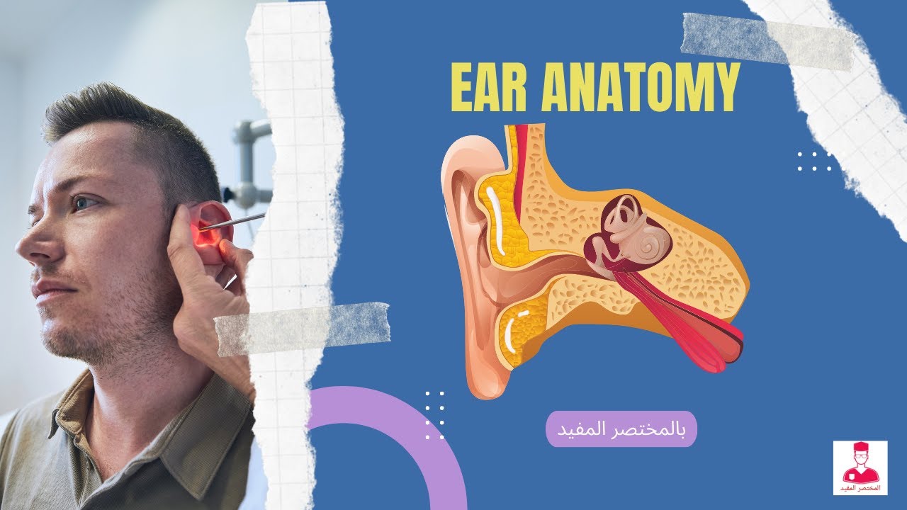
Added:Basic anatomical divisions of the ear (external, middle, and inner ear) and their general spatial relationships.
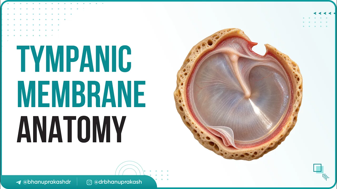
The anatomy and function of the tympanic membrane (eardrum) as the entry point to the middle ear.
Fundamentals of acoustics, specifically how sound waves travel through air as physical vibrations.
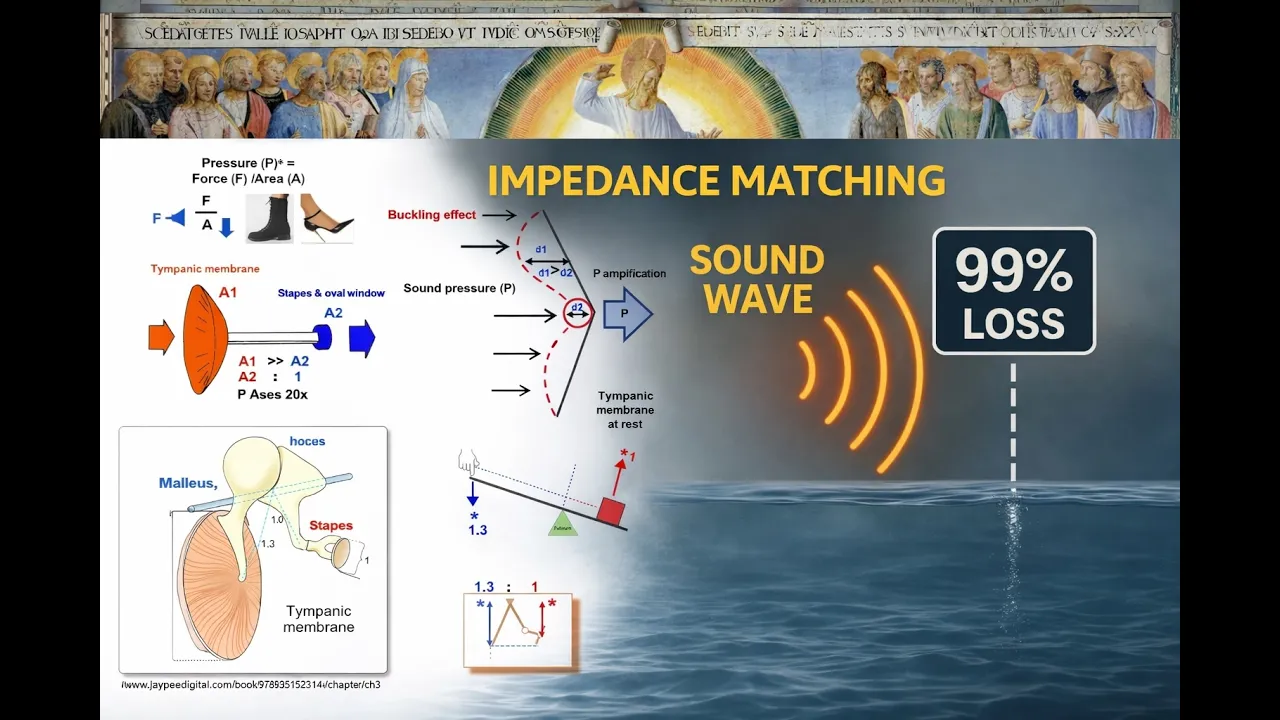
The physical mechanism of impedance matching, explaining how the ossicles amplify force to transfer sound energy from air to fluid.
Anatomy and physiology of the inner ear, specifically how the cochlea converts mechanical vibrations into electrical neural signals.
The acoustic reflex, including the protective roles of the tensor tympani and stapedius muscles against loud sounds.
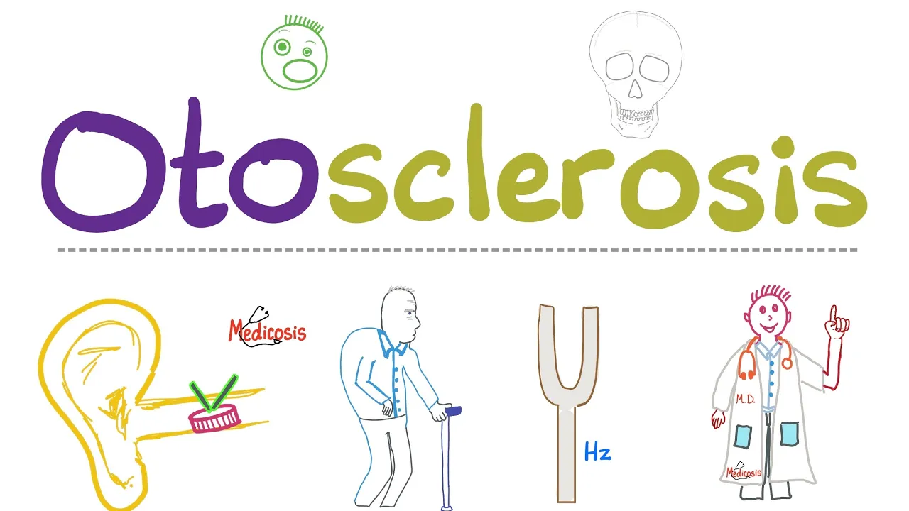
Clinical pathologies of the middle ear, such as otosclerosis, conductive hearing loss, and ossicular chain reconstruction.
66.5K views1.2Klikes9:26Original Release: 2018-10-30
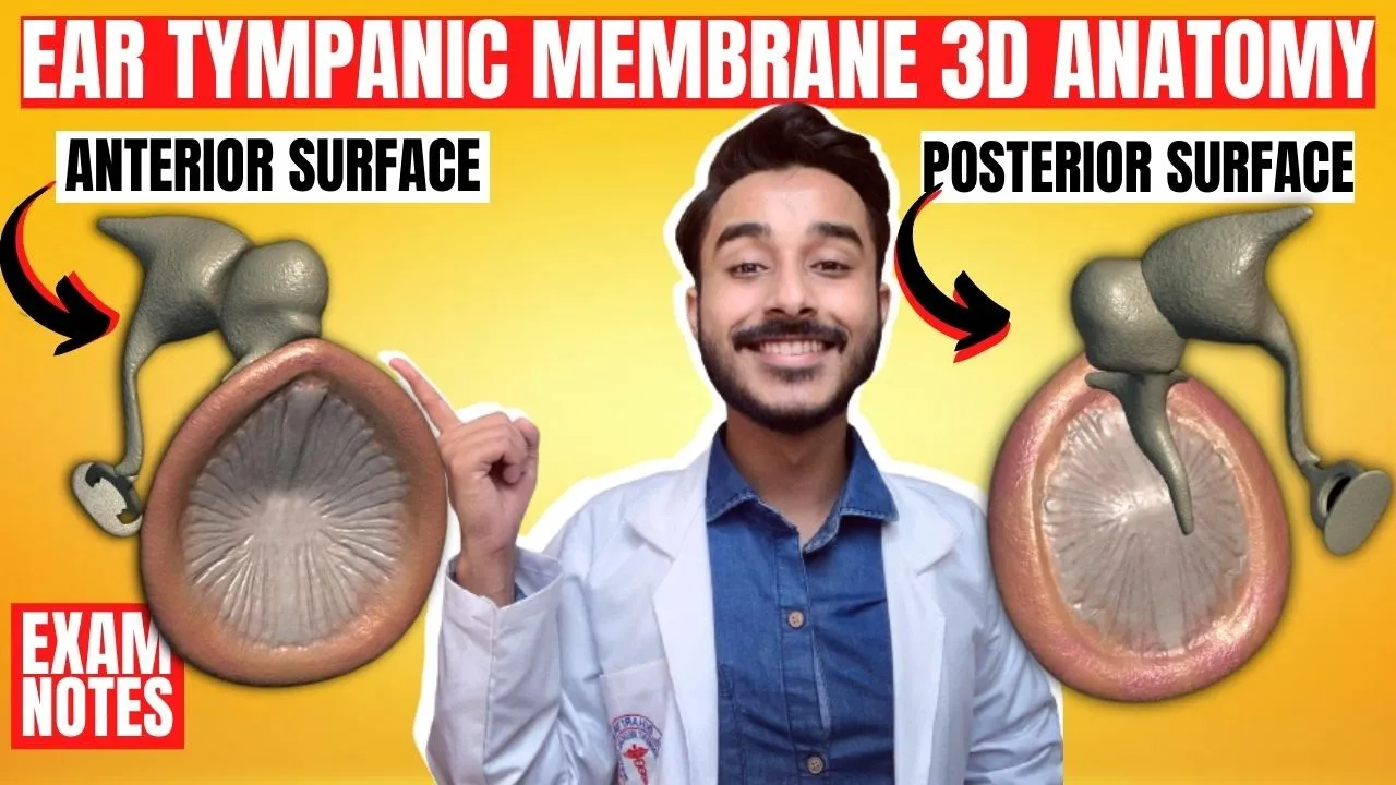
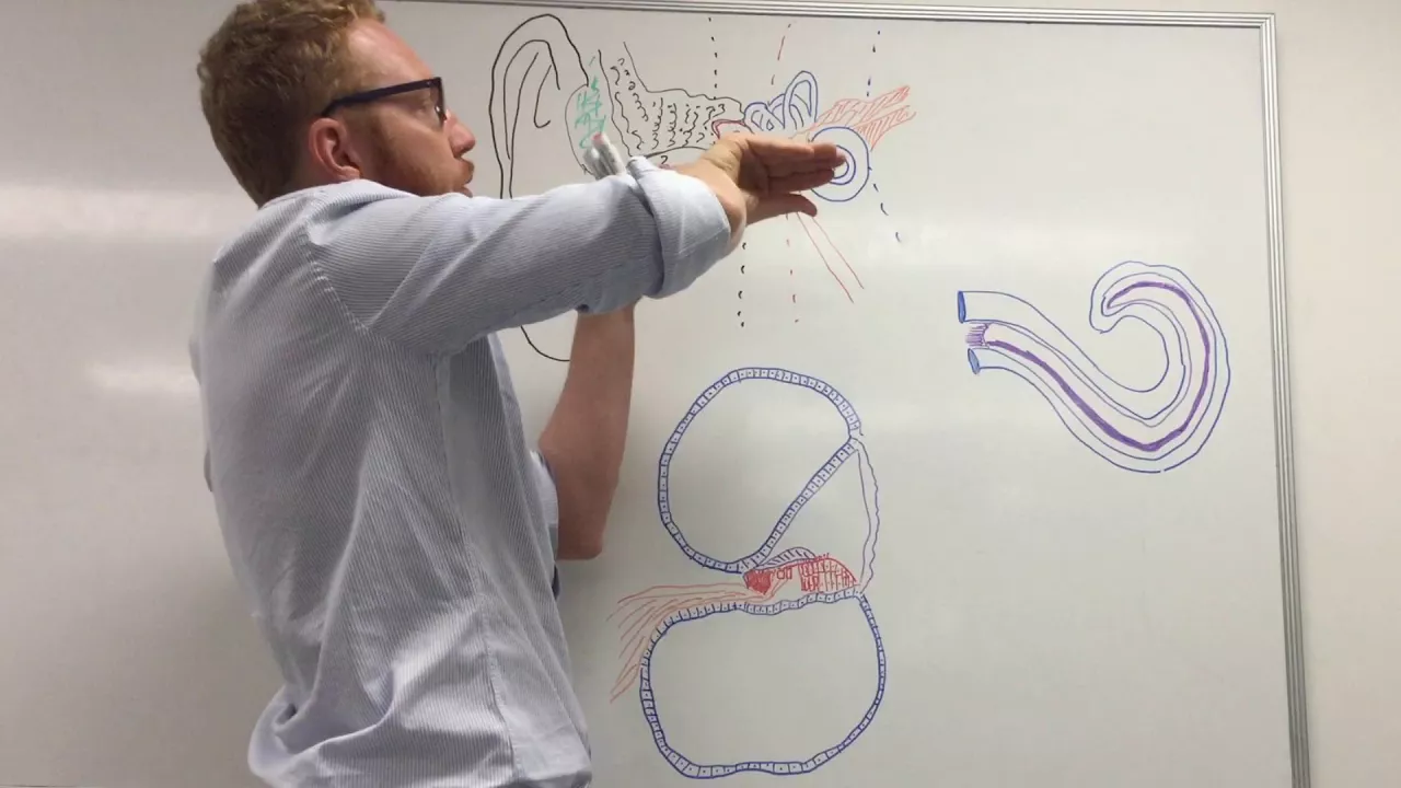
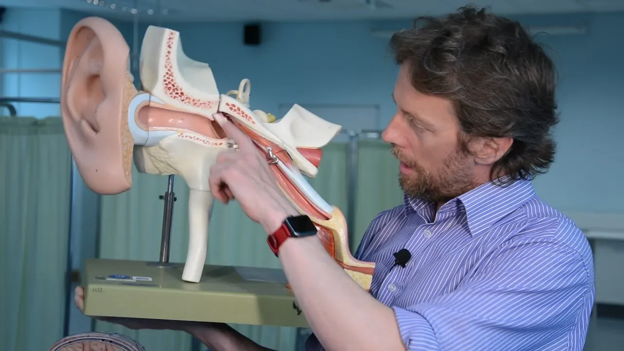
[Music] hello ho matin anetha mirrors and atomists people interested in and that's me health professional students waves we did a whole bunch of videos on the ear a while ago probably ages ago in YouTube terms and I promise that we look at the ossicles in more detail the ossicles being the tiny bones of the ear like the smallest bones in the body right so this is it I'm gonna briefly run through the parts of the tiny bones of the ear this is what I'm talking about right here's the external ear he's a fine joke tympanic tube in here there's well there's the tympanic membrane but on the other side that's the tympanic cavity or the middle in here if we take the bone off we can then see the components of the inner ear doodle that go and watch those videos slowly press pause rewind several times many cups of teeth and then you have a good understanding of that and ask me hopefully I've done a half-decent rather than a half-assed job but today we're going in here so so this is what I'm talking about right there's the tympanic membrane that's the outside of the tympanic membrane it's got a dome to it that's the apex of the dome there and what we can see on the other side is we can see the bone of the malleus so we've got three bones the malleus the incus and the stapes so if I take that out because we're not we're not interested in the other bits today we're just interested in these bits it's difficult to tell I've only got two of the bones on there ones you're the one it's way in there I have another model right so here's a tympanic membrane there are the three bones of malleus incus and stapes this these are the structures of the inner ear let you got a cochlear and the semicircular canals and stuff okay so let's look at this so this is the tip membrane we're looking at the external surface so soundwaves a boo-boom boo-boom boo-boom coming in from this direction right and can you see that it's got that concavity so that by the apex the tip of the concavity there is called the UM bow what a great word um bow and what we can see on the other side there is the is the malleus the first bone the hammer and that that is the the manubrium of the malleus or the the the handle of the of the malleus so is that is the handle of the malleus is embedded in the tympanic membrane is the tympanic membrane vibrates it deflects the malleus why the malleus bone moves so the whole bone which is kind of yeah it's got a big lumpy bill at the top of it right the whole bone deflects and causes all the other bones to move right if I take some of these other bits off well actually take what we can see there is we can see the malleus articulating with the incus the incus being the anvil so we have a hammer bashing against an anvil supposedly there's malleus running up there and then articulating with incus here the malleus then has a head and a neck and is that head this articulating with the incus and it has lateral and anterior processes and the lateral and anterior processes are difficult to see because they're there within this tissue up here so as part of the shape of the bone not helping to hold it in place right now the incus has a body a short cross and a long cross which means a long limb in a short limb and it has then that it has a lenticular process which is going to articulate with the third bone the stay piece here so that's why a piece is going to attach so can you see how we've got this kind of hinging arrangement and as the malleus is deflected incus is going to deflect as well and then it's gonna move the stay piece bone now it's the it's the short limb up here all right so we're in we're in like we're in a cavity right in the inner tympanic cavity and the roof of the cavities up here so it's it's the short limb which is an attachment point an anchoring point for the incus it's holding it in place as it is it as it moves and it's attached by a posterior ligament the joint between the incus and the malleus gets called the incude o'malley ollege ain't the stay piece bone is on the other part of the model so it's difficult to see right now but if i rotate the model you can see that characteristic shape there right there stay peas and it's named stay peas because it's shaped like a stirrup so I think stay peas is a Latin derivation but I'm sure Steelers were a later invention than that there's incus right this is lent the lenticular process of incus that then is going to articulate with stay peas at that point there so you can see how the vibrations of the the bones here are going to cause stay peas to move in and out and the reason stay peas moves is because the base of stay peas is sat upon the oval window of the cochlea all right so what it's going to do is as stay peas moves is going to cause the fluid inside the cochlea to vibrate and then the hair cells are going to detect those vibrations we talked all about that in the cochlear video going over go and have a watch so the base articulates with the oval window and causes pressure waves in in the fluid so stay peas has a head the neck it also has two limbs or two crura a posterior cross and an anterior or posterior lemon and Auntie real in and it has a base the julienne between incus and stapes does get called the in Kudo it's the PDL joint if you're really keen the reason stay piece has got this whole running through it is because during development the stupidy artery runs through here and that's involved in the development of these structures around here now as development continues the stupid ilari should disappear and then leaving that hole so those two pgl artery doesn't disappear so a persistence the PDL artery in the adult that would affect the movement of the stay peas bone there's also a step Adiel muscle that affects the movement of stapedius of the stay peas bone but we talked about things like that in the other videos go and check those out they're here the focus really is on the parts of the obstacles and that is it remember that the function of these bones the reason these bones exist is to is to take the the very low force of vibrations in the air that you're hearing now as I'm talking in your your speaker is vibrating the air and it's vibrating it's impaneling memory to take those vibrations and essentially amplify them so that we can transfer them from the medium of air to the medium of a fluid so going from a relatively large tympanic membrane and gaining some mechanical advantage through this series of bones so then the base of stay peas pushing on the the small relatively oval window can then cause hopefully pressure waves in the fluid within the cochlea which will cause the deflection of the membrane and the hair cells would pick that up but all that sort of cool stuff right okay there you go simple is right by the way right if you're if you're if you're steaming this one University if you know it's these things here right in the anatomy lab I've stuck QR codes and NFC tags on on the models right if you've got an Android phone or if you've got an Android phone with NFC you can probably go blip and it takes you to a little webpage tells you all about the model number guide and stuff if you haven't got an NFC phone you can use an iPhone or an Android phone just get a QR code app that just recognizes that Barco get a free one don't be a mug don't pay for them but to use the same webpage cool huh I haven't done it for the models but I'm game there see you guys next week [Music]
Up Next

8:51
How We Hear Different Pitches: Sound Frequency and the Cochlea
@InteractiveBiology
113.3K views•2011-02-22

50:12
Bacterial Communication: Quorum Sensing and Biofilm Formation Explained
@JHUAAP
12.6K views•2011-06-28

5:35
Enteric Nervous System Explained: The Gut's Brain | Neurobiology Lecture
@alumniu6029
438 views•2018-09-12

7:08
Bacteriophages: Earth's Deadliest Killers and Future Antibiotics
@kurzgesagt
34.6M views•2018-05-13
Related Study Plans & Knowledge Roadmaps
Structured learning paths in Biology
biology
RNAi: Biogenesis, RISC & Gene Silencing
01Molecular Biology Foundations: DNA to Protein
02Introduction to RNA Interference (RNAi)
03Molecular Biogenesis of miRNA and siRNA
+ 2 more learning modules
10h5 modules
Explore Roadmap
biology
Mycorrhizae: Signaling, Biophysics & Ecology
01Foundations of Mycorrhizal Symbiosis
02Molecular Signaling and Root Colonization
03Biophysics and Bioenergetics of Nutrient Exchange
+ 1 more learning modules
11h4 modules
Explore Roadmap
biology
Circadian Clocks: TTFL, SCN & Periphery
01Foundations of Molecular Biology & Gene Expression
02Introduction to Chronobiology and Actograms
03The Core TTFL: Molecular Architecture of the Clock
+ 3 more learning modules
14h6 modules
Explore Roadmap
biology
Population Genetics: Math, Drift & Selection
01Genetic & Probability Foundations
02The Hardy-Weinberg Principle and Baseline Models
03Mathematical Modeling of Natural Selection
+ 3 more learning modules
18h6 modules
Explore Roadmap