





















Gram-Positive bacteria have a thick peptidoglycan layer with lipoteichoic acids anchored to a single plasma membrane, while Gram-Negative bacteria have a thin peptidoglycan layer sandwiched between an inner plasma membrane and an outer membrane containing lipopolysaccharides (LPS/endotoxin); this structural difference affects how these bacteria respond to antibiotics and host immune systems.
Gram Positive vs Negative Bacteria Cell Walls
Added:Basic understanding of prokaryotic cell structure, specifically how bacterial cells differ from eukaryotic cells.
The general function of a cell wall in maintaining osmotic pressure and structural integrity in microorganisms.
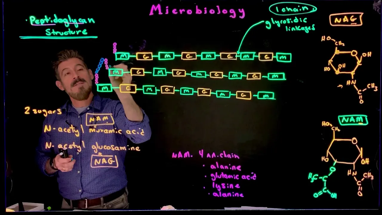
An introduction to peptidoglycan (murein) as the primary structural polymer unique to bacterial cell walls.
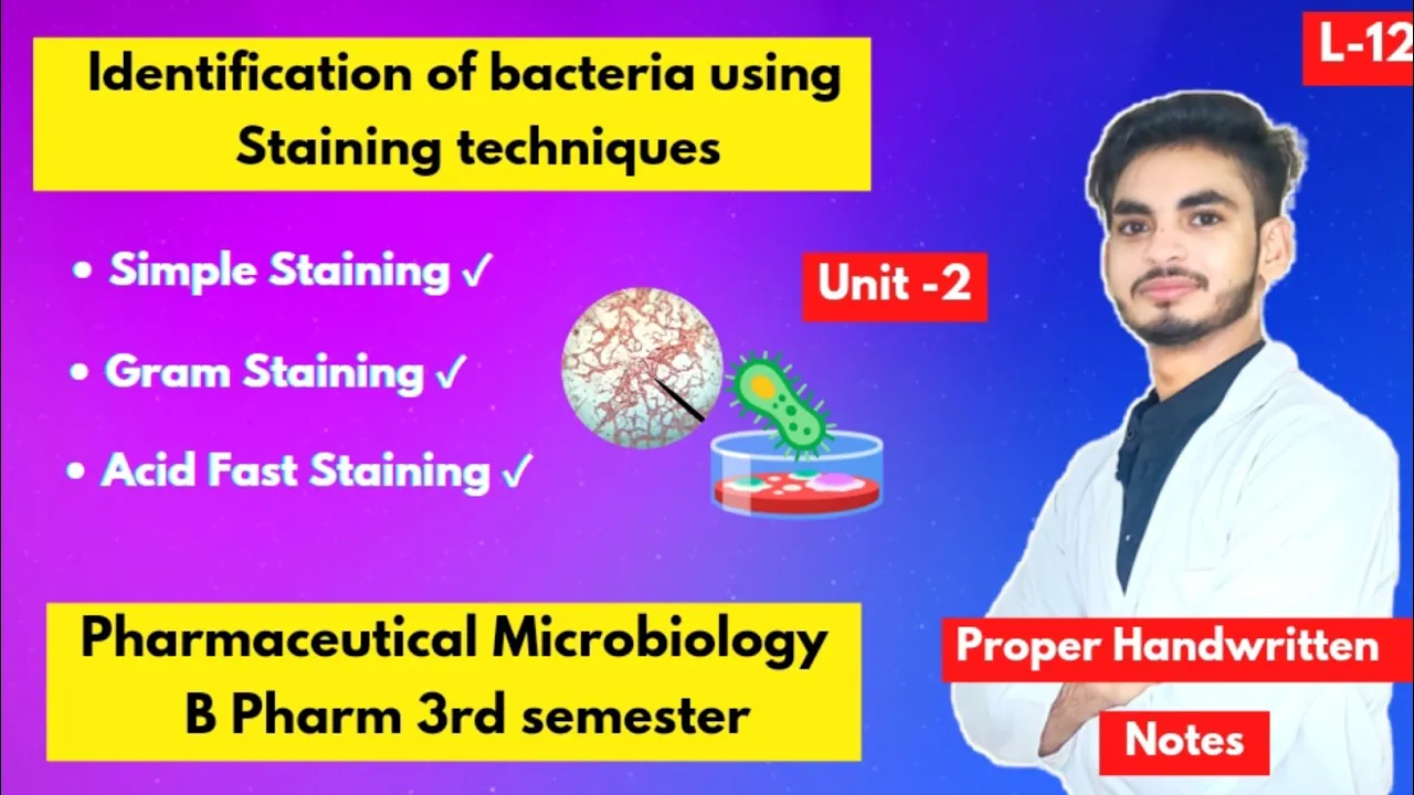
The purpose and basic concept of the Gram staining technique as a classification tool in microbiology.
The step-by-step chemical mechanism of the Gram stain procedure and how physical differences in the cell walls retain specific dyes.
How differences in cell wall structure affect antibiotic susceptibility, particularly regarding beta-lactam antibiotics like penicillin.
The clinical significance of Lipopolysaccharide (LPS) in Gram-negative bacteria and its role as an endotoxin causing immune response and septic shock.
Study of atypical bacteria that do not conform to standard Gram classification, such as Acid-fast bacteria (Mycobacterium) and wall-less bacteria (Mycoplasma).
452.4K views5Klikes5:49Original Release: 2014-01-16
Hello and welcome to Biology Professor.
Today, we will be talking about one way that you can distinguish most types of bacteria.
Most bacteria fall into one of two categories.
Gram-Positive or Gram-Negative So first, let's talk about the Gram-Positive type of bacteria.
The way they are distinguished is between differences in their cell walls.
With the Gram-Positive cell wall, Tte two most important features are a thick layer of peptidoglycan and one membrane.
Let's look at this in a little bit more depth.
This thick layer of peptidoglycan is important for structure of the cell and for helping the bacteria cell maintain its shape.
Then you have this inner wall zone that is between the thick peptidoglycan layer and the single membrane.
Within this plasma membrane, you have two main components.
These are the lipids which are shown here, a lipid bilayer, and also several different types of proteins.
I'll mark these with an asterisk (*) each.
So these are all proteins that serve different functions within that plasma membrane.
Finally there is one other distinguishing characteristic that you see in Gram-Positive cell walls and this is what I have indicated here with these blue lines.
These are called lipoteichoic acids and these anchor through the peptidoglycan layer onto lipids in this lipid bilayer.
So these are lipoteichoic acids.
And now if we look these are some common examples of Gram-Positive bacteria. That I am sure you have heard of Staphylococcus aureus can actually be part of the normal human bacterial flora.
Actually, a majority of humans have staph in their nose and on the skin.
But it can also cause disease.
Specifically some strains of Staph. aureus can cause food poisoning and they can also cause infections of the skin and of the respiratory tract.
There is also Bacillus anthracis which causes Anthrax.
Clostridium tetani which is responsible for Tetanus.
and Streptococcus mutans this is usually associated with cavities in the mouth. So, this is the structure of a Gram-Positive cell wall.
So now we are going to continue our conversation about the difference between Gram-Positive and Gram-Negative cell walls.
This time looking at Gram-Negative cell wall.
We still see some of the same components. There is still a plasma membrane.
It's still composed of a lipid bilayer and it still contains proteins.
That I will label here with an asterisk (x).
There is still a layer of peptidoglycan, but you can see that compared to Gram-Positive peptidoglycan layer, in Gram-Negative cells, this peptidoglycan layer is much, much thinner.
and also unique to Gram-Negative cell walls, this peptidoglycan layer is anchored to an outer membrane, so a second lipid bilayer, with additional proteins.
Now in the outer membrane there are some proteins that are called porins.
These porins allow for passive diffusion of things from the outside into the space between the two membranes, which is called the periplasmic space.
These porins allow most things to diffuse through as long as the can fit.
There is nothing specific about that.
The actual selective permeability of the cell is still controlled by this inner plasma membrane.
Now another very important feature of Gram-Negative cell walls is what is on the very outside of the this outer membrane.
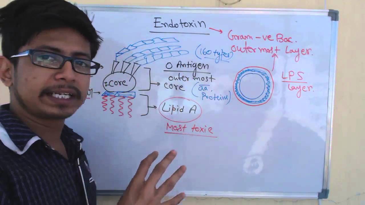
LPS, or lipopolysaccharides, this a component that is only found on the outside of Gram-Negative cell walls.
and that has very important consequences when you have infections of Gram-Negative bacteria in the blood.
This LPS, you will also hear it referred to as endotoxin.
for exactly that reason.
Now lets look at some examples of Gram-Negative bacteria.
You have probably heard of a lot of these.
E. coli is one that we hear about a lot as a pathogen because it can cause food poisoning.
But it is actually a normal commensal part of the human flora.
So it is not always pathogenic.
There is also Helicobacter pylori, which causes ulcers and stomach cancer.
Vibrio cholerae, which causes cholera.
Yersinia pestis, which causes plague.
This is the bacteria that is responsible for the historically famous Black Death.
Also, Salmonella enterica, which is another common cause of food poisoning.
So that concludes our explanation of the differences between Gram-Positive cell walls and Gram-Negative cell walls.
If you are interested in more information about how these two types of bacteria are distinguish between when diagnosing illnesses or when studying them in the laboratory, please see my video on the Gram stain procedure and also thank you for watching.
Up Next

5:50
Quorum Sensing in Vibrio fischeri: A Molecular Overview
@ferreiramario73
791 views•2018-04-01

50:12
Bacterial Communication: Quorum Sensing and Biofilm Formation Explained
@JHUAAP
12.6K views•2011-06-28

5:35
Enteric Nervous System Explained: The Gut's Brain | Neurobiology Lecture
@alumniu6029
438 views•2018-09-12

7:08
Bacteriophages: Earth's Deadliest Killers and Future Antibiotics
@kurzgesagt
34.6M views•2018-05-13
Related Study Plans & Knowledge Roadmaps
Structured learning paths in Biology
biology
RNAi: Biogenesis, RISC & Gene Silencing
01Molecular Biology Foundations: DNA to Protein
02Introduction to RNA Interference (RNAi)
03Molecular Biogenesis of miRNA and siRNA
+ 2 more learning modules
10h5 modules
Explore Roadmap
biology
Mycorrhizae: Signaling, Biophysics & Ecology
01Foundations of Mycorrhizal Symbiosis
02Molecular Signaling and Root Colonization
03Biophysics and Bioenergetics of Nutrient Exchange
+ 1 more learning modules
11h4 modules
Explore Roadmap
biology
Circadian Clocks: TTFL, SCN & Periphery
01Foundations of Molecular Biology & Gene Expression
02Introduction to Chronobiology and Actograms
03The Core TTFL: Molecular Architecture of the Clock
+ 3 more learning modules
14h6 modules
Explore Roadmap
biology
Population Genetics: Math, Drift & Selection
01Genetic & Probability Foundations
02The Hardy-Weinberg Principle and Baseline Models
03Mathematical Modeling of Natural Selection
+ 3 more learning modules
18h6 modules
Explore Roadmap