![Anatomical Position and Directional Terms [Anatomy MADE EASY]](https://i.ytimg.com/vi/t6-ueqFK1IE/maxresdefault.jpg)


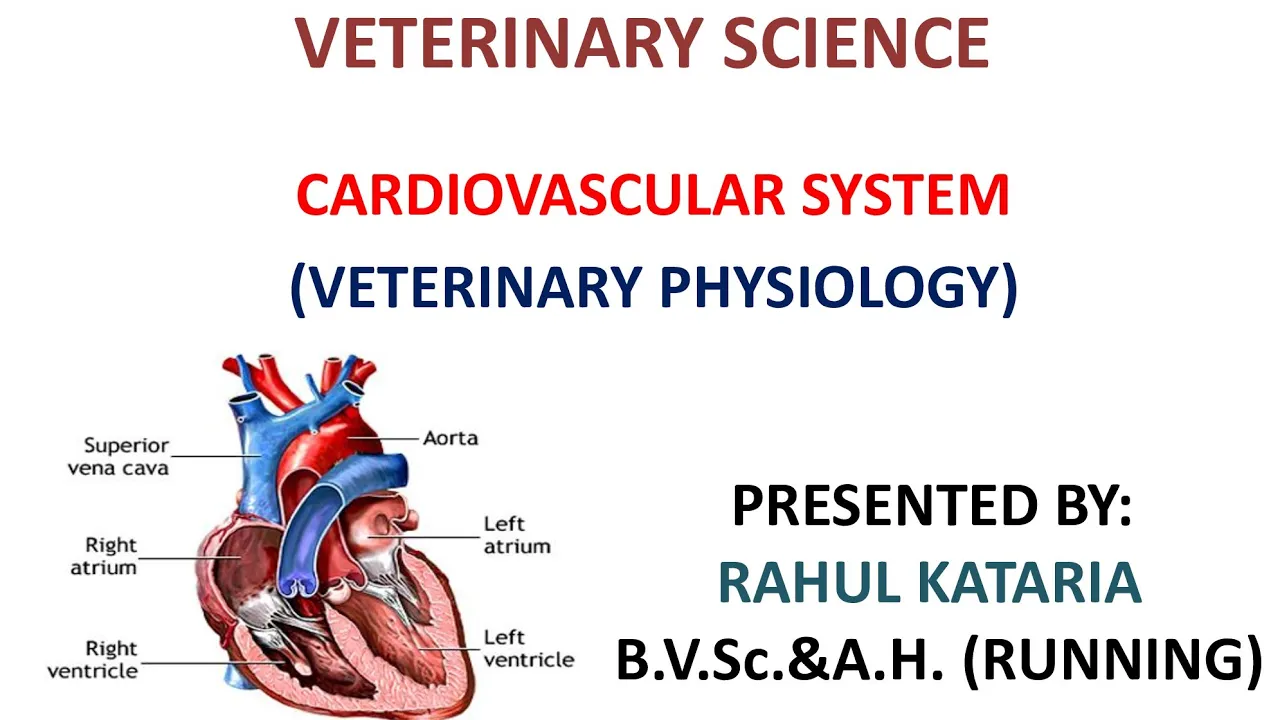






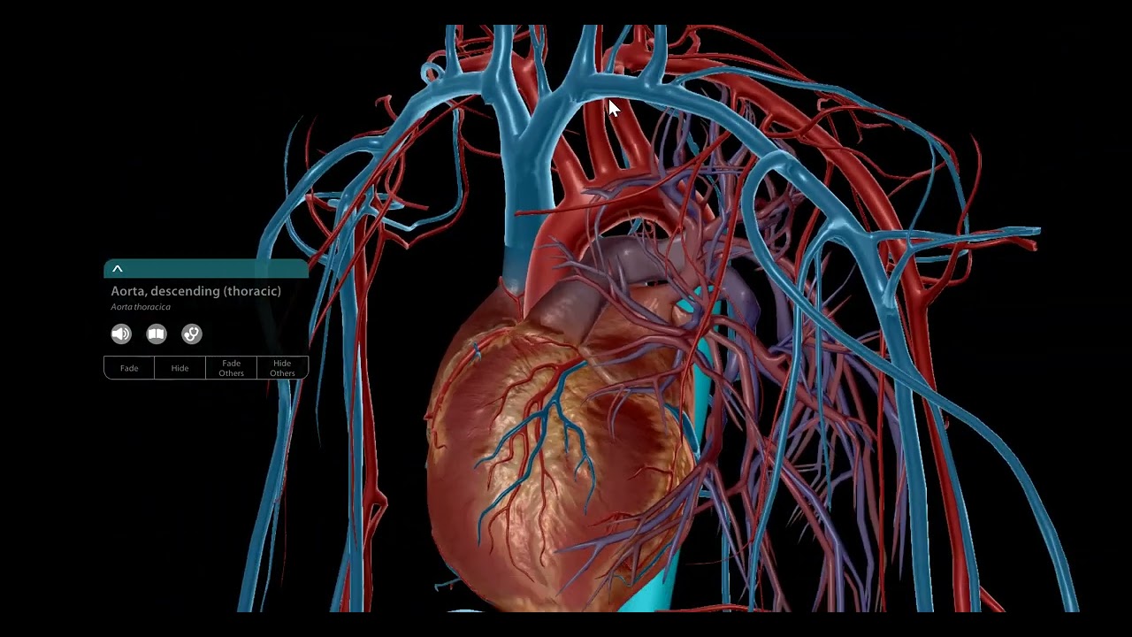


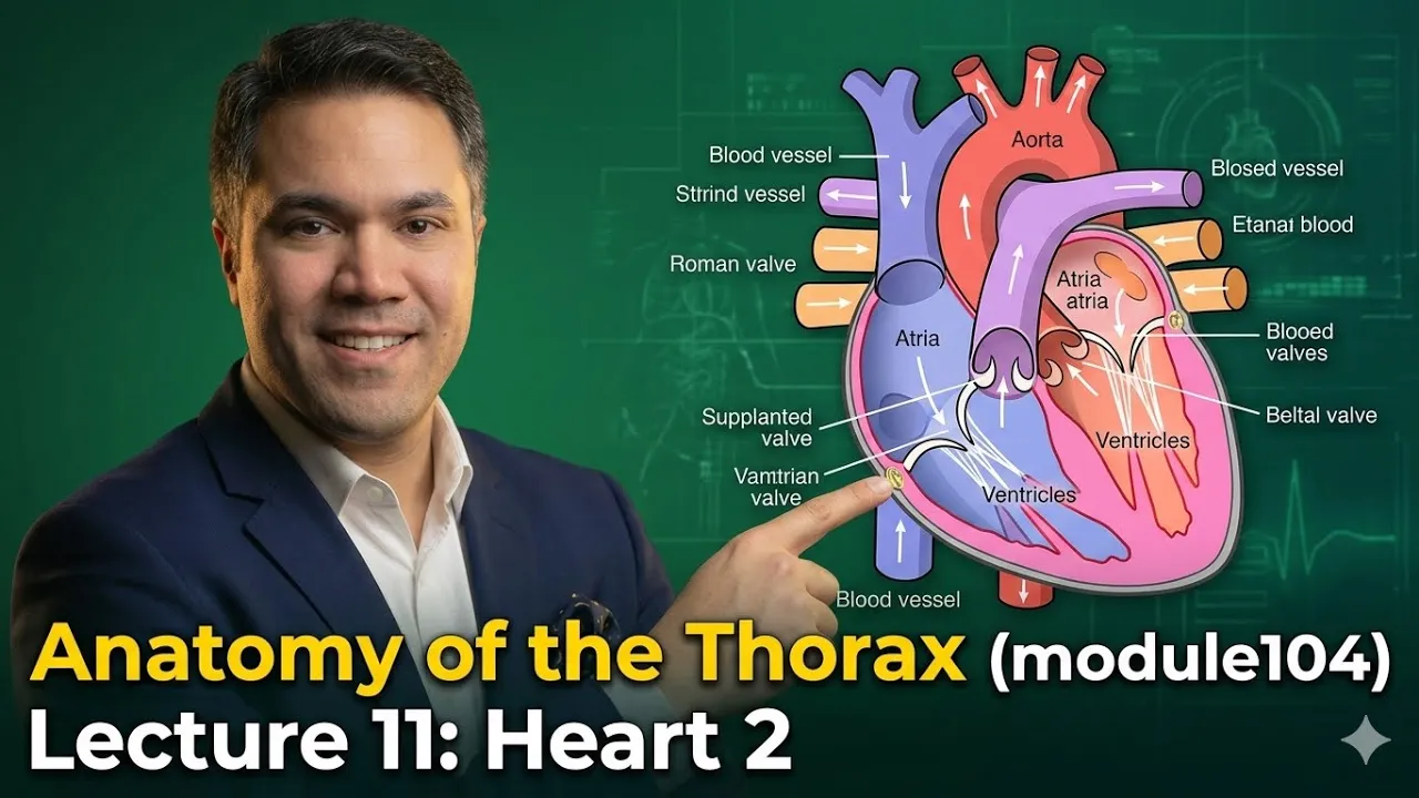
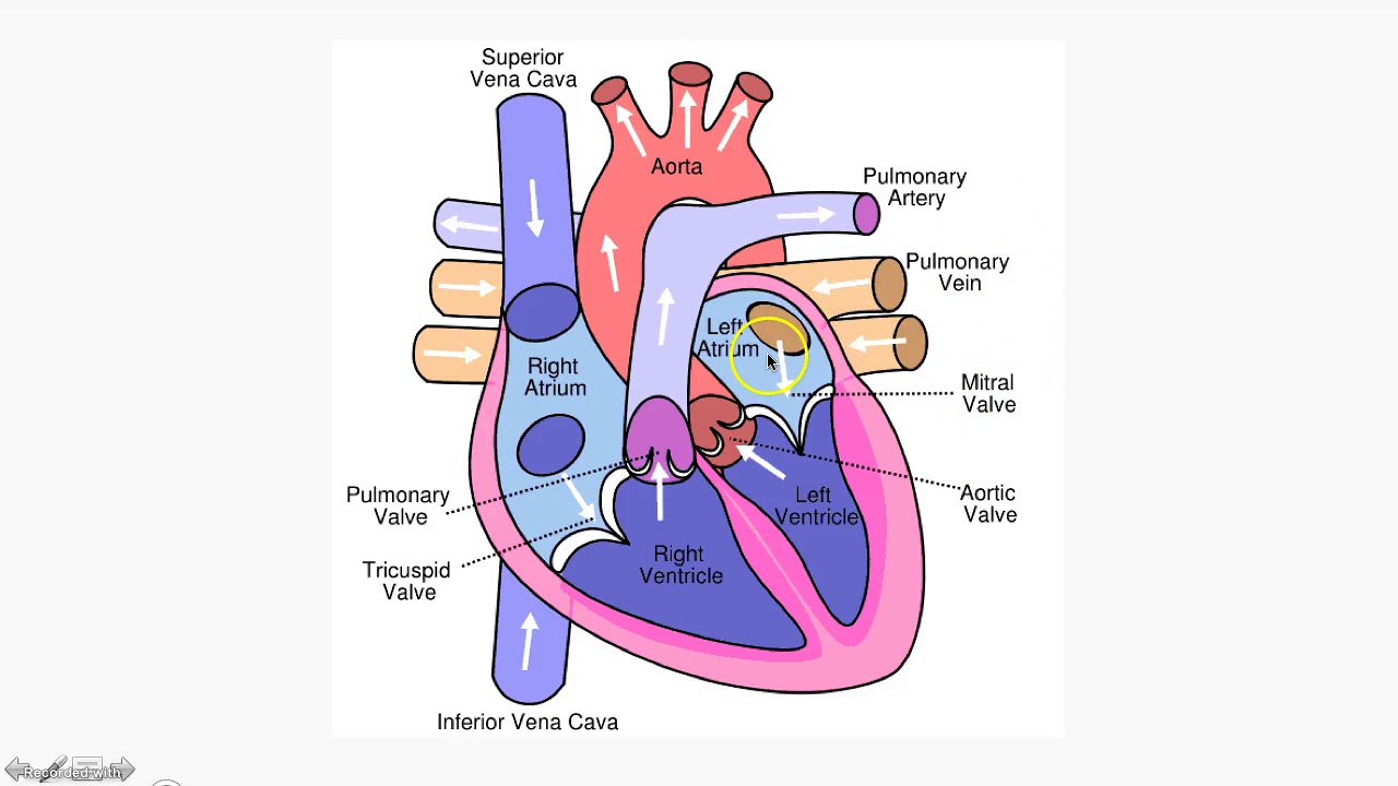





The heart is a muscular organ roughly the size of a fist, weighing about 300 grams, that beats 60-80 times per minute and pumps 5-6 liters of blood per minute throughout life; it is enclosed in a double-walled pericardial sac consisting of a fibrous outer layer and serous inner layer, with the visceral pericardium adhering to the heart surface and containing pericardial fluid for lubrication; the heart has four chambers (right atrium, right ventricle, left atrium, left ventricle) connected by four valves (tricuspid, pulmonary, mitral/bicuspid, aortic) that regulate blood flow, with the right side pumping deoxygenated blood to the lungs via pulmonary circulation and the left side pumping oxygenated blood to the systemic circulation through the aorta; coronary circulation supplies oxygenated blood to the heart muscle itself via the right and left coronary arteries, which branch from the aorta, and drains deoxygenated blood through cardiac veins into the coronary sinus and ultimately to the right atrium.
Heart Anatomy Tutorial (Pericardium, Chambers, Valves, Coronary Circulation)
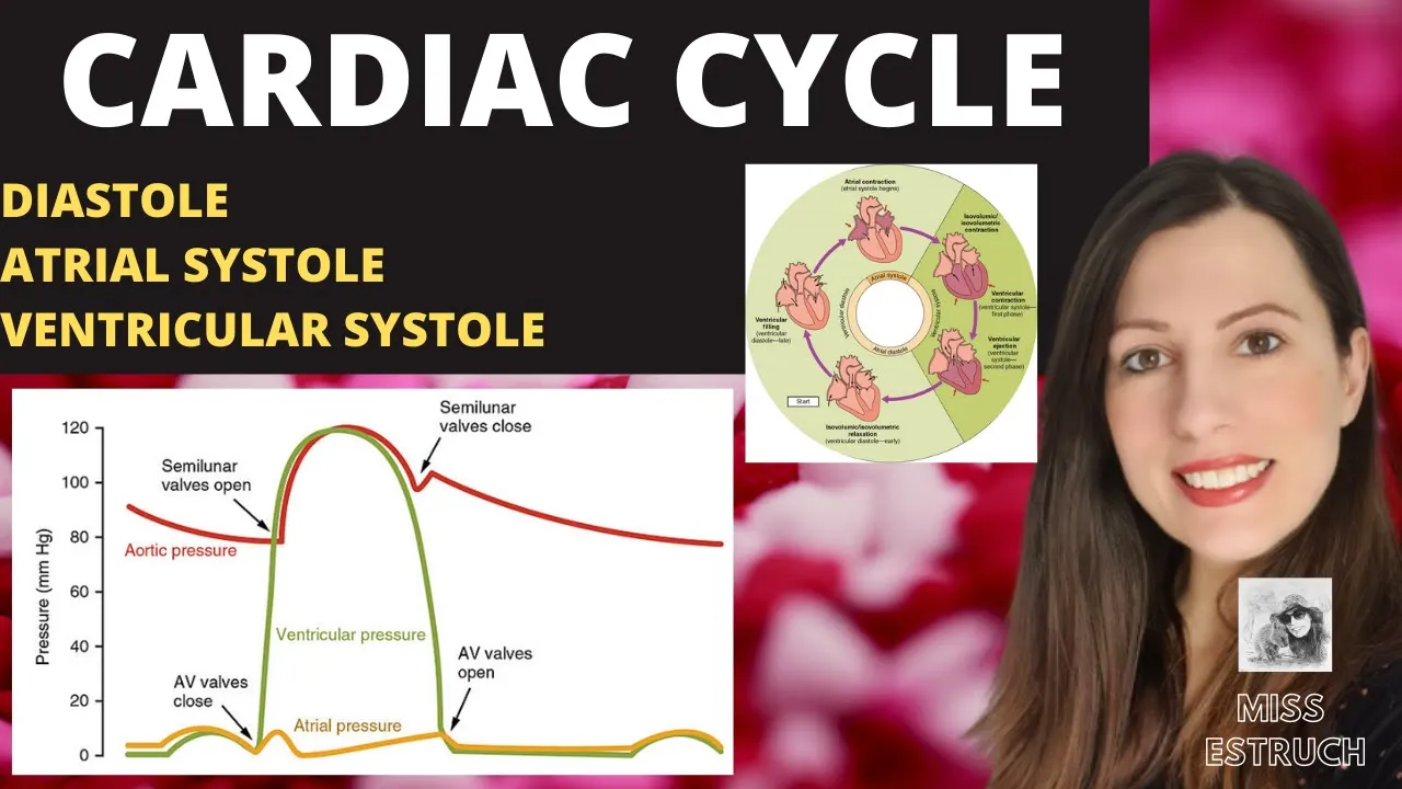
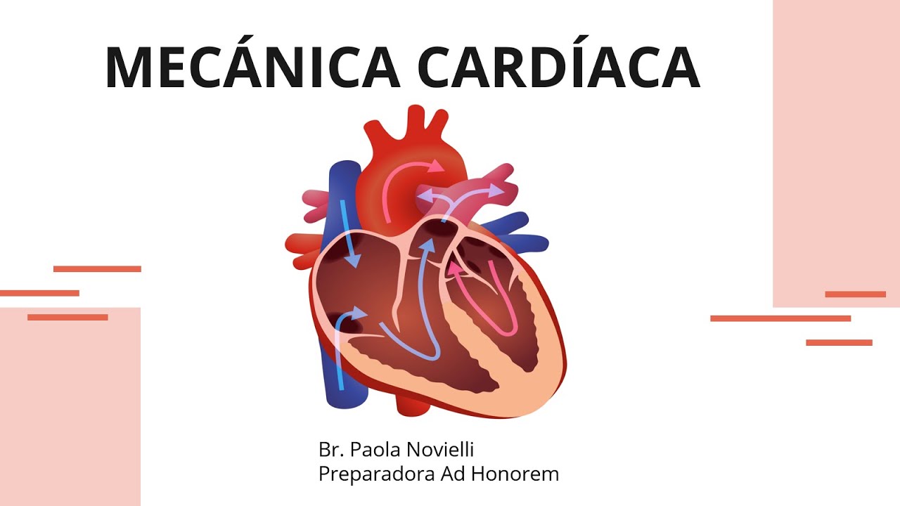
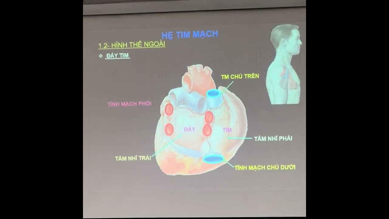
Added:okay we're now going to do an overview of heart anatomy and answer the what questions what are the layers and functions of the pericardial sac and what are the chambers valves and great vessels of the heart what are the primary coronary arteries and cardiac veins hello everyone my name is dr. Morton and I'm the noted anatomist okay so the human heart is roughly the size of a fist and it weighs about 300 grams and beats 60 to 80 times a minute throughout your entire lifetime and pumps five to six liters of blood throughout your body it is a very remarkable organ so the first thing we're going to talk about is the pericardial sac which is really the packaging of the heart and the pericardial sac is as double-walled sac that contains the heart and the roots of the great vessels a Horta pulmonary trunk and so forth now the prado Park car diem has two layers of fibrous layer and a serous layer and the visceral pericardium adheres to the outside of the heart okay so here we have the an illustration I did actually in graduate school and there in green is showing the pericardial sac which is the sac encasing the heart and the outside of the heart is known as the fibrous layer of the parietal pericardium just like any serous membrane it's got a prattle layer but this is made of fibrous connective tissue dense irregular collagenous connective tissue now let's take a scalpel and cut the SAC open like this she now will see that the internal surface of the parietal pericardium is the serous layer is where it says serous pride a layer the part of the pericardium this is what makes serous fluid whereas the external fibrous layer of the prattle pericardium is dense irregular collagenous connective tissue and then if we now take a look at a cross-section there's the fibrous layer there's the serous layer and then the part that's adhered to the heart that's the visceral pericardium let's do this again let's take a cross-section however of the pericardial sac like this and show it so this is a cross-section let's get ourselves oriented there's our lungs that are flanking either side of the heart and the heart is contained within a pericardial sac and the outside is the fibrous pericardium shown in green this is see regular CT and this is what prevents the heart from over feeling it doesn't stretch a whole lot so it allows and it also fixes the heart in the mediastinum now the serous pericardium has two layers a parietal layer that forms the wall on the inside of the fibrous layer and then the visceral layer that's adhered to the heart but notice that if we zoom in the parietal layer and the visceral layer they just are continuous with each other the heart is like a fist pushing in on a balloon this serous pericardium so if we now take a look at the parietal pericardium it consists of two things the outer fibrous layer and the inner serous layer of the Prado pericardium and then the part that's adhered to the heart that's the visceral pericardium all right now the pericardial space is between the parietal and visceral pericardial like that and it what this is the fluid the pericardial space is filled with pure cardio fluid and that's what lubricates the heart so let's do this one more time in this coronal section where if we take that little bit and blow that up and what we see is the following there is the endocardium which is intimately associated with the blood in the chambers and then there's the myocardium or the heart muscle which pumps blood and then there's the visceral pericardium that's on the outside this serous membrane but if you take the serous membrane you actually have a lot of adipose tissue so if you take the visceral pericardium plus the adipose tissue that's what is formed the epicardium but often we just use those terms synonymously then outside of the visceral pericardium is the pericardial space that's filled with pericardial fluid now for some reason this illustration that from my textbook it looks like that's blood and that's not blood the pericardial space is filled with pericardial fluid which is like water then you have the parietal pericardium the serous layer and the parietal pericardium the fibrous layer and together that just simply is the parietal pericardium there at one okay now let's talk about heart chambers now this coronal section or not coronal but basically you can see the different chambers of the heart and there are the chambers and great vessels and valves of the heart so we're going to talk about them except instead of using a cadaver we're going use a nice basic black-and-white illustration so the superior vena cava or SVC is the vein that delivers deoxygenated blood from all tissues above the diaphragm to the right atrium in other words any veins that are above the diaphragm blood will eventually from those veins end up in the SVC now the inferior vena cava or IVC it delivers deoxygenated blood from all tissues below the diaphragm into the right atrium in other words all veins below the diaphragm but all the blood within all the veins below the diaphragm will eventually end up in the IVC then there's the structure called the coronary sinus it delivers deoxygenated blood from the myocardium via the coronary circulation into the right atrium and so there in blue is the opening of the coronary sinus that delivers blood into the right atrium so if we look at this posterior view of the heart there is the coronary sinus draining over and the blood dumps into that right atrium there okay so now the right atrium is Minh to one of these chambers of the heart is to receive all the oxygen and your blood from the block body from the SVC the IVC and from the coronary sinus and if we open up the right atrium we see a structure on the interatrial septum that can that contains the structure called the fossa ovalis and the fossa ovalis is the remnant the fetal remnant of the foramen ovale which shunted blood from the right atrium to the left atrium to bypass the lungs in a fetus the right atrium forms the right border of the heart like this so there we have the right atrium and notice that the whole right side of the heart is primarily formed by the right atrium now the floor of the right atrium is this valve and that's our tricuspid valve and that's next the tricuspid valve I remember this one because it tried to be right and also try our eye is the ri right AV valve some ways to remember this so here's a superior view of the heart with the atria removed and the yellow arrow shows the two three cusps that make the tricuspid valve another name for the tricuspid valve is the right AV or right atrial ventricular valve so tricuspid valve right AV valve right atrial ventricular valve all synonymous Thank You anatomist ray now the tricuspid valve opens to let blood flow from the right atrium through that valve into the right ventricle then it closes during systole to prevent backflow of blood into the right atrium hold that thought I'm going to come back to it the right ventricle is the is the biggest chamber on the right side of the heart and it pumps deoxygenated blood into the pulmonary trunk on route to the pulmonary arteries on route to the lungs to get oxygen and the right ventricle forms the anterior border of the heart so if we look at this picture there's an anterior view and have outlined the right ventricle it's the chamber that forms primarily the front of the heart that touches the ribcage and sternum so let's now take a little detour from the heart chambers and I'll talk about heart valves there's really two types of valves atrioventricular valves and semilunar valves we're going to start with the atrioventricular or AV valves so here we've got this little blow-up I'm going to take that and show in this little illustration there's the atrium above the AV valve and the ventricle below the AV valve and then coming out projection from the oh and there's the tricuspid valve in this case okay now the papillary muscles are really extensions of the myocardium and the papillary muscles are connected to the tricuspid and bicuspid valves via chordae tendineae or heart strings dense connective tissue so in this cadaver picture there is a ventricle there is a papillary muscle that's chordae tendineae attached to the tricuspid valve so in this illustration we're going to show the function where the atrium is above the AV valve the ventricles below the AV valve and then there's an AV valve there's a chordae tendineae and there's the papillary muscles and so during diastole okay so the function is the AV valves enable blood to flow from the atrium into the ventricle during diastole then during systole when the ventricles contract blood is pumped from the ventricle up and so the AV valves prevent blood from a GERD changing back from the ventricle into the atrium by slamming shut like that in making whenever the valves close like slamming the door it makes a sound we say s1 for first heart sound the lub in the lub dub now the chordae tendineae ensure that the AV valve leaflets do not prolapse into the atrium you see that now watch what happens she do you see that so what happens then is when the blood forces those AV valves shut and the blood then continues to balloon up because these vowels are connective tissue and so like parachutes they'll balloon up but the chordae tendineae prevent these valves from the leaflets from prolapsing into the atrium see that now without them meaning the chordae tendineae regurgitation occurs so watch we take away one of those chordae tendineae and when the blood pushes up against the valves it then the valve leaflet prolapse --is and blood can move from ventricle to atrium causing a lot of problems okay so now the pulmonary valve is next one we're going to talk about and so the pulmonary valve in this cross section with the atria removed and a superior view is the most anterior heart valve and it opens to let blood flow from the right ventricle through the pulmonary valve into the pulmonary trunk and pulmonary arteries and ultimately going to the lungs to get oxygen and the pulmonary valve closes during diastole and prevents backflow of blood into the right ventricle so let's talk about polycythemia lunar valves now are their function and so the ventricle is below and the pulmonary trunk or aorta are above the semilunar valves and there's a semilunar valve so watch or happens during systole the semilunar valves enable blood to flow out of the ventricles and then during diastole prevent blood from regurgitating back into the ventricles from here to here and whenever valves close like slamming a door it makes a sound we say s2 or the second heart sound or the dub in the lub-dub lub-dub pulmonary arteries transport deoxygenated blood from the right ventricle to the lungs like that and so this is something you want to put a little caveat a little star a little happy face look Canadian flag because pulmonary artery's are the only arteries in an adult that transports deoxygenated blood the only one fetus is different we'll talk about that another time and so the pulmonary arteries and I always remember arteries for away so if this is a for a way and so if you forget or get this mixed up always remember that the arteries take blood away from the heart and why are we sending blood from the heart away from the heart to the lungs why are we sending it to the lungs because we need to get rid of co2 and we need to get oxygen and so the blood going to away from the heart to the lungs is rich in co2 and we're going to get oxygen pulmonary arteries right there all right so the middle of the top picture shows the heart and then you got the right and left lung and then watch pulmonary arteries send through the left and right pulmonary arteries to the left and right lungs to get oxygen so here we have the trachea and bronchial tree and we're gonna take a look and blow up this little bronchial and see that oxygen is inhaled through this bronchial and goes into these things called the alveolar sacs and alveolar sacs are these terminal air spaces where gas exchange is going to occur because pulmonary arteries are bringing co2 rich blood through these pulmonary capillaries and so we're at these pulmonary capillaries this is where gas exchange occurs where the pulmonary capillary blood is rich in co2 and poor in oxygen where the alveolar sacs within their space it's rich in oxygen and poor in co2 so we have a high to low concentration co2 diffuses into the alveolar space and oxygen diffuses into the pulmonary capillaries you then exhale co2 and the blood exiting the pulmonary capillaries in the pulmonary veins is rich in OH - it's now oxygenated blood going back to the left atrium of the heart so here we have the pulmonary veins bringing oxygen-rich blood back to the left atrium so pulmonary veins transport oxygenated blood from the lungs to the left atrium and like those arrows showed and this is the only vein and an adult that transports oxygenated blood put a little happy face or like a poo emoji at this bugsier whatever to remember pulmonary veins are different than the systemic veins in the blood that they transport in the house that Jack built and in this posterior view of the heart we see the left atrium and then you have two pulmonary veins and left and two pulmonary veins in the right dumping blood into the left atrium if you hear like yelling everything's cool I just have a teenage son that's above me in floor and he keeps yelling and playing with their dog weenie so I keep trying to record I'm just gonna keep going so if you hear it everything's cool at the Morton household all right now on the left atrium there's our left atrium it receives oxygenated blood from the lungs as we talked about she like that and it forms the most posterior border of the heart and so here we have this posterior view and there is that left atrium it's the most posterior and it's what actually touches the esophagus in the thoracic cavity now the bicuspid valve gets its name because it has one two cusps as shown here in the superior view but it's also called the mitral valve why is it called the mitral valve because if we take this heart and you can see this mitral valve and turn it upside down like this and we look at this religious hat it's called a mitre and the mitre early anatomist said okay that kind of looks like our valve so they called it the mitre valve or mitral valve but that wasn't where they ended they said well instead of just the bicuspid valve cuz there's two customer mitral valve cuz it looks like a mite or why don't we also called the left AV valve because it's the atrial ventricular valve on the left and I just remember mitral l4 left okay the bicuspid mitral or left AV valve opens to let blood flow from the left atrium to the left ventricle and so it closes during systole to prevent backflow of blood into the left atrium so here we have watch Shing Shing it prevents blood from going from the left ventricle back into the left atrium and with that bicuspid valve shuts it makes the s1 or first heart sound now the left ventricle receives oxygenated blood from the left atrium and pumps it into the aorta like that through the aortic valve and notice the thickness of the myocardium in the left ventricle and the thickness of the myocardium of the right centrical now we take a look at a cross-section of the heart look at the thickness difference we're talking about you know 10 to 20 millimeters of mercury pressures with the right ventricle pushes up against to open the pulmonary valve where you're looking at 80 to 120 millimeters of mercury pressure for the left ventricle to push against to open the aortic valve much thicker myocardium as a result that mean arterial pressure in systemic vessels arteries is bigger than pulmonary arteries now the left ventricle forms the left border and apex of the heart so I mean look at this anterior and posterior view of the heart and we outline the left ventricle that's what forms primarily the left border and the pointy apex of the heart the or Dec valve opens to let blood flow from the left ventricle into the aorta and so in this cross-section there's air air a or tek valve which is behind or posterior to the pulmonary valve and we have coming off the left cusps the left coronary artery and the right cusps the right coronary artery and so the aortic valve closes during diastole and prevents backflow of blood into the left ventricle so here we have the aorta above and the left ventricle below and watch ching-ching during diastole blood flows and slams the semilunar aortic valve shut it makes the s2 sound or the second heart sound the dub in lub dub we're now going to talk about the aorta and how the order delivers blood to systemic and coronary circulation x' and so here's well I'm actually not going to talk too much about it other than and that's good ever there's the order this really big artery and it is the thickest elastic muscular artery it's considered an elastic heart because it's got so much like 50% elastic tissue and 50% smooth muscle in the tunica media takes a lot of pressure and that's what distributes blood to all systemic arteries and arterioles let's now talk about coronary circulation so what is the current coronary circulation and why do we need it here's this coronal section of the heart there's oxygen in the left ventricle and there's myocardium we would think that oxygen could just diffuse and supply this myocardium but it doesn't work that way because the blood is moving too fast at too high of pressure so what is the coronary circulation it's blood the heart pumping blood into the heart and the heart delivering blood back to the heart now what we mean by that is that the left ventricle pumps oxygenated blood into the ascending aorta which then gives rise immediately to left and right coronary arteries which then supply the heart meaning the myocardium the actual heart muscle tissue and then the heart muscle tissue gives us deoxygenated blood back to the heart via cardiac veins into the right atrium so blood goes from the heart through coronary arteries to the heart and through cardiac veins back to the heart it's a circulation or circulatory system so the major arteries first one is the right coronary artery or RCA it supplies the right side of the heart and so there's this yellow arrow showing the right coronary artery coursing what's called the coronary sulcus which cores of the right crown of the heart and so here's a superior view of the heart and the atria been removed and then I'm outlining the aortic valve and then highlighting the right cusps of the right of the aortic valve and then there's the opening giving rise to the lumen to the right coronary artery and so the right coronary artery the word coronary means like a crown so if you think of someone's head and you put a crown on their head and you go oh where the crown moves around the head that's what's like the coronary artery it courses in this coronary sulcus like it's a crown okay the right coronary artery supplies right side of the heart and it also gives rise to some other branches like the sinoatrial node 'el artery or the SA nodal artery which supplies the SA node which is the pacemaker of the heart and so we now take a look at this posterior view the posterior descending artery or PDA supplies the posterior interventricular septum and so there's the left ventricle and right ventricle and the PDA is demonstrated on the posterior view we see it and then the anterior view I see it ghosted is if you're looking through like Superman looking through the heart now the Natomas also call this the posterior interventricular artery because it's on the posterior part of the interventricular septum now the left side of the heart we're in time with the left coronary artery or LCA it supplies the left side of the heart it's actually quite small and of course in the coronary sulcus but that much like it's really short and it technically itself doesn't supply anything but it does supply the heart via its branches so here's a superior view there's the aortic valve and the aorta and there's the opening and that's it that's the left coronary artery see that a little bit like a centimeter long but it gives rise to two major branches the el-ad and the LC X let's talk about those two branches first here's our left coronary artery and the first branch is called the left anterior descending artery or el-ad they're left because it's coming off the left coronary artery anterior because it's on the anterior part of the heart and descending because it descends on the front of the heart and it supplies the anterior interventricular septum because there's the right and left ventricle and it's coursing in the septum between them as well as the apex of the heart and because it's between the two ventricles on that interventricular septum on the front we also call it the anterior interventricular artery but you'll probably talk about know it as the L ad for the rest of your career now the left coronary artery also gives rise to the left circumflex or L cx4 left circumflex artery nough supplies the left lateral wall and so in this superior view there's that less circumflex he notices coursing in that coronary sulcus or coronary groove now we got to get the blood back to the heart as we do it through venous system and there's a coronary sinus and this is what drains all the tissues of the myocardium of the heart and so there's our coronary salt a coronary sinus part of me now the great cardiac vein drains the same cardiac territory as the el-ad so we look at the anterior part of the heart and there's the great cardiac vein coursing around and and joins in the coronary sinus then there's the middle and small cardiac veins they drain the same territory as the right coronary artery so there's our middle cardiac vein on the back of interventricular septum and there's our small cardiac vein along the right margin of the heart and so the coronary sinus drains all the heart so there's a coronary sinus and so it drains and collects all the venous blood from the great middle and small cardiac veins and then it dumps into the right atrium and brings their blood deoxygenated blood to the right atrium like that so here we have the aorta giving rise to the right coronary artery and the left coronary artery which branches into the el-ad and also right along the led paralleling is the great cardiac vein and then there's our left circumflex coursing around the back and then here we have the right coronary artery giving rise to the posterior descending artery which is at the same area as the middle cardiac vein and then there's our coronary sinus now I would mention here which is an interesting thing is that anatomist for some reason said we're gonna call all the arteries that supply the heart coronary arteries and all the veins that drain the heart cardiac veins even though that the technically could use in this anonymously but we don't when we refer the arteries of the heart their coronary arteries and we refer to the veins of the heart their cardiac veins except for the biggest one we call the coronary sinus primarily because it's coursing in the back of the coronary groove okay let's now do heart anatomy in a nutshell so here we have deoxygenated blood in the right atrium which then flows through the tricuspid valve into the right ventricle and the right ventricle pumps this deoxygenated blood through the pulmonary valve into the pulmonary arteries taking blood away from the heart to the lungs and we go to the pulmonary capillaries in the lungs because we need to exchange co2 with oxygen now the oxygen-rich blood exits the pulmonary capillaries in the pulmonary veins this is now oxygenated blood and this oxygenated blood from the pulmonary veins flows into the left atrium the pulmonary circulation is blood flowing from the heart to the lungs back to the heart right ventricle pulmonary capillaries left atrium that's pulmonary circulation now the deoxy the oxygenated blood parmi oxygenated flows through the mitral valve into the left ventricle and the left ventricle pumps as oxygenated blood through the aortic valve into the aorta and systemic arteries and this oxygenated blood flows to all these millions of tissues and capillaries and we call these systemic capillaries because they're there in the systems of the body like musculoskeletal system nervous system osteology skeletal system digestive system and endocrine system and so forth so these systemic capillaries we go there because we need to deliver oxygen to the systemic tissues and then we also are then receiving the seat the carbon dioxide from these tissues so the blood exiting systemic capillaries into the stomach veins is deoxygenated blood and this deoxygenated blood goes back to the heart so the systemic circulation is the blood pumping from the heart to systemic tissues back to the heart that's what's meant by systemic circulation and we go back to the right atrium back to the very beginning a very good place to start good movie you have not seen a very good movie okay and that my friends is heart anatomy in a nutshell [Music] [Music]
Up Next
Physiology of Hearing: Sound Journey Explained
@doctorbhanuprakash
257.7K views•2024-02-27

Bacterial Communication: Quorum Sensing and Biofilm Formation Explained
@JHUAAP
12.6K views•2011-06-28

Enteric Nervous System Explained: The Gut's Brain | Neurobiology Lecture
@alumniu6029
438 views•2018-09-12

Bacteriophages: Earth's Deadliest Killers and Future Antibiotics
@kurzgesagt
34.6M views•2018-05-13
Related Study Plans & Knowledge Roadmaps
Structured learning paths in Biology