














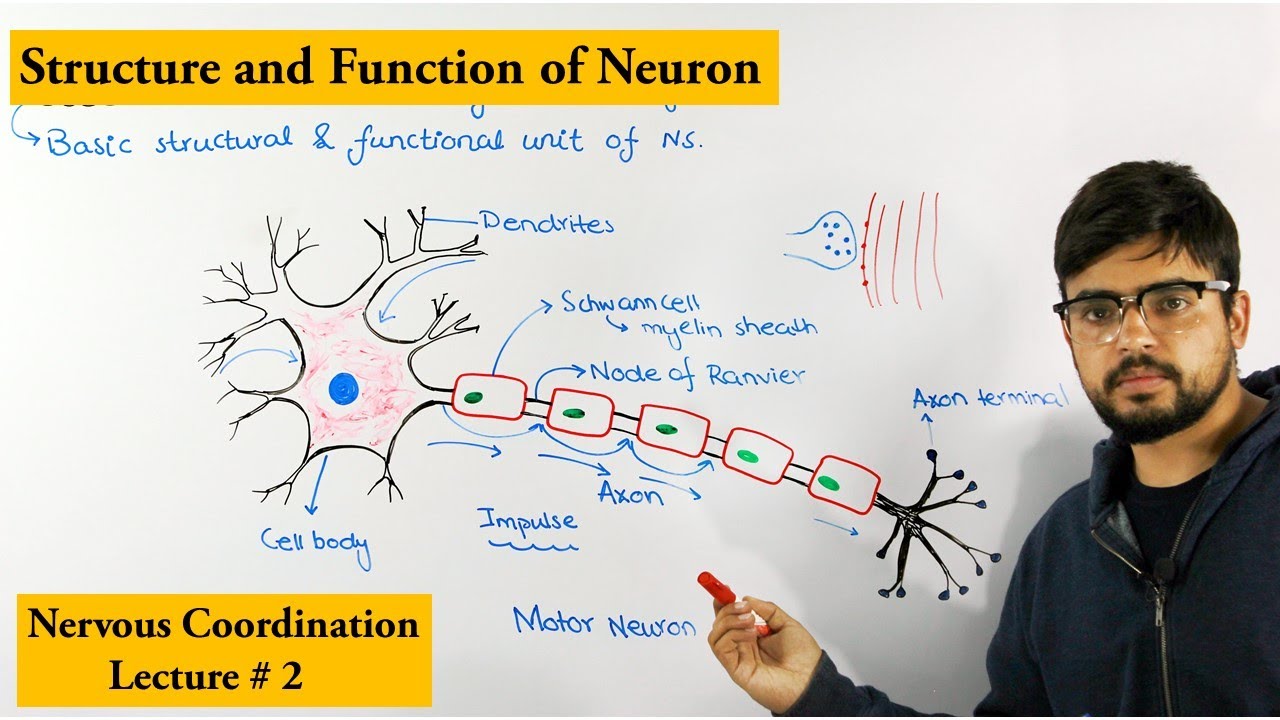





A neuron consists of four major parts: dendrites (which receive signals), the soma or perikaryon (the cell body containing the nucleus and Nissl bodies), the axon (which transmits signals away from the cell body), and axonal terminals (which connect to other neurons); the axon is surrounded by Schwann cells that create the myelin sheath, with gaps between them called nodes of Ranvier, and the entire structure is covered by endoneurium connective tissue.
Neuron Anatomy Explained: Soma, Axon, Myelin Sheath | Nervous System Model
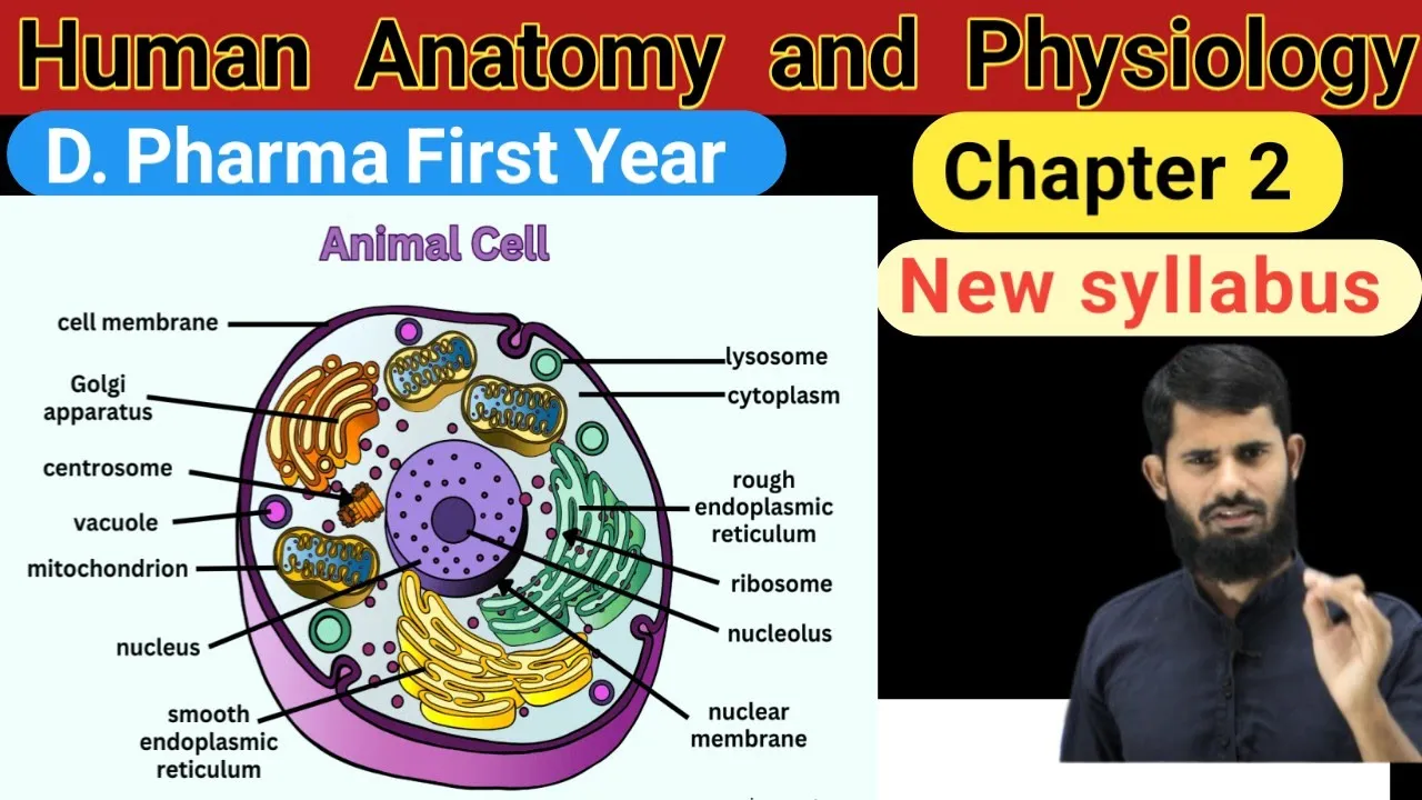
Added:Basic cell biology, including cell structure, organelles, and the function of the cell nucleus.
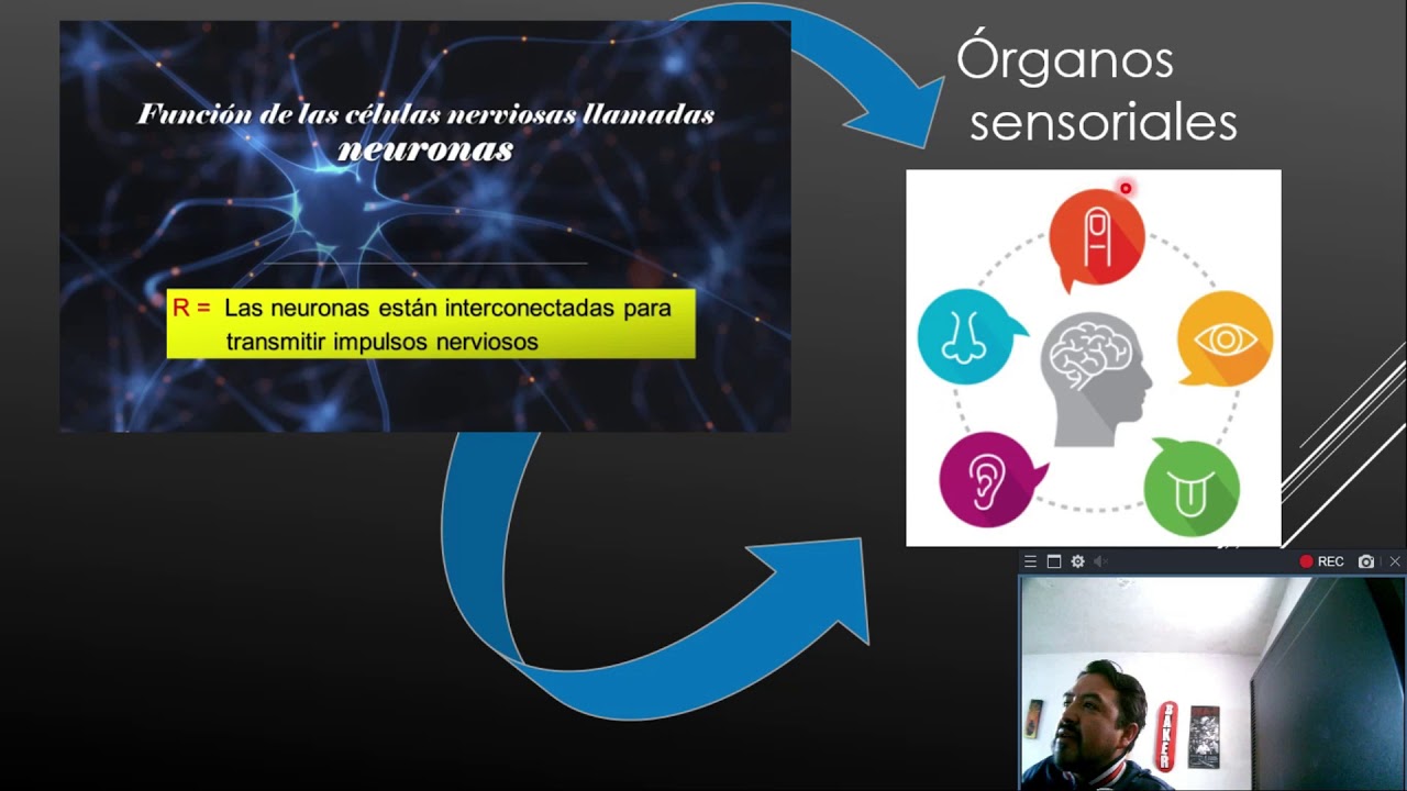
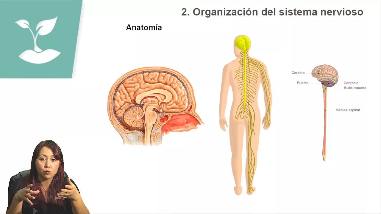
An introductory understanding of the nervous system's role in coordinating bodily functions and sensory perception.
The conceptual understanding that the nervous system communicates using electrical and chemical signals.
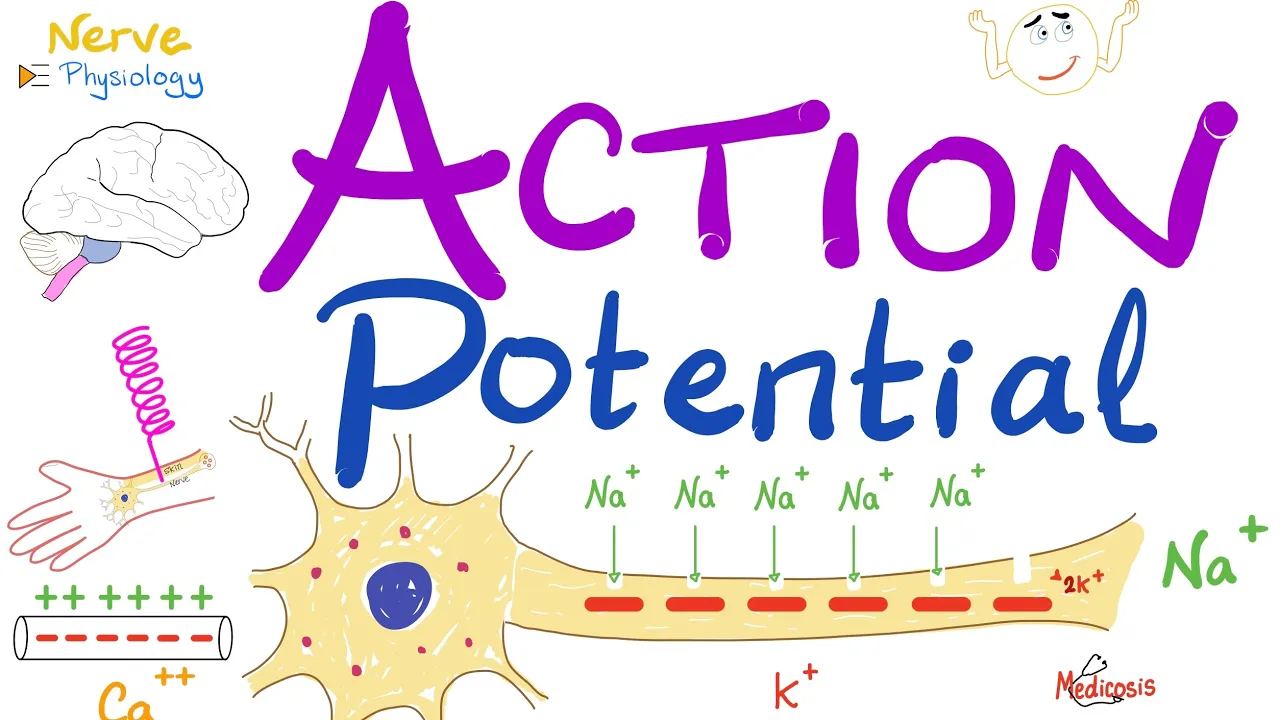
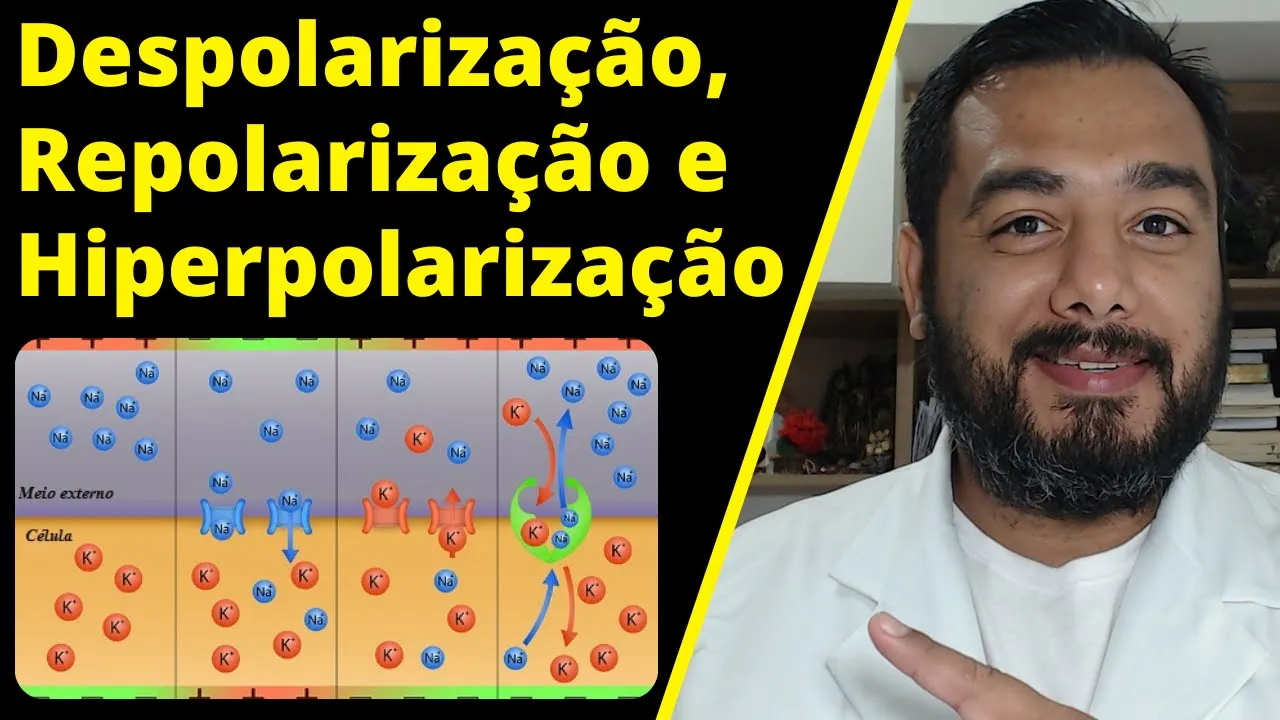
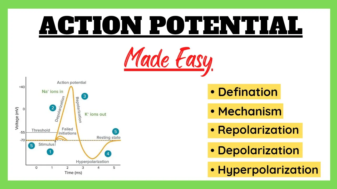
The physiological process of the Action Potential, including depolarization, repolarization, and hyperpolarization.
Saltatory conduction, explaining how myelin sheaths and nodes of Ranvier increase the speed of electrical nerve impulses.
Synaptic transmission, focusing on how chemical neurotransmitters bridge the gap (synapse) between neurons.
The role of glial cells (such as Schwann cells and oligodendrocytes) in supporting neurons and creating the myelin sheath.
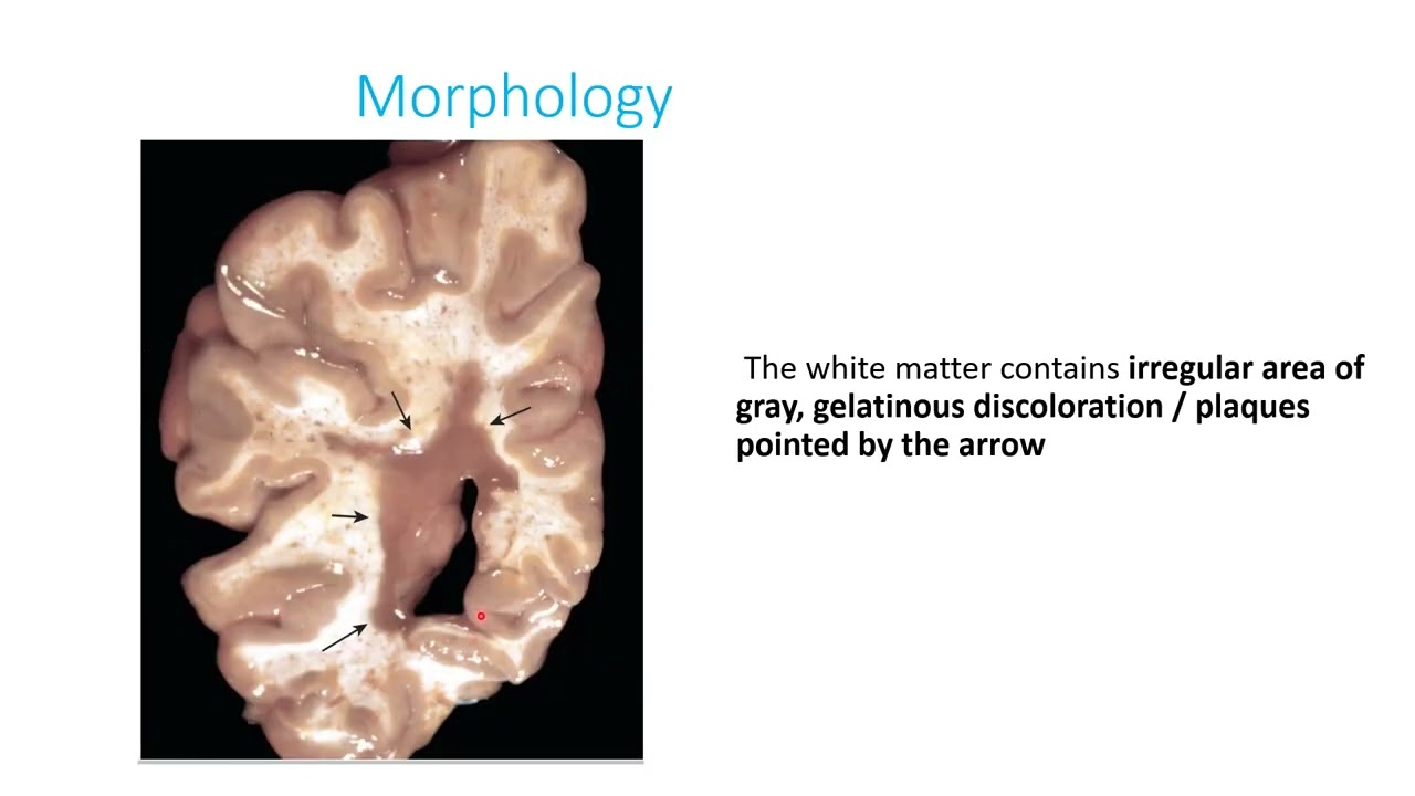
Clinical applications and neuropathology, such as how demyelination leads to disorders like Multiple Sclerosis (MS).
164.6K views1.4Klikes6:35Original Release: 2013-09-27
here's a Neuron model as we take a look at these little parts of the neuron let's get oriented first over here on this side would be the body as well as we come down this would be the distal portion or the axon so let's begin to focus on some of the structures that we can see here on the body well first things first we can kind of just say that that whole thing is the body or also known as the Soma Soma means body or also known as the paric Carion so the paricon people have defined it as oh that's the area around the nucleus which would be the body so that's why we call it that so it could be the paricon or the Soma most people call it the Soma if I were to lift this off they're just trying to illustrate that yes it's three-dimensional but if I were to cut this off you can see that it looks very similar to a normal cell you got the nucleus you got all these endoplasmic reticulum with the goldi body kind of here hanging out and then all these little red dots those are all the ribosomes making all our wonderful proteins so let's kind of get a couple things under our belt before before we move on if we broke out the neuron into four major parts you can divide them into the things that come off the body which are called dendrites the body itself the Soma or pararon the axon that goes all the way down and then the part that we actually don't see is when the axon then splits off into its axonal terminals or synaptic terminals or Bhutan things like that whatever you want to call but that part is not on this model we have these three dendrites ax or Soma and then the axon all right let's take a look at some of the other additional structures that connect this if a neuron is in space it is useless I mean neurons are made to carry information and it's just like saying if I have like a video cable or computer cable and it's just sitting on my desk it doesn't connect to a computer it doesn't connect to a TV doesn't connect to anything it's useless so any kind of neuron has to be connected to something else whether it's a muscle whether it's the brain or something something has to be connected and here is demonstrated with plenty of other connections so you can kind of see these other little things that hang off and touching these dendrites are from other neurons Making Connections passing information so these are our axonal terminals or synaptic Terminals and then in between would be our synapse synapse is just space between the cells all right let's get to the nitty-gritty of the anatomy here so if we already got the dendrites the Soma the axon well what else can we see we can see the nucleus the GOI body and then this rough endoplasmic reticulum these are called nissle bodies when somebody kind of looked at it they saw wow there's so many kind of ribosomes hanging out from there well that person was Mr nissle and he says they're my bodies so I would call them nissle bodies I don't know if he had that accent or not but regardless that is nissle bodies like the stuff that you can kind of see on a microscope they they look like that they're the ribosomes with the endoplasmic reticulum kind of hanging out from there so in addition to that all these dendrites come together in the Soma and then there's only one axon coming out so this part right here you kind of see a cutway leading into this very very long axon this would be considered this triangle portion called the axon hilock or the beginning of the axon beginning of the axon is the axon hillock most sensitive part of the neuron specifically when we say sensitives it just has the most uh channels so that things can be stimulated there and voila you can stimulate an action potential all the way down from there you can kind of see the axon coming down down down with the axonal fibros coming through there so but if you notice this is not just the axon there's some cells that surround the axon with its cell membrane well these types of cells their name is called neurolemmocytes a lot of people call them now Schwan cells because Mr Schwan actually discovered them and from there he called them Schwan cells so Schwan cells a lot easier to say than neurolemmocytes but regardless that is what they are neurolemmocytes and they create what we call a m sheath and the m and sheath is all these little if you can kind of see all these little kind of ridges and all these extra uh amounts of cell membranes like so many of them this is the myin sheath and some people can get confused you know when I point to this is it is it a cell is it the M sheath well I make this analogy if you if you know of a character that wraps your his arms around somebody that just kind of wraps and wraps and wraps and wraps I could point to this say what is this covering and you would look at it and you'd be like well if it's a person well that's that's his arms and I say well who is that well then you would say well that's that's the that's that person's name so in the same sense if I pointed as what's this covering all these little layers you would say that's a milin sheath but if I said what cell is that you would say oh that's that's a schanel Mr you know Mr schanel hello so that is the difference between the mil and sheath which is actually the layers of the plasma membrane of the M sheath you can't separate them just like if I said oh so you talking about his arms or him they're the same thing I can't separate them because they're attached and they're part of each other and that's what we find here this is a Schwan cell with the milin sheath on it and then as it comes over you can kind of see Schwan cells are kind of short so that they have these little gaps this is what we call the no node arier French Mr rier uh kind of found these where there's gaps in between and you'll also find they're also called neurop fibro uh gaps or neuroi nodes uh so these are the nodes of MVA and so and then the lastly is this connective tissue covering and this should extend all the way but they kind of cut it off the rest this is called the enduum connective tissue it sounds a lot like endomysium if you think of muscles but this is endon neurum connective tissue sheet to give it some strength and stuff like that for the actual axon so let's review really quickly so this is our neuron we have four major parts to it and the parts of the neuron itself is the dendrites the Soma or pararon and then the axon and then the axonal terminals which you can kind of see here from this other side from there you can see the peric Carion also has these nissle bodies that has all these you know ribosomal endoplasmic reticulum when it bends into the axon this is the axon hilock and then the axon is surrounded by a myin sheath which is the cell membrane of a cell called the Schwan cell and then you have multiple of them so you have gaps in between called the nodes of ramier or neuro fibral nodes and then you have a connective tissue covering called the endon nuum
Up Next
24:27
Hodgkin-Huxley Model: Action Potential Biophysics Explained
@danielrobb9165
29.4K views•2020-04-12

50:12
Bacterial Communication: Quorum Sensing and Biofilm Formation Explained
@JHUAAP
12.6K views•2011-06-28

5:35
Enteric Nervous System Explained: The Gut's Brain | Neurobiology Lecture
@alumniu6029
438 views•2018-09-12

7:08
Bacteriophages: Earth's Deadliest Killers and Future Antibiotics
@kurzgesagt
34.6M views•2018-05-13
Related Study Plans & Knowledge Roadmaps
Structured learning paths in Biology
biology
RNAi: Biogenesis, RISC & Gene Silencing
01Molecular Biology Foundations: DNA to Protein
02Introduction to RNA Interference (RNAi)
03Molecular Biogenesis of miRNA and siRNA
+ 2 more learning modules
10h5 modules
Explore Roadmap
biology
Mycorrhizae: Signaling, Biophysics & Ecology
01Foundations of Mycorrhizal Symbiosis
02Molecular Signaling and Root Colonization
03Biophysics and Bioenergetics of Nutrient Exchange
+ 1 more learning modules
11h4 modules
Explore Roadmap
biology
Circadian Clocks: TTFL, SCN & Periphery
01Foundations of Molecular Biology & Gene Expression
02Introduction to Chronobiology and Actograms
03The Core TTFL: Molecular Architecture of the Clock
+ 3 more learning modules
14h6 modules
Explore Roadmap
biology
Population Genetics: Math, Drift & Selection
01Genetic & Probability Foundations
02The Hardy-Weinberg Principle and Baseline Models
03Mathematical Modeling of Natural Selection
+ 3 more learning modules
18h6 modules
Explore Roadmap