











![[LIVE] Learn Genetic Engineering - Part 1: How does it work?](https://i.ytimg.com/vi/E_TAHxPBqKY/hqdefault.jpg?v=63839add)

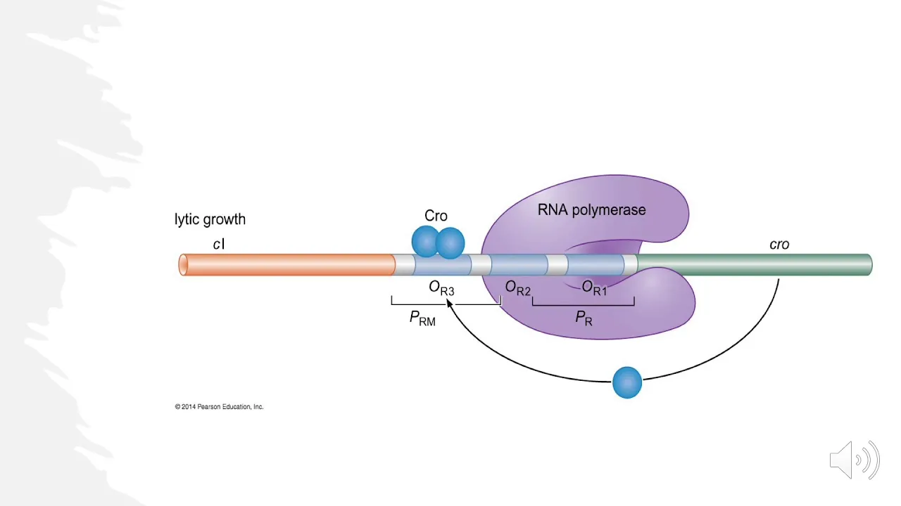









Bacteriophages (phages) are viruses that specifically infect bacteria, discovered by Felix d'Herelle and Frederick Twort between 1915-1918; they were identified using the plaque assay, which revealed their particulate nature, and later visualized by electron microscopy showing their characteristic head-and-tail structure with double-stranded DNA genomes. Phages propagate through two life cycles: the lytic cycle where they kill the host cell after replicating, or the temperate cycle where they integrate into the bacterial chromosome as prophages and replicate alongside the host without immediate cell death. Phages exhibit remarkable abundance in nature, with approximately 10^31 viral particles in Earth's biosphere, making them the majority of all biological entities and representing an enormous reservoir of genetic diversity and potential new genes.
Bacteriophages: Nature's Most Abundant Viruses | Graham Hatfull
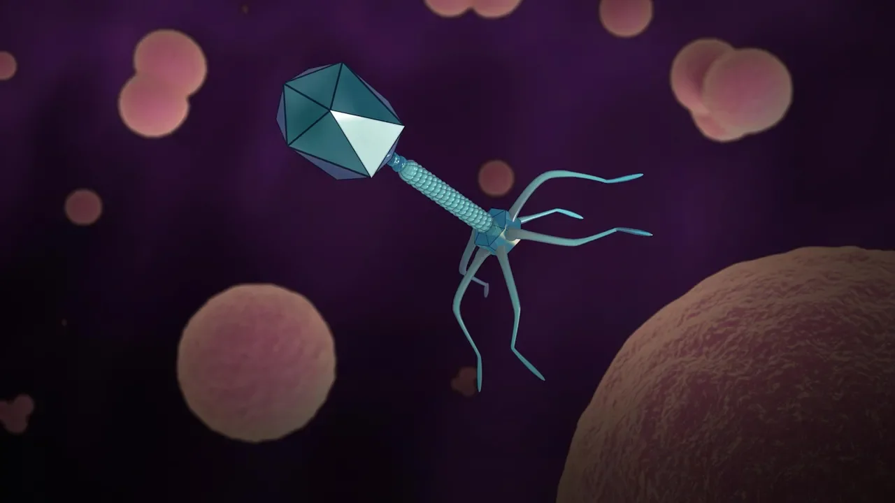
Added:hello my name is Graham hatful uh I'm a professor at the University of Pittsburgh uh and a howto's Medical Institute professor and we're going to talk today about bacteria phases their genes and their genomes um first of all in part one um I'd like to just discuss what bacteria fases are um some of their biological properties uh and how they were discovered so bacteria fases are viruses that infect bacterial h so just as we and our some of our many of our animal cousins uh are infected by viruses that are specific for us bacteria also have viruses that infect them and they're called bacteria phases or phases for short they were discovered or co-discovered by um Felix derell and Frederick twart um in between 1915 and 1918 and they were discovered as a agents that when added or present in a culture of growing bacteria were capable of uh of killing the bacteria and essentially um having a a bacteriocidal uh effect it was really um Felix derell who developed an assay called the plaque test um where he was able to um take samples of uh bacteria phases uh that were able to kill bacteria and he made dilutions increasingly greater series series of dilutions of the bacteria face sample and then plate that out in the presence of the bacterial host on solid me media using like Petri dishes as shown here um and what he saw was was that when he diluted out the sample sufficiently he could see individual areas of killing um as you can see here uh with smaller and large areas where the cells are dead uh and where the viruses have grown these are called plaques and each one of these individual plaques has arisen by a single particle which was capable of infecting a cell and as the bacteria grew across the surface of the AAR dish um the virus um propagated itself multiplied uh until in in the plaque here there may be perhaps uh a million or 10 million or more individual Fage particles this plaque test was really important because um they could tell from looking at uh cultures of or looking at lates of bacteria phases that there was nothing to see in there um they could filter the samples they knew they still had the Infectious property um but the tube looked completely clear there was nothing to see and when they placed these particles um with this capability in the light microscope um there was nothing to be seen so this these were mysterious entities um but Felix derell showed conclusively that they really are particulate in nature it was somewhat later in the 19 late 1930s and 194s that the electron microscope was uh was developed which has a level of resolution Way Beyond the light microscope uh and uh was able to show for the very first time what viruses including bacteria fases actually looked like and I'll show you some pictures in a minute um but this just shows a stylized example of what one of those fages look like um at the very top here you can see is a structure that is referred to as the head sometimes it's called the capsid that is attached to this uh longer structure here here which is the tail and the DNA or the genetic information that the virus carries in order to uh to multiply itself the instructions for its own replication are carried here uh in the virus head and so um we can see and we now know uh of our understanding of what this particular uh virus and these types of viruses look like is that this structure here at the very bottom which is the tip of the tail that is the region that recognizes and binds to the outside of the bacterial host during the process of infection the structures that we see principally constructed or made up of protein components that stuff stays on the outside of the cell and the DNA or the genetic information um is what passes from the head through the tail into the cell and then reprograms that cell in order to make more copies of the virus by electron microscopy um we can see and we now know that a very large proportion of these uh naturally found viruses are in an order which are referred to as the cordoes of which these are primarily phages that contain Tails as I just described and shown in the examples here and they contain double stranded DNA so there's lots of different types of viruses in different shapes but the vast majority that you find in nature fall into this particular uh order and uh these have names according to the types of tails that they have um these are the myoid and they have contractile tails and so the tail actually contracts like a syringe when the DNA is injected into the back bacterial host these are called the pooid um which have little short stubby Tails uh and these ones on the right here are the cipher verid uh and these have long uh non-contractile and flexible Tails so these are the main forms of these viruses there are however a variety of number um of different types of viruses that have different shapes and have indeed different types of uh DNA or RNA genomes within them so we can think about the various steps that are involved in the propagation of a bacteria phase during this process known as litic growth um the Fage starts by absorbing to the outside of the cell the DNA is injected inside the cell in this process of penetration there is a set of early proteins uh which are encoded by the Fage but which uses the host Machinery to express them viral DNA is replicated to make lots more copies of the virus DNA and then these late proteins are expressed um that make uh the structures that I just showed you uh in the electron microscope the the capsid structure gets assembled the tails get assembled and then in the process of DNA packaging the DNA is stuffed into those heads until the heads are full the tails are attached and the very final step of this process um the bacterial cell is going to lice um with enzymes encoded by the Fage genome to break open the outside of the cell and the progyny viruses typically 50 to 100 New progeny virus viruses will be released and to go on and repeat this cycle whenever they find a another bacterial host so while many phages go through um a litic uh growth cycle and that is essentially simply how they reproduce themselves that's not the only uh type or form of growth cycle that fages can enjoy and there's a large class perhaps even a majority of bacteria fases that enjoy what is referred to as a temperate um life uh State um what that means is that when a temperate Fage infects its bacterial host there are two possible alternative outcomes one of those outcomes is litic growth um in which they propagate themselves in exactly the same way as I described to you in the previous slide and normally that occurs perhaps 80 or 90% of the times that the host cell is infected by a temperate phage 10 to 20% of the time there's an alternative outcome and that alternative outcome is referred to as logy in lyny the litic genes the genes that are required to propagate itself and and lice and kill the cell they're all switched off they are repressed and the Fage DNA establishes itself um so that it can be propagated in the long term within the subsequent uh growth cycles of the bacteria host and that's usually although not always accomplished by integration of the Fage DNA into the bacterial chromosome so the Fage genome itself becomes Incorporated and becomes a part of the bacterial chromosome and gets rep gets replicated just like all of the other bacterial genes do so we can think of logy as a form of parasitism um where the Fage is simply going along for the ride in what is otherwise um a very healthy cell uh it's basically it's a free ride U through many many generations of bacterial growth so lysogens uh tend to be stable um but they are not uh going to they don't have to uh be uh remain in that state forever uh they can go through many many many rounds of of of growth however uh as a process that happens either spontaneously or can be reduced by D DNA damage such as UV light um lysogens can leave this uh this uh this the the comfort of that life cycle and they can be induced into full litic growth there's a relatively easy way to distinguish between phages that are temperate and those that are litic and can only undergo the litic uh growth cycle by the types of plaques that you see on AAR plates litic fages form simply just clear plaques as shown in the bottom right hand corner here where all of the cells within that infected area have been killed killed by Fage infection over on the left at the bottom here you can see what temperate fages look like they form turbid plaques you see plaques because there's cells that are being killed through litic growth and propagation of the virus but there's that important key subset of cells that uh that in which lysogeny is established and those uh those lysogens are able to survive and to grow and they can grow perfectly happy even though they're bathed within this um density of bacteria phased particles so the fact that lysogens can actually grow quite happily even though there's lots of viruses around to infect them um is referred to as super infection immunity lysogens are immune to Super infection by a phage of the same or a closely related type um this slide just shows an example o of how that can be uh studied and what that looks like in the lab um at the top left here are plaques of a turbid uh Fage a temperate Fage growing on a lawn of bacteria using either a toothpick or a wire um you can go and pick cells from the very center of one of those plaques streak it out on an agar plate as shown at the bottom left here um in order to generate single colonies each grown up from a single cell that will have come from the turbit plaque and then these individual colonies can in turn be tested for their immune phenotypes as shown in the top right in this example with we're looking at um we're comparing a non-genic strain with a lysogen and in the top we've just spotted on dilutions of a bacteria phas sample and you can see that these serial dilutions um from the most going down to the least uh number of particles um gives you the tighter I the number of particles per milliliter of um uh of of sample on the non logen but when you compare it with exactly the same dilutions of that particular phage onto a lysogen uh of itself so this particular Fage is called Giles on the right is the Giles lysogen and you can see that there is essentially very little infection except perhaps a little bit of clearing at the very highest concentration of Fage at the top left hand corner of that panel and a control Fage in this case below it showing L5 um which which is also a temperate phage but it does not share immunity with Gils and therefore you see essentially the same number of plaques on the lysogen uh of Gils and the non logen and therefore bacteria phases can be grouped together according to their immune specificities and it can be done just by comparing uh the infectivity of a whole set of phages on lysogens and non- lysogens so this is an important parameter uh uh that enables us to group together fages that may be similar by sharing these types of uh parameters I mentioned that phage DNA can can integrate itself into the host chromosome and this is a common feature of temperate bacteria phases they do it by a process which is referred to U as sight specific re combination this process is catalyzed IED by an enzyme which is called Integra um or int for short the Integra is encoded by the Fage genome and integr catalyzes re combination between two specific sites or segments of DNA one is the so-called Fage attachment site or at P for short and the other is a specific site within the bacterial chromosome which is which is called the attachment site for the bacterium or at B integration results in the formation of what is called a proas an integrated phase DNA it's become part of the chromosome and it is now the propas DNA is flanked by the left and right attachment sites referred to as uh ATL and at R respectively uh this reaction um does use um host functions quite commonly um integr works together with a host protein a bacterial protein called integration host factor or IHF for short um and you'll recall that I told you that lysogens can undergo spont spontaneous or or induced induction into lter growth which means that um there has to be a process for this to come back out again a biological reversal of this overall reaction that's called excision and excision is again catalyzed by Integra and there is a second phage encoded protein called exis or XIs for short um and XIs is a protein that essentially dictates and determines the directionality of how uh these reactions will occur in the absence of exis you do integration in the presence of exis then the integr um is uh only capable of doing the the excision reaction a few years ago um uh people started thinking about how many bacteria fases there really were out there in the biosphere and what they did was they developed a technique called epif florescence where they could take a sample let's say of seawater which is easy to get um get seawater add a sample of a Dye which binds to the nucleic acids place that sample under the under fluorescence microscope and simply uh look and count for the viruses and other components that you see um in that kind of experiment this is an example of what they saw there's a very large number of what you can see as small green fluorescent Dots here those are all of the viruses that are present and there's a smaller number a fewer number of these large brighter spots um which are larger objects and they are the bacteria and so what it was possible to do was to Simply to count how many virus-like particles are present in these samples and and when that was done uh it was clear uh that the viral population is indeed absolutely vast when you measure there's about 10 ^ of 6 to 10 ^ of 7 viral particles per Mill and this number seems to be reasonably steady no matter where you look um it's true uh in seawater if you look in coastal samples if you look in Oceanic samples or the surface or the Deep um and there's a similar abundance or a related degree of abundance in terrestrial samples as well it is it is believed and so because we can measure the number of particles present in small samples and we can multiply the amount of uh seawater and the amount of uh terrestrial components and when we do that we can conclude that the the biosphere contains a total of 10 to the^ of 31 um virus particles the vast majority of which are bacteria fases this is an incredible number this number would suggest that there's more bacteria phas particles in the biosphere than all other biological entities added together phages are in fact the majority of all biological IC things in the biosphere they're not only abundant uh this appears to be a very Dynamic population as well um you can see from the fluorescence patterns in this slide um but it's true in in most samples that have been examined that the ratio of bacteria phas particles to bacteria is about between 5:1 and 10:1 and that's important because it means that um that the bacteria are likely to constantly be subjected to infection by the bacteria phases um in their natural environments and in fact there are ecological studies that estimate the number of viral infections per second um that occur on a global scale and that number is estimated to be between 10 the^ of 23 and 24 um which is just an incredible number of uh of activity a dynamic population indeed um these numbers would suggest that the entire Fage population turns over every four or five days it is a large and a stunningly Dynamic um set of um biological items not surprisingly perhaps um the viral population is extremely diverse when we look at the genetic level um currently um uh a number far short of 10 the^ of 31 Fage particles have been subjected to DNA sequencing perhaps about 650 or so uh and from these genomes we can look and we can see how similar or different they are to each other and and what we learned from this is that indeed there's many many different types of sequences and these genomes appear to uh Harvest and to contain large numbers of geneses which are unlike any other genes that we've seen before so we can conclude then that bacteria phases represent the majority of all biological entities uh in the in the biosphere they're a dynamic population constantly infecting bacterial hosts and and generating more coppers of themselves the bacteria must be struggling to maintain their survival through resistance uh to these infections and um I think a compelling argument can be made that this population has probably also been evolving for a very long time perhaps two three perhaps even four um billion years extending right back to the very early days of when life evolved and and and finally fages um appear to uh represent the largest unexplored reservoir of of new genetic information uh in in the biosphere if you want to discover new genes perhaps with new functions perhaps with new structures um the bi the bacteria phase population uh I think we would argue is exactly where you should start to look in part two we'll look in some more detail at the genetic structures of U bacteria phase genomes uh and and see how that's given us some insights into how these genomes have evolved
Up Next
Virus Reproduction: Lytic and Lysogenic Cycles Explained
@missangler
87.7K views•2022-02-08

Bacterial Communication: Quorum Sensing and Biofilm Formation Explained
@JHUAAP
12.6K views•2011-06-28

MicroRNAs: Gene Regulation Explained by David Bartel | iBiology
@scicommlab
233.1K views•2014-03-24

Bacteriophages: Earth's Deadliest Killers and Future Antibiotics
@kurzgesagt
34.6M views•2018-05-13
Related Study Plans & Knowledge Roadmaps
Structured learning paths in Biology