Fluent aphasia, also known as Wernicke's aphasia, is a type of aphasia characterized by fluent but meaningless speech with poor comprehension; individuals speak with effortless flow and grammatical structure but produce words that lack coherent meaning, often creating nonsensical phrases while remaining unaware of their communication difficulties.
Fluent Aphasia (Wernicke's Aphasia): Speech & Comprehension Impairments Explained
Added:Deep Dive
Basic neuroanatomy, specifically the location and function of the temporal lobe and Wernicke's area in the dominant hemisphere of the brain.

Cerebrum | Structure & Function
7.2K
The frontal lobe contains the primary motor cortex (pre-central gyrus) for voluntary movement, motor association cortex for movement planning, prefrontal cortex for personality and memory, and Broca's area (dominant hemisphere) for speech production. The temporal lobe processes auditory information through the primary auditory cortex (basic sound detection) and auditory association cortex (sound identification). Wernicke's area (dominant hemisphere) handles language comprehension, distinguishing it from Broca's area which controls speech output.
Click to watch →

Auxiliando a identificar a Área de Broca e Área de Wernicke
2.7K
Broca's area (motor speech area) is located anterior to the central sulcus in the posterior inferior frontal lobe, while Wernicke's area (language comprehension area) is located in the superior temporal gyrus above the Sylvian fissure; both areas follow the general brain organization where motor functions are anterior and sensory functions are posterior, with the left hemisphere being dominant for language in most individuals.
Click to watch →
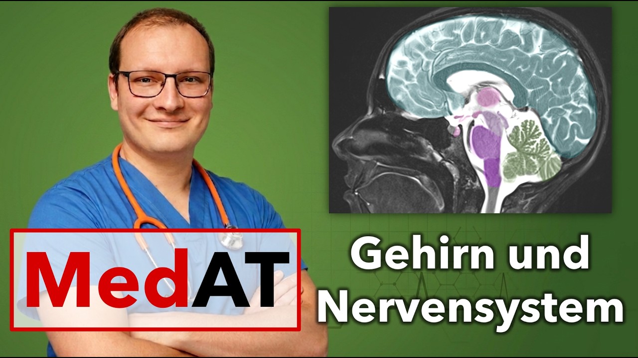
Nervensystem für Matura und MedAT - Einführung für Biologie und Medizin
5.8K
In most people, language areas are located in the left hemisphere. Wernicke's area (temporal lobe) receives and interprets language—patients with damage here (Wernicke's aphasia) cannot understand spoken or written language and produce meaningless speech despite grammatical structure. Broca's area (frontal lobe) produces language—patients with damage here (Broca's aphasia) cannot speak, write, or gesture but can understand language and copy letters. The dominant hemisphere refers to which contains language areas, not which arm is dominant.
Click to watch →

360 Degree Series | Most Imp. MCQ’s #116 | Staff Nurse | AIIMS | GMCH | DMER | Siddharth Sir
18.7K
Wernicke's area is located in the temporal lobe (superior temporal gyrus) and is responsible for language comprehension. Damage to Wernicke's area causes receptive aphasia, where patients cannot understand spoken or written language. They may produce fluent but meaningless speech and are often unaware of their communication deficits.
Click to watch →
Cerebrum &Sensory cortex
290
Wernicke's area is located in the superior temporal gyrus of the dominant hemisphere (usually the left hemisphere) and is responsible for language comprehension. It helps us understand spoken and written language by processing the meaning of words and sentences. Broca's area is located in the inferior frontal gyrus of the dominant hemisphere and is responsible for speech production. It helps us produce spoken language by coordinating the movements of the muscles involved in speech.
Click to watch →
Deep Dive
The core distinction between receptive language (understanding spoken or written communication) and expressive language (producing speech).
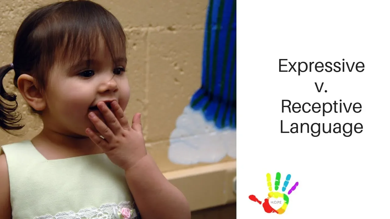
Receptive versus Expressive Language
18.6K
Receptive language refers to the ability to understand and follow another person's language (such as selecting a card when asked 'which one says ribbit' or bringing a pencil when told 'go get me a pencil'), while expressive language refers to the ability to respond or give a vocal response using one's own language (such as naming an object when shown a picture or describing its color).
Click to watch →
Receptive Language Milestones by 18 Months | teachmetotalk | Laura Mize | Speech Therapy Toddlers
6.2K
This segment explains the critical distinction between receptive language (what children understand) and expressive language (what children can say). Children always understand words before they can use them to communicate. The 12-18 month period features a receptive language explosion that sets up the expressive language explosion that follows. Many parents and physicians miss receptive language delays because they focus on talking spurts and attribute lack of understanding to behavior rather than language comprehension.
Click to watch →
Receptive Language Milestones by 12 Months | Laura Mize | teachmetotalk.com
6.8K
Receptive language refers to what a child understands or comprehends from the words they hear, also known as auditory comprehension skills. It represents the input piece of communication, contrasting with expressive language as the output piece. A critical distinction is that receptive language problems automatically lead to expressive language problems because children must understand words before they can use them meaningfully. This connection is essential for parents and professionals to understand, as receptive language delays often underlie apparent 'late talking' issues. When a child falls behind in receptive language, they are not just a late talker—more developmental concerns may be present.
Click to watch →

برنامج هي|اسباب و عوامل تأخر النطق عند الاطفال و كيفية علاجها مع "د/سهام حسن" اخصائي نفسي و تخاطب
315.9K
There is a difference between receptive language (اللغة الاستقبالية) and expressive language (اللغة التعبيرية). Receptive language refers to a child's ability to understand what others say and follow instructions. Expressive language refers to a child's ability to communicate their needs and thoughts verbally. A child may understand everything but struggle to express themselves, which is a common developmental stage.
Click to watch →

Otizm mi Dil Gecikmesi mi? Gecikmiş Dil ve Konuşma ile Otizm Arasındaki Etkileşime Dair 7 Fark.
239.7K
In typical development, children's receptive language (understanding) skills are generally stronger than their expressive language (speaking) skills. Children with speech delays typically understand more than they can say. Children with autism may show different patterns, where their understanding abilities may not be as strong as expected, and their speech may include echolalia (repeating words heard) without clear communicative intent.
Click to watch →
Deep Dive
The general definition of aphasia as an acquired language impairment, and how cerebrovascular accidents (strokes) can cause localized neurological deficits.
Stroke Nursing (CVA) Cerebrovascular Accident Ischemic Hemorrhagic Symptoms Treatment tPA
2.4M
Stroke can produce various neurological deficits affecting communication and function. Aphasia encompasses receptive aphasia (inability to understand spoken language affecting Wernicke's area), expressive aphasia (inability to produce speech despite comprehension affecting Broca's area), mixed aphasia, and global aphasia (complete communication failure). Additional deficits include dysarthria (slurred speech from weak muscles), apraxia (inability to perform voluntary movements despite intact muscle function), agraphia (loss of writing ability), alexia (loss of reading comprehension), agnosia (inability to recognize familiar objects or people), and hemianopia (loss of half the visual field in each eye).
Click to watch →

MayaHenryEditedLectureSeq
101
Aphasia is an acquired language impairment occurring after brain injury, typically in the left hemisphere, affecting production and comprehension while sparing intelligence, memory, and personality. Most cases result from stroke, with 180,000 new cases annually in the U.S. and one million people living with aphasia. The average age of onset is 68. Aphasia is not a disease but a symptom that can result from stroke, traumatic brain injury, tumors, or neurodegenerative diseases. People with aphasia retain their intelligence and memories despite communication difficulties.
Click to watch →

Kelas Online UKDI Feb 2024 Neurologi
8.7K
Aphasia is a language disorder caused by brain damage. Motor aphasia (Broca's area, 44-45) causes inability to speak but preserved comprehension and repetition. Sensory aphasia (Wernicke's area, 22) causes inability to understand but preserved speech and repetition. Global aphasia causes inability to speak or understand. Transcortical motor aphasia (supplementary motor area) causes inability to speak but preserved comprehension and repetition. Transcortical sensory aphasia (supplementary sensory area) causes inability to understand but preserved speech and repetition. Conduction aphasia (arcuate fasciculus) causes inability to repeat but preserved comprehension and speech. Stroke is classified as ischemic (80%) or hemorrhagic (20%). Ischemic stroke presents with focal deficits without headache; hemorrhagic stroke presents with headache, vomiting, and decreased consciousness. Localization: Carotid territory strokes (80%) cause contralateral motor/sensory deficits and aphasia; vertebrobasilar territory strokes (20%) cause vertigo, visual disturbances, and consciousness changes. Treatment for ischemic stroke includes alteplase (first-line within 3 hours) or aspirin; antihypertensives if BP >220/120. Treatment for hemorrhagic stroke includes antihypertensives and surgical intervention (craniotomy).
Click to watch →
Topic- Neurology |MRCP PART-1 ( TEST-1) | Dr. Mukesh Bhatia | DBMCI
58.7K
Aphasia (language disturbance) localizes to the left hemisphere (in most right-handed individuals). Broca's area (inferior frontal gyrus) causes expressive aphasia (difficulty producing speech), while Wernicke's area (superior temporal gyrus) causes receptive aphasia (difficulty understanding speech). Both areas are supplied by the middle cerebral artery. The key teaching point is that left MCA stroke causes aphasia, while right MCA stroke does not.
Click to watch →

Doctor explains APHASIA - definition, symptoms, causes, investigations...
108.1K
Aphasia is a language disorder caused by brain damage, typically from stroke, head injury, tumors, or neurological diseases, affecting the left hemisphere's language centers; it manifests in different forms including fluent Wernicke's aphasia (meaningless speech with poor comprehension), non-fluent Broca's aphasia (effortful speech with preserved comprehension), and global aphasia (severe communication impairment); diagnosis involves MRI/CT scans and evaluation by speech-language pathologists to assess language production and comprehension abilities.
Click to watch →
Deep Dive
The difference between language disorders (affecting meaning, grammar, and symbols) and motor speech disorders like dysarthria or apraxia.

EL HABLA en TÚ DE CIENCIAS Y YO DE LETRAS con @Linguriosa y @Lahiperactina COMPLETO 1x11
2.2K
This segment distinguishes between speech disorders (affecting physical production) and language disorders (affecting comprehension and expression). Dysarthria involves weakened speech muscles, causing slow, slurred speech, and can result from ALS, stroke, or Parkinson's. Aphasia affects language processing: Broca's aphasia impairs speech production while preserving comprehension, while Wernicke's aphasia impairs comprehension while speech remains fluent but nonsensical. The hosts discuss Bruce Willis's aphasia from frontotemporal dementia and note that these conditions can be rehabilitated through therapy.
Click to watch →
Transtornos Motores da Fala - Juliana Rocha
139
Speech-language pathologists must focus on core competencies: language disorders, motor speech disorders, and related conditions. Clients span from infants to adults, and childhood apraxia of speech persists into adulthood. Clinicians must distinguish between language disorders (language delay, Specific Language Impairment, disorders with known etiological factors) and motor speech disorders (motor speech delay, childhood apraxia of speech, phonological disorder, developmental verbal dyspraxia). Speech involves multiple interconnected elements including cognitive, motor, and social components. A child with motor speech disorder may develop secondary language difficulties because they cannot test new words, leading to reduced vocabulary and grammatical difficulties.
Click to watch →

روانشناسی کودکان استثنایی دکتر پیری پارت 10 و پایانی
1.1K
Speech and language disorders include: aphasia (acquired language impairment from brain damage), dysarthria (motor speech disorder affecting articulation), and apraxia of speech (difficulty planning and coordinating speech movements). Aphasia has two main types: receptive aphasia (Wernicke's) - damage to Wernicke's area in the left temporal lobe, causing inability to understand language; and expressive aphasia (Broca's) - damage to Broca's area in the left frontal lobe, causing difficulty producing speech. Motor speech disorders include dysarthria (difficulty articulating words due to muscle weakness), apraxia of speech (difficulty planning and coordinating speech movements), and ataxia (affects coordination and speech timing). These disorders can result from neurological damage.
Click to watch →
What is a Speech Disorder? (Apraxia of Speech and Dysarthria)
203.7K
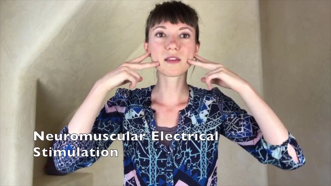
Speech disorders are diagnosed as either apraxia of speech (a motor planning disorder where the brain knows what to say but cannot execute the motor plan consistently, causing variable articulation errors) or disarthria (a physical disorder involving muscle weakness, decreased range of motion, or paresis affecting speech production); treatment approaches vary accordingly, with apraxia requiring motor coordination exercises and disarthria typically addressed through strengthening exercises, vocal therapy, massage, and neuromuscular electrical stimulation like VitalStim.
Click to watch →

Avaliando a Linguagem Infantil - Dia 01
10.2K
Childhood apraxia of speech is a motor speech disorder that affects the planning and programming of speech movements. Children with apraxia produce inconsistent speech errors and have prosodic difficulties. Motor speech disorders include childhood apraxia of speech, dysarthria, and childhood motor speech delay. Developmental Language Disorder (DLD) is an idiopathic condition characterized by persistent difficulty in developing language, with a prevalence of approximately 8.5%.
Click to watch →
Prerequisite Knowledge
- Concept 01Basic neuroanatomy, specifically the location and function of the temporal lobe and Wernicke's area in the dominant hemisphere of the brain.
- Concept 02The core distinction between receptive language (understanding spoken or written communication) and expressive language (producing speech).
- Concept 03The general definition of aphasia as an acquired language impairment, and how cerebrovascular accidents (strokes) can cause localized neurological deficits.
- Concept 04The difference between language disorders (affecting meaning, grammar, and symbols) and motor speech disorders like dysarthria or apraxia.
Subsequent Learning
- Step 01A comparative study between Fluent (Wernicke's) and Non-fluent (Broca's) Aphasia, mapping out their distinct clinical profiles.
- Step 02Diagnostic tools and assessment protocols used by speech-language pathologists to evaluate the type and severity of comprehension deficits.
- Step 03Rehabilitation methods and speech therapy interventions tailored for Wernicke's aphasia, such as Treatment for Wernicke's Aphasia (TWA).
- Step 04The concept of anosognosia (the lack of awareness of one's own deficit), which frequently accompanies fluent aphasia, and its impact on therapy.
- Step 05Neuroplasticity and the mechanisms of language recovery and cortical reorganization in post-stroke patients.
Greetings
Current Plans
Cruise Context
iPad Activity
Daily Life
Golf & Hobbies
Helping Others
Farewell
Greetings
0:07Playing Section
- 1
Megan and Byron exchange brief pleasantries.
- 2
Byron expresses happiness and complements Megan.
The Dual-Stream Model and Distributed Network Perspective
The traditional view of Wernicke's aphasia relies on the classical localizationist model, which attributes fluent but empty speech and comprehension deficits strictly to damage in Wernicke's area. However, modern cognitive neuroscience, particularly through Hickok and Poeppel’s Dual-Stream Model, challenges this paradigm. Research shows that language comprehension is not localized to a single region but is supported by a highly distributed, bi-hemispheric network. Deficits in fluent aphasia often arise from disruptions in the 'ventral stream' (mapping sound to meaning) or the 'dorsal stream' (mapping sound to articulation), rather than a loss of a localized 'comprehension center.' Furthermore, patients with Wernicke's aphasia exhibit highly heterogeneous symptoms, suggesting the condition is not a single unitary deficit, but a complex combination of phonological, semantic, and auditory working memory impairments across widespread temporal, parietal, and frontal networks.
Deep Dive
A comparative study between Fluent (Wernicke's) and Non-fluent (Broca's) Aphasia, mapping out their distinct clinical profiles.
comparaison entre l'aphasie de BROCA et l'aphasie de WERNICKE
57.4K
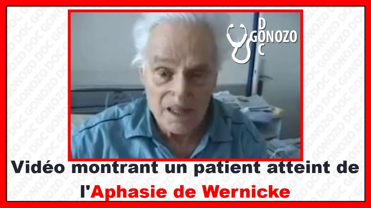
Broca's aphasia is an expressive (non-fluent) language disorder caused by damage to Broca's area in the inferior frontal gyrus of the dominant hemisphere, characterized by difficulty producing speech and finding words (anomia), while Wernicke's aphasia is a receptive (fluent) language disorder caused by damage to Wernicke's area in the posterior superior temporal gyrus, characterized by fluent but incomprehensible speech and the patient's false belief that they are being understood.
Click to watch →

Hematology & Neurology by Dr. Dilip Kumar | Medicine Saturdays | Cerebellum Academy
50K
Aphasia is a language disorder from dominant hemisphere lesions. Classification: (1) Fluent aphasia (Wernicke's, transcortical sensory, conduction, nominal) - preserved fluency; (2) Non-fluent aphasia (Broca's, transcortical motor, global) - impaired fluency. Key features: Wernicke's = fluent, impaired comprehension, impaired repetition; Broca's = non-fluent, preserved comprehension, impaired repetition; Transcortical = intact repetition (distinguishes from Wernicke's/Broca's); Conduction = isolated repetition loss; Global = all functions lost. Nominal aphasia = isolated naming loss (early Alzheimer's).
Click to watch →

Aphasia: Wernicke's vs Broca's - Clinical Anatomy | Kenhub
189.4K
Aphasia is a language disorder caused by brain damage, primarily affecting speech production and comprehension; Broca's aphasia (expressive aphasia) results from damage to Broca's area (Brodmann areas 44-45) in the frontal lobe, causing difficulty expressing thoughts while comprehension remains intact; Wernicke's aphasia (receptive aphasia) results from damage to Wernicke's area (Brodmann area 22) in the temporal lobe, causing fluent but meaningless speech with impaired comprehension; treatment primarily involves speech and language therapy to restore communication abilities.
Click to watch →
BANQUEO | Neurología - ENAM 2021
11.5K
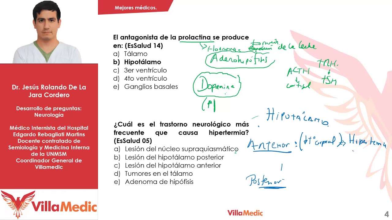
Broca's aphasia (expressive aphasia) involves difficulty producing speech (non-fluent) with preserved comprehension. Patients can understand but cannot articulate words properly. Wernicke's aphasia (receptive aphasia) involves difficulty understanding language with fluent but meaningless speech. Patients produce neologisms (invented words) and are unaware of their deficits. Both involve impaired repetition and naming.
Click to watch →
Aphasia (Broca vs. Wernicke vs. Conduction vs. Global vs. Transcortical)
14.2K
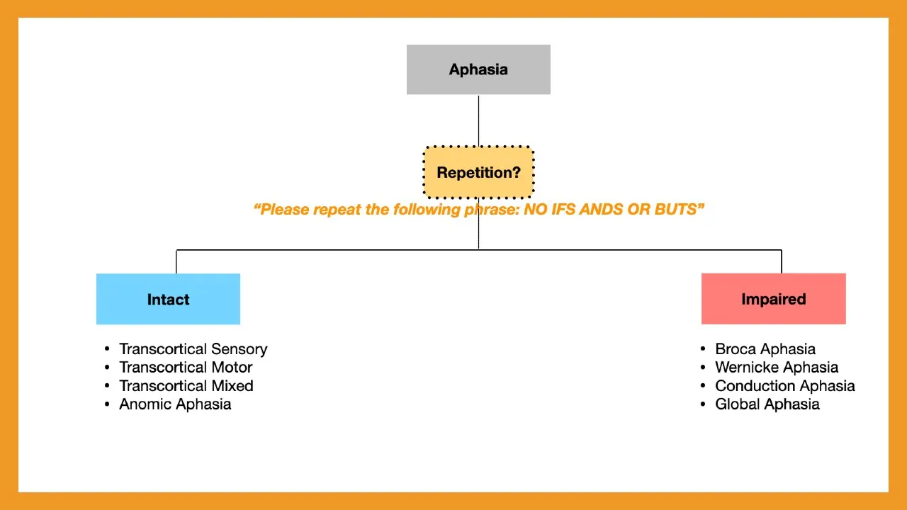
Three main non-repetitive aphasia types exist. Broca's aphasia (inferior frontal gyrus, BA 44/45) is non-fluent with intact comprehension; patients know what they want to say but cannot produce words ('broken' speech). Wernicke's aphasia (posterior superior temporal gyrus, BA 22) is fluent but with impaired comprehension; patients speak fluently but with word salad and neologisms ('wordy' speech). Conduction aphasia (arcuate fasciculus damage) preserves both fluency and comprehension but impairs repetition due to disrupted connection between Broca's and Wernicke's areas, causing phonological errors and self-correction attempts.
Click to watch →
Deep Dive
Diagnostic tools and assessment protocols used by speech-language pathologists to evaluate the type and severity of comprehension deficits.

Screening and analysis of specific language impairment | AISC
254
Speech-language pathologists use several diagnostic tools: Picture pointing tests assess picture recognition and interaction. Puppet/toy play observation evaluates spontaneous communication. Storytelling tasks analyze word choice and grammatical structure. Clinical Evaluation of Language Fundamentals (CELF) and grammar/phonological screening tests involve repeating words to identify specific speech problems. These comprehensive assessments typically take 2-3 hours.
Click to watch →

Evaluación de Lenguaje y Habla: Proceso y Herramientas
18.2K
The complete evaluation process includes: anamnesis/history taking, applying instruments, scoring, analyzing and interpreting results, integrating information, drawing conclusions, communicating results, and establishing intervention plans. Simply applying and scoring instruments is insufficient. Evaluation instruments include: quantitative (numerical scores) vs qualitative (descriptive) tools, standardized (compares to reference population) vs non-standardized (descriptive), formal (protocolized) vs informal (less structured), tasks (activities), protocols (step-by-step procedures), tests (single evaluation situations), and batteries (sets of tests). Parental reports complement direct evaluation. The choice of reference population is critical - comparing across different populations yields misleading results. Language assessment tools serve different purposes: screening tests (PLI, PLON) provide quick initial assessment, diagnostic batteries (CELF, PL-5) provide comprehensive detailed assessment, and specific tests evaluate particular skills (vocabulary, morphosyntax, pragmatics). Early development assessment uses inventories (MacArthur-Bates CDI) and global batteries (McCarthy, Bayley, Brigance, Portage) for young children. Comprehensive language batteries (CELF, PL-5) evaluate comprehension, expression, pragmatics, and literacy. Morphosyntax assessment evaluates grammatical development through tests like Aguado's Test of Morphosyntax. Pragmatics assessment evaluates language use in social contexts through tools like the Edmonton Narrative Norms Instrument (ENNI). Diagnostic differentiation tests (TAMIS) help distinguish between language delay and language disorder. Speech and articulation assessment evaluates phonetic and phonological processes through tests like Goldman-Fristoe and TEPROSIF. Functional language assessment analyzes spontaneous language samples (conversation, play, storytelling) to evaluate communication in natural contexts.
Click to watch →

Вебінар на тему: Клініко-педагогічна класифікація порушень мовлення
946
Speech-language pathology diagnosis includes several aspects: Sound production analysis (correctness and frequency of individual sounds and their combinations in words and phrases). Language comprehension (ability to understand words and phrases addressed to the child and reactions to proposed tasks). Phonemic hearing (ability to distinguish phonetically similar sounds). Syntax and grammar (how the child constructs sentences and uses grammatical structures). Communicative skills (how the child interacts with others using speech). Breathing (depth of inhalation and exhalation, presence of synchronous movements during breathing, nasal breathing). Static and dynamic organ of articulation (ability to obtain and switch positions). The diagnostic process begins with analyzing the child's behavior starting from the corridor. The specialist observes how the child enters the office, whether they can independently put on shoes, and how they interact with adults. For young children, the specialist offers to move to a carpet with toys to assess color recognition, geometric figure recognition, understanding of addressed speech, ability to assemble objects as needed, and counting from one to five. The specialist also checks speech-motor memory by giving instructions like 'first give me red, then green' or 'give me red, give me yellow, give me not yellow and not red' while placing several objects in front of the child. Phonemic hearing is assessed using buttons that sound differently. Size concepts are assessed using cards showing objects of different sizes. Animal sounds are assessed using cards showing different animals. Geometric figures are assessed using cards where the child must identify and match shapes. Visual-rhythmic rows are created using cubes or other materials. Storytelling is assessed using picture cards where the child must tell a story about characters. Geometric pyramids are used to assess phonemic hearing.
Click to watch →

Как провести диагностику неговорящего ребенка. Курсы для родителей и специалистов в описании.
34.5K
The diagnostician assesses speech comprehension through multiple methods: (1) Non-speech sound differentiation using pictures with corresponding sounds, (2) Color identification and two-step instructions for children 3+, (3) Object-based instruction following like 'feed the dog with pepper,' and (4) Sound imitation using a microphone to overcome speech negativism. These tests determine if the child has reached the nominal level of speech understanding, which is essential before speech can be initiated.
Click to watch →

Episódio 4: De Fono pra Fono: Neuro
153
Language assessment must evaluate comprehension (what the patient understands) and expression (what the patient can produce). Comprehension assessment considers context, gestures, and facial expressions. Expression assessment evaluates oral and written communication abilities, including word-finding difficulties (anomia) where patients know what they want to say but cannot retrieve the word. Standardized protocols like Boston Diagnostic Aphasia Examination provide quantitative assessment with scoring, but should not be the sole focus. Qualitative assessment examines how patients use cues, what strategies they employ, and the effort required. The choice between protocol-based and qualitative approaches depends on patient condition and clinical context.
Click to watch →
Deep Dive
Rehabilitation methods and speech therapy interventions tailored for Wernicke's aphasia, such as Treatment for Wernicke's Aphasia (TWA).

Интервью с логопедом. Мозговые механизмы нарушения речи.
170
Wernicke's area damage causes fluent but meaningless speech characterized by 'word salad' - disconnected words that don't form coherent sentences. Children substitute similar-sounding words, explain words by function rather than meaning, or produce incomprehensible speech. This differs from motor speech disorders where children struggle to produce sounds. Rehabilitation approaches differ fundamentally: for production disorders, therapists stimulate spontaneous speech through interactive activities; for Wernicke's aphasia, the approach is to calm and reduce pathological speech flow. Age affects recovery patterns, with younger children lacking pathological word salad and older children compensating better through accumulated experience.
Click to watch →

05 - IDDSI level 7, head/neck cancer, chest x-ray reports, VFSS checklist, and Wernicke’s aphasia
128
Wernicke's aphasia (fluent or receptive aphasia) results from damage to the left posterior temporal region (Wernicke's area), which processes word meaning. Patients have difficulty understanding speech, produce jargon-filled speech, and typically have poor awareness of their deficits. Two main treatment approaches exist: the context-based approach and structured exercises. The context-based approach uses authentic communication contexts to improve self-awareness, recognition of successful/unsuccessful communication attempts, and self-monitoring. The Visual Scanning and Language Task progresses through four steps: spoken word to picture matching, reading comprehension, oral reading, and auditory comprehension. Items are removed from practice once mastered across two consecutive sessions.
Click to watch →

Речевая реабилитация и восстановление функций после инсультов и черепно-мозговых травм
10.3K
Wernicke's aphasia involves difficulty understanding complex grammatical constructions and sentences. Patients produce fluent but meaningless speech and often lack awareness of their errors. The condition results from temporal lobe damage. Rehabilitation focuses on improving phonemic perception through exercises with similar-sounding words (e.g., distinguishing 'бочка' from 'почка'), developing analytical reading skills, and working on semantic understanding. The video emphasizes that Russian language complexity—with its extensive case system—makes this particularly challenging. Patients may have trouble understanding simple instructions and require repeated explanations.
Click to watch →
Wernicke's Aphasia and Broca's Aphasia
186.2K
Aphasia is a language disorder caused by brain damage (such as stroke or head injury) that affects speech production, comprehension, reading, and writing; Broca's aphasia is characterized by non-fluent speech with good comprehension where patients struggle to produce words, while Wernicke's aphasia involves fluent but meaningless speech with poor comprehension, requiring different therapeutic approaches such as cueing for Broca's and visual action therapy or gesture-based communication for Wernicke's.
Click to watch →
CASO CLÍNICO: AFASIA WERNICKE
445
Wernicke's aphasia is a language disorder caused by damage to Wernicke's area in the posterior superior temporal gyrus of the left hemisphere, characterized by fluent but meaningless speech with comprehension deficits, paraphasias, and neologisms; rehabilitation can lead to improvement in object naming, reading comprehension, and repetition of common words over time.
Click to watch →
Deep Dive
The concept of anosognosia (the lack of awareness of one's own deficit), which frequently accompanies fluent aphasia, and its impact on therapy.

El TRASTORNO de BRUCE WILLIS | PSICÓLOGA LO EXPLICA
35.3K
Anosognosia is a condition where patients are unaware of their own deficits. In aphasia, patients often do not realize they have lost language abilities, similar to how a computer doesn't complain when a function is uninstalled.
Click to watch →
ANOSOGNOSIA: Signs, Symptoms, Causes (Bipolar Disorder & Denial)
33.5K
Anosognosia significantly impacts treatment outcomes for bipolar disorder. Patients with this condition have decreased chances of proper diagnosis because they cannot recognize their own problems. They also show reduced treatment compliance, including refusing medication or therapy. This leads to increased hospitalizations, arrests, incarcerations, and sometimes violent incidents. The condition can be cyclic, coming and going in varying waves of intensity similar to bipolar disorder itself.
Click to watch →

NOW IT'S MAGA THAT CAN'T FIGURE OUT WHAT'S WRONG WITH TRUMP | Countdown with Keith Olbermann
99.1K
Anosognosia is a brain injury disease that makes it impossible for a person to recognize they have a disease, injury, stroke, or Alzheimer's. It is caused by various things, often by stroke. Patients deny or appear unaware of deficits such as paralysis or blindness. To casual observers, anosognosiac patients may appear quite normal and even bright and witty, and when not on the subject of their disability, they are quite rational.
Click to watch →

The Unraveling of Jared Lee Loughner
85.7K
Anosognosia is the lack of awareness of one's own deficits or illness. Approximately half of patients with schizophrenia do not recognize their symptoms or deficits, similar to how stroke patients may not realize they have lost motor function if damage also affects brain regions responsible for bodily awareness. This unawareness extends to recognizing that their speech is disorganized or that their thoughts are unclear. Anosognosia contributes to treatment resistance because patients do not believe they need help.
Click to watch →
What is Anosognosia?
54.1K
Anosognosia is the single largest reason why people living with schizophrenia or bipolar disorder refuse medication or refuse to seek treatment. When individuals do not believe there is anything wrong with them or that they have an illness, it becomes extremely difficult for them to understand the necessity of taking medications or seeking professional help.
Click to watch →
Deep Dive
Neuroplasticity and the mechanisms of language recovery and cortical reorganization in post-stroke patients.
"Neural mechanisms of language reorganization", Dorothee Saur
759
Language recovery after stroke follows a three-phase model involving early network breakdown, subacute reorganization, and chronic normalization, with key mechanisms including diaschisis (functional disconnection of remote brain regions), recovery of spared left-hemisphere language areas, and recruitment of right-hemisphere homologous regions; subacute language activation patterns predict chronic recovery outcomes, and non-invasive brain stimulation can induce compensatory plasticity within and across language networks.
Click to watch →

Neurorecovery and Neuroplasticity of Aphasia
425
After stroke-induced aphasia, language recovery occurs through neuroplasticity mechanisms where undamaged brain regions reorganize to assume functions of damaged areas; this reorganization involves both increased activation in non-language regions and enhanced connectivity between spared language network nodes, and can be enhanced through combined language therapy and non-invasive brain stimulation such as transcranial direct current stimulation (tDCS).
Click to watch →

Dynamics of language reorganization after left temporo-parietal and frontal stroke
1.4K
This longitudinal fMRI study reveals that language recovery after left hemisphere stroke follows a sequential pattern: initial global network disturbance (diaschisis) in areas distant from the lesion, followed by reactivation of domain-general networks (dorsal lateral prefrontal cortex and insula), then gradual restoration of preserved left hemisphere language networks, with lesion-homologous activation occurring only in frontal stroke patients and not in temporal-parietal stroke patients.
Click to watch →

Rehabilitación en etapa aguda post ACV
1.1K
Stroke rehabilitation relies on neuroplasticity, the brain's ability to reorganize after injury. Three key mechanisms include: (1) Cortical reorganization where damaged functions migrate to non-lesioned areas, starting contralateral and returning to perilesional regions; (2) Structural and functional plasticity involving dendritic and axonal regeneration beginning around day 3 post-stroke; (3) Genetic and epigenetic changes. In animals, reorganization peaks weeks 1-4; in humans, it begins week 1 but continues beyond month 1, explaining why rehabilitation must extend beyond the acute phase.
Click to watch →

Advances in predicting language recovery after stroke: Precision rehab - Swathi Kiran, PhD - BU
287
Aphasia recovery involves dynamic neural reorganization detectable through fMRI and DCM analysis. Naming therapy induces increased bilateral cortical activation with left hemisphere normalization and right hemisphere compensation. The middle frontal gyrus, traditionally non-language, drives recovery processes in aphasic patients. Connectivity shifts depend on lesion location—superior frontal damage increases right hemisphere connectivity while ventral damage heightens left frontal modulation. These findings reveal that recovery mechanisms involve distributed network reorganization rather than simple left-right compensation.
Click to watch →
Greetings
Current Plans
Cruise Context
iPad Activity
Daily Life
Golf & Hobbies
Helping Others
Farewell
Greetings
0:07- 1
Megan and Byron exchange brief pleasantries.
- 2
Byron expresses happiness and complements Megan.
The Dual-Stream Model and Distributed Network Perspective
The traditional view of Wernicke's aphasia relies on the classical localizationist model, which attributes fluent but empty speech and comprehension deficits strictly to damage in Wernicke's area. However, modern cognitive neuroscience, particularly through Hickok and Poeppel’s Dual-Stream Model, challenges this paradigm. Research shows that language comprehension is not localized to a single region but is supported by a highly distributed, bi-hemispheric network. Deficits in fluent aphasia often arise from disruptions in the 'ventral stream' (mapping sound to meaning) or the 'dorsal stream' (mapping sound to articulation), rather than a loss of a localized 'comprehension center.' Furthermore, patients with Wernicke's aphasia exhibit highly heterogeneous symptoms, suggesting the condition is not a single unitary deficit, but a complex combination of phonological, semantic, and auditory working memory impairments across widespread temporal, parietal, and frontal networks.
Basic neuroanatomy, specifically the location and function of the temporal lobe and Wernicke's area in the dominant hemisphere of the brain.
The core distinction between receptive language (understanding spoken or written communication) and expressive language (producing speech).
The general definition of aphasia as an acquired language impairment, and how cerebrovascular accidents (strokes) can cause localized neurological deficits.
The difference between language disorders (affecting meaning, grammar, and symbols) and motor speech disorders like dysarthria or apraxia.
A comparative study between Fluent (Wernicke's) and Non-fluent (Broca's) Aphasia, mapping out their distinct clinical profiles.
Diagnostic tools and assessment protocols used by speech-language pathologists to evaluate the type and severity of comprehension deficits.
Rehabilitation methods and speech therapy interventions tailored for Wernicke's aphasia, such as Treatment for Wernicke's Aphasia (TWA).
The concept of anosognosia (the lack of awareness of one's own deficit), which frequently accompanies fluent aphasia, and its impact on therapy.
Neuroplasticity and the mechanisms of language recovery and cortical reorganization in post-stroke patients.
4.5M views38.4Klikes1:30@TactustherapyOriginal Release: 2015-09-08
Megan: "Hi Byron! How are you?" Byron: "I'm happy. Are you pretty? You look good."
Megan: "What are you doing today?" Byron: "We stayed with the water over here at the moment and talk with the people for them over there. They're diving for them at the moment, but they'll save in the moment held water very soon, for him, with luck, for him."
Megan: "So we're on a cruise and we're about to get to Juneau..." Byron: "We will sort right here and they'll save their hands right there for them."
Megan: "And what were we just doing with the iPad?" Byron: " Uhh.. right at the moment they don't show a darn thing. Ha ha!"
Megan: "With the iPad, that were we doing? Like here?"
Byron: "I'd like my change for me and change hands for me. It would happy. I would talk with Donna sometimes. We're out with them. Other people are working with them and them.
I'm very happy with them. This girl with verly good. And happy and I play golf and hit up trees. We play out with the hands. We save a lot of hands on hold for peoples, for us. Other hands. I don't know what you get, but I talk with a lot of hand for him.
Sometime. Am I talk of anymore to saying." Megan: "Alright, thank you very much!"
Byron: "Thank you very much, I appreciate it, and I hope the world lasts for you."
Megan: "Thank you, it's been a pleasure. Bye-bye!" Byron: "Have a good day!"
Up Next
3:30
Conduction Aphasia: Arcuate Fasciculus | USMLE Review
@allornonelaw
2.7K views•2014-04-20

48:47
Graphic Medicine: Comics for Collaborative Healthcare Communication
@nationalpatientadvocate
189 views•2023-12-04

10:28
Neuroanatomy: Central and Peripheral Nervous System Divisions Explained
@AKLECTURES
136.2K views•2014-09-20

3:05
Stages of Labor and Vaginal Birth | Childbirth Animation
@nucleusmedicalmedia
52.1M views•2017-08-18
Related Study Plans & Knowledge Roadmaps
Structured learning paths in Medicine
medicine
Abnormal LFTs: Diagnostic Workup & Pathology
01Liver Anatomy and Basic Physiology
02Introduction to Liver Function Tests (LFTs)
03Hepatocellular vs. Cholestatic Patterns
+ 3 more learning modules
12h6 modules
Explore Roadmap
medicine
Analyzing Blood Smears: RBC, WBC & Platelets
01Introduction to Blood Components & Smear Preparation
02Normal Morphology of Blood Cells
03Erythrocyte Pathologies & Morphological Variants
+ 2 more learning modules
12h5 modules
Explore Roadmap
medicine
Mechanical Ventilation: Waveforms & Alarms
01Anatomy and Physiology of Breathing
02Introduction to Mechanical Ventilation & Core Modes
03Ventilator Settings and Waveform Interpretation
+ 2 more learning modules
12h5 modules
Explore Roadmap
medicine
Neuro Exam: Localization & Stroke Syndromes
01Foundations of Neuroanatomy and Vascular Supply
02The Neurological Examination: Step-by-Step
03Principles of Neurological Localization
+ 3 more learning modules
18h6 modules
Explore Roadmap