Conduction aphasia results from damage to the arcuate fasciculus, the fiber bundle connecting Broca's area (Brodmann area 44 in the frontal lobe) with Wernicke's area (Brodmann area 22 in the superior temporal gyrus); its defining characteristic is that patients comprehend words correctly but cannot produce them for speech or writing, and they exhibit significant difficulty repeating statements.
Conduction Aphasia: Arcuate Fasciculus | USMLE Review
Added:Deep Dive
Anatomy and functions of Broca's area (inferior frontal gyrus) and Wernicke's area (superior temporal gyrus).
Central Nervous System Physiology Rapid Revision 🧠🧠🧠
137.6K
Broca's area (Brodmann area 44, located in the inferior frontal gyrus) is responsible for speech production. Damage causes expressive aphasia (Broca's aphasia) where patients know what they want to say but cannot express it properly. Wernicke's area (Brodmann area 22, located in the superior temporal gyrus) is responsible for speech comprehension. Damage causes fluent aphasia where patients speak fluently but their speech lacks meaning.
Click to watch →
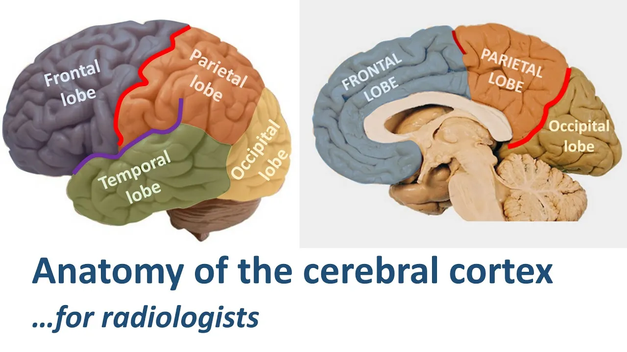
Radiological anatomy of the cerebral cortex... made easy.
71.3K
Broca's area (motor speech center) is located in the triangular and opercular parts of the inferior frontal gyrus, which has a distinctive M-shape on sagittal images due to its position along the anterior and ascending branches of the Sylvian fissure. Wernicke's area (receptive speech center) is classically described behind Heschl's gyrus on the superior temporal gyrus, though modern studies suggest it is an association area between parietal and temporal lobes. The temporal lobe contains superior, middle, and inferior temporal gyri separated by superior and middle temporal sulci. The basal surface contains lateral occipital temporal gyrus (fusiform gyrus), medial occipital temporal gyrus (para hippocampal gyrus), and hippocampus. Heschl's gyrus contains the primary auditory cortex and appears as a small knob-like structure on top of the superior temporal gyrus.
Click to watch →

NEET PG | NF100 E62: Aphasia made easy | Dr Nikita Nanwani
24.4K
Broca's area is located in the inferior frontal gyrus (frontal lobe) and is the motor/expressive speech area. Wernicke's area is located in the superior temporal gyrus (temporal lobe) and is the sensory/comprehension speech area. A mnemonic to remember: 'BEFI' (Broca's) stands for 'Expressive' and 'ST' (Superior Temporal) stands for 'Sensory'. Broca's is associated with the letters B, E, F, I in the alphabet, while Wernicke's is associated with S, T, U, V, W.
Click to watch →
Revise Physiology PYQ Edition With Dr Kamal K V for upcoming fmge exam.
4.1K
Broca's area (inferior frontal gyrus) controls motor speech production. Damage causes motor aphasia: non-fluent speech but preserved comprehension. Wernicke's area (superior temporal gyrus) controls language comprehension. Damage causes Wernicke's aphasia: fluent but meaningless speech with impaired comprehension. Both areas are located in the dominant hemisphere (usually left).
Click to watch →

MRB 2024 Assistant Surgeon - High Yield Discussion - Physiology with Dr. Prem Balaguru
9.9K
Wernicke's area (inferior temporal gyrus, MCA inferior division) is responsible for comprehension. Broca's area (inferior frontal gyrus, MCA superior division) is responsible for motor planning for articulation and speech fluency. Wernicke's aphasia causes comprehension deficits with fluent but meaningless speech. Broca's aphasia causes fluency deficits with preserved comprehension.
Click to watch →
Deep Dive
The basic vascular supply of the dominant cerebral hemisphere, specifically the divisions of the middle cerebral artery (MCA).

Blood Supply of the Nervous System
3.2K
The middle cerebral artery (MCA) passes laterally to the optic chiasm and runs in the depth of the lateral fissure. At the insula, it divides into multiple terminal branches. The MCA gives off central branches called lenticulostriate arteries that supply the basal ganglia (caudate nucleus and lentiform nucleus) and the internal capsule. Occlusion of these arteries can cause contralateral hemiplegia because the internal capsule contains important motor pathways. The MCA supplies the superior lateral surface of the cerebral hemisphere (except the upper 1 inch supplied by ACA and lower 1 inch supplied by PCA), the lateral part of the orbital surface, the temporal pole, and the insula. It supplies the motor and sensory areas for the face, arm, and hand on the homunculus, as well as the language areas (Broca's and Wernicke's areas) in the dominant hemisphere.
Click to watch →
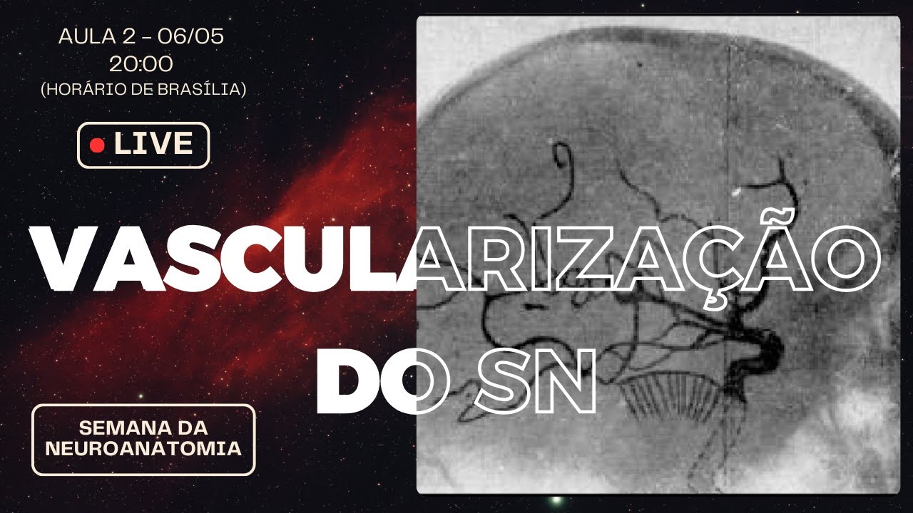
Vascularização do Sistema Nervoso
629
The middle cerebral artery (MCA) is divided into 5 segments: M1 (horizontal/sphenoidal), M2 (insular), M3 (short/insular contour), M4 (opercular), and M5 (cortical). The MCA supplies the lateral surface of the cerebral hemisphere, including the primary motor cortex, primary somatosensory cortex, Broca's area (motor speech), Wernicke's area (language comprehension), and auditory areas. The MCA is the most commonly affected artery in ischemic stroke.
Click to watch →

Middle cerebral artery
3.2K
The middle cerebral artery (MCA) is one of three major paired cerebral arteries, arising from the internal carotid and supplying the lateral cerebral cortex, anterior temporal lobes, and insular cortices. It connects to the anterior cerebral and posterior communicating arteries but is not part of the Circle of Willis. The MCA divides into four segments: M1 (sphenoidal/insular segment) perforates the basal ganglia; M2 (insular/Sylvian segment) may bifurcate or trifurcate into trunks; M3 (opercular segments) extends laterally; M4 (terminal/cortical segments) irrigate the cortex. The frontal lobe receives blood through lateral frontobasal arteries (inferior frontal gyrus), prefrontal arteries (middle frontal gyrus), pre-Rolandic artery (posterior frontal gyri), and Rolandic arteries (central sulcus regions, bifurcating in 72% of cases).
Click to watch →

75 Blood supply of the cerebral hemisphere الدكتور أحمد كمال cxt
305
The cerebral hemisphere receives blood supply from three major arteries: the middle cerebral artery (MCA) supplies the lateral surface including frontal, parietal, and temporal lobes; the anterior cerebral artery (ACA) supplies the medial surface including frontal and parietal lobes; and the posterior cerebral artery (PCA) supplies the occipital lobe and inferior temporal lobe. Understanding this vascular distribution is essential for localizing neurological deficits and stroke syndromes. The MCA is the largest branch of the internal carotid artery and supplies approximately 80% of the cerebral cortex.
Click to watch →

MCA, 뇌졸중환자 치료
1.8K
The Middle Cerebral Artery (MCA) is the most commonly damaged artery in stroke patients. The MCA has three main branches: lateral lenticulostriate arteries (supplying deep structures like internal capsule and thalamus), superior division (supplying frontal lobe including Broca's area), and inferior division (supplying temporal lobe including Wernicke's area). The MCA supplies the lateral surface of the cerebral hemisphere, including frontal, parietal, and temporal lobes. The superior division supplies Broca's area (motor speech), while the inferior division supplies Wernicke's area (sensory speech). The MCA also supplies the motor and sensory cortices contralaterally, with the face and arm more commonly affected than the leg due to somatotopic organization.
Click to watch →
Deep Dive
Fundamental neurological definitions of fluent versus non-fluent speech production.

COMO MELHORAR A GAGUEIRA - PARTE 1
111K
Fluency is the ability to speak without physical or mental effort, producing natural, spontaneous speech. Seven key characteristics define fluent speech: (1) Hesitation - brief pauses to think or reconstruct thoughts; (2) Reformulation - rephrasing statements using markers like 'quer dizer' or 'ou melhor'; (3) Fluent silent pause - strategic pauses between sentences for breathing and melodic variation; (4) Speech rate - individual speed within normal range; (5) Smoothness - relaxed, tension-free vocal emission; (6) Grammatical ability - correct use of grammatical elements for rich sentences; (7) Semantic complexity - sophisticated vocabulary without vague terms. These characteristics distinguish fluent from disfluent speech.
Click to watch →

Lec 13: Language in the brain
737
Broca's aphasia is called non-fluent aphasia because of the difficulty in producing speech. Wernicke's aphasia is called fluent aphasia because patients produce speech without any deficit in fluency, though the speech lacks meaning.
Click to watch →

A Look at Aphasia, the Silent Disorder
1.4K
Aphasia is generally grouped into two major categories: fluent versus non-fluent types. Fluent aphasia (sometimes called receptive aphasia) involves conversation that sounds flowing but words don't make sense, with normal rhythm and intonation but impaired comprehension. People with fluent aphasia may not recognize their own speech errors and may not demonstrate frustration despite making substantive errors. Non-fluent aphasia involves hesitant speech with long pauses between words, difficulty naming objects, and problems with grammar and syntax.
Click to watch →

Higher Mental Functions
6.4K
Fluent aphasia produces more than 100 words per minute with semantic paraphasias (using wrong words for concepts) and empty content. Non-fluent aphasia produces less than 50 words per minute with literal paraphasias (similar-sounding word substitutions) and telegraphic speech. Broca's aphasia is non-fluent; Wernicke's aphasia is fluent.
Click to watch →

How to Retrain Your Brain to Understand Speech Again | Mercola Cellular Wisdom
375
Aphasia manifests differently depending on injury location. Fluent aphasia (Broca's aphasia) produces speech that sounds very smooth and rhythmic with normal pacing, but words may lack meaning or become jumbled into what people call 'word salad,' with poor comprehension. Non-fluent aphasia involves physical struggle to get words out—speech is effortful and consists of short phrases—with relatively better comprehension than fluent aphasia. Both types are still impaired by the 80-250 millisecond glitch in neural processing.
Click to watch →
Deep Dive
The physiological pathway of normal language processing from auditory perception to vocal articulation.
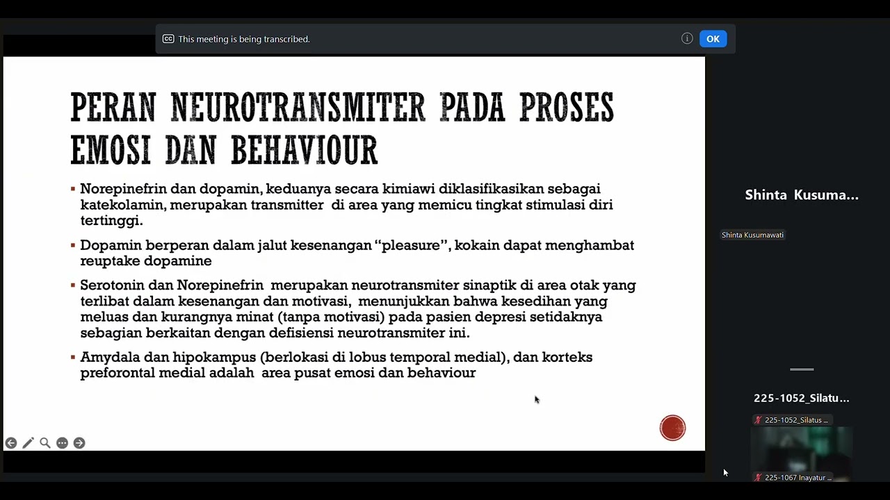
Kuliah 10 : Fisiologi Behaviour, Emotion, Learning, and Memori (Dr. dr. Shinta Kusumawati, Sp. N)
172
Auditory language processing follows this pathway: sound stimuli are converted to electrical signals, pass through the brainstem to the thalamus, then to the primary auditory cortex. From there, information goes to the angular gyrus, then to Wernicke's area for comprehension. Comprehended information is then sent to Broca's area for speech production.
Click to watch →

Function of specific associated area,right and left hemisphere function,physiology of language
235
The language production pathway involves sound information reaching the primary auditory area, then to Wernicke's area for comprehension, then to Broca's area for speech planning, and finally to the motor cortex to activate speech muscles. The language comprehension pathway involves visual information reaching the visual cortex, then to the angular gyrus for converting written text to spoken representations, then to Wernicke's area for comprehension, and finally to Broca's area and motor cortex for verbal response.
Click to watch →

PSYCH 105 - Chapter 9 - Part 2
399
Language processing follows a pathway: sound enters through ears to the primary auditory cortex, then information is sent to Broca's area which decides how to produce sounds, and commands are sent to the primary motor cortex to activate mouth movements. Damage to any part of this pathway disrupts language production.
Click to watch →

Nervous system S12 | Cortical associated areas
1.4K
Language processing involves two main pathways: auditory (sound → primary auditory cortex → Wernicke's area → arcuate fasciculus → Broca's area → motor cortex) and visual (visual input → visual cortex → angular gyrus → Wernicke's area → arcuate fasciculus → Broca's area → motor cortex). Both pathways converge at Wernicke's area for comprehension and proceed to Broca's area for speech production. The arcuate fasciculus connects comprehension and production centers, enabling the transfer of information between them. This demonstrates the neural basis of language understanding and expression.
Click to watch →

Language Aphasia Dysarthria
1.5K
Language processing follows specific neural pathways. For comprehension: auditory signals travel from primary auditory cortex to auditory association cortex to Wernicke's area; visual signals travel from primary visual cortex to visual association cortex to Wernicke's area. For production: signals travel from Wernicke's area through the arcuate fasciculus to Broca's area, then to the motor cortex, and finally to articulator muscles. Healthy nerves and muscles are essential for proper speech production.
Click to watch →
Prerequisite Knowledge
- Concept 01Anatomy and functions of Broca's area (inferior frontal gyrus) and Wernicke's area (superior temporal gyrus).
- Concept 02The basic vascular supply of the dominant cerebral hemisphere, specifically the divisions of the middle cerebral artery (MCA).
- Concept 03Fundamental neurological definitions of fluent versus non-fluent speech production.
- Concept 04The physiological pathway of normal language processing from auditory perception to vocal articulation.
Subsequent Learning
- Step 01Differential diagnosis of transcortical aphasias (transcortical motor, sensory, and mixed) and how they differ from conduction aphasia regarding repetition.
- Step 02Clinical bedside assessment techniques for language, specifically testing repetition using standardized phrases (e.g., 'No ifs, ands, or buts').
- Step 03The study of other white matter tract disconnection syndromes in neurology, such as alexia without agraphia.
- Step 04Speech-language pathology rehabilitation strategies and neuroplastic recovery patterns for patients with arcuate fasciculus lesions.
Lesion Site
Core Symptoms
Lesion Site
0:11Playing Section
- 1
The lesion is located in the arcuate fasciculus.
- 2
These fibers connect Broca's area to Wernicke's area.
- 3
Localization also includes the parietal lobe or insula.
The Cortical Dysfunction and Dual-Stream Model
While the classic Wernicke-Geschwind model taught for the USMLE attributes conduction aphasia strictly to a disconnection of the arcuate fasciculus, modern cognitive neuroscience challenges this view. Contemporary research and neuroimaging indicate that conduction aphasia is often caused by cortical lesions rather than pure white matter tract damage, specifically localizing to the left temporoparietal junction (area Spt). According to the Dual-Stream Model of speech processing, conduction aphasia is better understood as a deficit in auditory-motor integration rather than a simple transmission failure along a single fiber pathway. Studies show that selective damage to the arcuate fasciculus does not reliably produce conduction aphasia, and many patients with the condition have intact white matter tracts but cortical damage. This shifts the understanding of conduction aphasia from a simple 'disconnection' syndrome to a complex breakdown in a cortical network responsible for translating acoustic speech signals into motor articulatory plans.
Deep Dive
Differential diagnosis of transcortical aphasias (transcortical motor, sensory, and mixed) and how they differ from conduction aphasia regarding repetition.
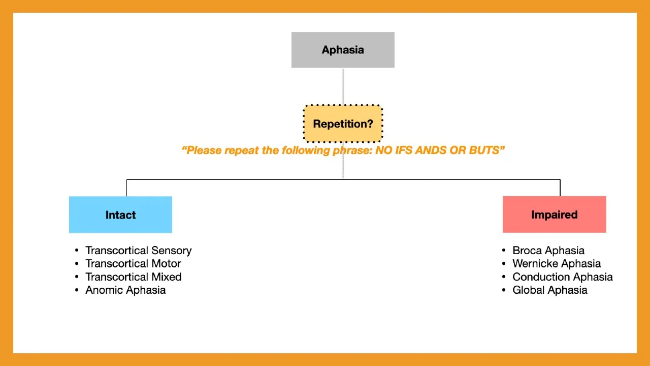
Aphasia (Broca vs. Wernicke vs. Conduction vs. Global vs. Transcortical)
14.2K
Aphasia is classified into two main categories based on repetition ability: impaired repetition aphasias (Broca's, Wernicke's, conduction, and global aphasia) and transcortical aphasias (with intact repetition). Broca's aphasia is non-fluent with intact comprehension (due to inferior frontal gyrus damage), Wernicke's aphasia is fluent with impaired comprehension (due to posterior superior temporal gyrus damage), conduction aphasia involves fluent speech with intact comprehension but impaired repetition (due to arcuate fasciculus damage), and global aphasia involves complete impairment of all language functions. Transcortical variants (motor and sensory) preserve repetition while mimicking Broca's or Wernicke's patterns respectively.
Click to watch →
NEET PG | NF100 E62: Aphasia made easy | Dr Nikita Nanwani
24.4K
Aphasias are classified by patterns of impairment in fluency, comprehension, and repetition: Broca's aphasia (motor) shows impaired fluency with preserved comprehension; Wernicke's aphasia (sensory) shows impaired comprehension with preserved fluency; conduction aphasia shows impaired repetition with preserved fluency and comprehension; transcortical aphasias (motor, sensory, mixed) show preserved repetition due to intact arcuate fasciculus; global aphasia shows impairment in all functions; anomic aphasia shows only impaired naming. The key diagnostic approach is to first assess repetition to distinguish transcortical from other aphasias, then evaluate fluency and comprehension to identify the specific type.
Click to watch →

Aphasia | Types (Broca’s, Wernicke’s, Global), Causes, Signs & Symptoms, Diagnosis, Treatment
116.8K
Global aphasia represents the most severe form with bilateral lesions causing comprehensive language failure. Conduction aphasia preserves speech and comprehension but impairs repetition, with patients recognizing and attempting to correct errors. Transcortical aphasia subtypes differ by lesion location: transcortical sensory aphasia (around Wernicke's area) preserves repetition; transcortical motor aphasia (around Broca's area) preserves comprehension and repetition despite non-fluent speech; mixed transcortical aphasia combines features of both. Anomia (angular gyrus lesion) uniquely preserves speech, comprehension, and repetition while causing only word-finding difficulties. Aphasia diagnosis relies on clinical assessment including cranial nerve exams and imaging (CT, MRI, carotid ultrasound). Excluded conditions include developmental disorders, motor speech disorders, and psychiatric conditions. Treatment focuses on speech-language therapy as the mainstay, supplemented by acute stroke management, anticoagulation for atrial fibrillation, surgical intervention for masses, and antivirals for encephalitis. Recovery prognosis correlates with younger age, smaller lesions, better general health, and left-handedness potentially offering bilateral language compensation.
Click to watch →
Cognitive Neurology and Classic Concepts
2.4K
Transcortical aphasias (border zone infarcts) occur when proximal artery segments receive good blood supply but distal portions do not, sparing the arcuate fasciculus and preserving repetition. Transcortical motor aphasia resembles Broca's aphasia but with preserved repetition. Transcortical sensory aphasia resembles Wernicke's aphasia but with preserved repetition. Conduction aphasia results from arcuate fasciculus lesions—patients have fluent speech but cannot repeat phrases accurately. Alexia without agraphia results from left posterior cerebral artery involvement affecting the left occipital cortex and splenium of the corpus callosum—the right visual cortex cannot access the language areas on the left side.
Click to watch →
Split brain and disconnection syndromes - Prof. S.R. Chandra
1.4K
Conduction aphasia results from arcuate fasciculus damage connecting Broca's and Wernicke's areas. Patients have fluent speech with paraphasic errors and severely impaired repetition, yet preserved comprehension and expression. They may repeat numbers but not words. Transcortical aphasias occur when speech areas become isolated from cortical association areas: motor transcortical aphasia preserves repetition while resembling Broca's aphasia, and sensory transcortical aphasia preserves repetition while resembling Wernicke's aphasia. These syndromes demonstrate that repetition and comprehension can be dissociated from expression, revealing the modular nature of language processing.
Click to watch →
Deep Dive
Clinical bedside assessment techniques for language, specifically testing repetition using standardized phrases (e.g., 'No ifs, ands, or buts').
Aphasia (Broca vs. Wernicke vs. Conduction vs. Global vs. Transcortical)
14.2K
The key clinical assessment for aphasia diagnosis is evaluating whether the patient has intact repetition. Clinically, this is tested by asking the patient to repeat a phrase without any qualifiers (e.g., 'no ifs, ands, or buts'). If the patient can correctly repeat the phrase, they have intact repetition, suggesting transcortical aphasia. If they cannot repeat the phrase, they have impaired repetition, indicating Broca's aphasia, Wernicke's aphasia, conduction aphasia, or global aphasia. This distinction forms the foundation for categorizing aphasia types.
Click to watch →
Cranial Nerve Exam (#1): Pat LaFontaine & Dr. James Kelly | Unintentional ASMR | Soft-spoken Edition
509.3K
The examiner tests verbal repetition and language skills by having the patient repeat a phrase ('No ifs, ends, or buts') and then recall the three objects and three words previously shown. This assesses the patient's verbal repetition ability and short-term memory for verbal information.
Click to watch →

MENTAL STATUS EXAMINATION
3K
The MMSE tests language through naming objects, repetition of complex phrases like 'No ifs, ands, or buts,' and following written commands. The phrase 'No ifs, ands, or buts' is particularly sensitive for detecting language disorders because it has meaning but is difficult for the nervous system to repeat accurately. Writing ability is assessed by asking patients to write a complete sentence, which tests both language and motor function. Visual-spatial skills are tested by having patients copy overlapping pentagons, which assesses the parietal lobe's function in visual-spatial processing and the motor system's ability to reproduce visual information.
Click to watch →

Video Atlas of the Detailed Neurologic Examination 2
16K
The 'no ifs and no buts' test is a classical neurological assessment of language function. The phrase has meaning in English but is extremely difficult for the nervous system to repeat accurately. This test, attributed to Norman Geschwind, is sensitive but not specific for language disorders. It tests the ability to repeat complex phrases that require linguistic processing, making it useful for detecting subtle language abnormalities that might not be apparent in simpler conversation.
Click to watch →
Head to Toe Assessment
2.8K
Language function assessment includes evaluating comprehension, repetition, and naming abilities. The assessor should ask the patient to describe their day (comprehension), give simple commands like closing and opening eyes (following instructions), repeat phrases like 'no ifs, ands, or buts' (repetition), and identify objects shown (naming). Speech should be fluent and accurate, and comprehension should be correct.
Click to watch →
Deep Dive
The study of other white matter tract disconnection syndromes in neurology, such as alexia without agraphia.
Split brain and disconnection syndromes - Prof. S.R. Chandra
1.4K
The brain exhibits functional asymmetry where the left hemisphere specializes in language, speech production, and semantic processing, while the right hemisphere handles spatial processing, facial recognition, and emotional recognition; when the corpus callosum is surgically sectioned or damaged, it creates disconnection syndromes such as alexia without agraphia (inability to read despite intact writing ability), pure word deafness (inability to comprehend spoken words despite hearing intact), conduction aphasia (fluent speech with impaired repetition), and tactile agnosia (inability to name objects by touch), demonstrating that normal brain function requires interhemispheric integration of specialized cortical regions connected by white matter tracts.
Click to watch →

Disconnect Syndromes - Transcortical Apraxia & Alexia without Agraphia
3K
Disconnect syndromes occur when lesions in white matter fiber tracts disrupt communication between brain regions; transcortical apraxia results from anterior cerebral artery lesions blocking the corpus callosum, preventing verbal commands from reaching the opposite motor cortex while independent movement remains intact, whereas alexia without agraphia occurs from posterior cerebral artery lesions damaging the splenium and left visual cortex, causing inability to comprehend written words despite preserved visual input and writing ability due to disconnection between right visual cortex and left language areas.
Click to watch →

CORE CLINICAL NEUROLOGY PART - 8,CEREBRAL CORTEX INTRODUCTION
113
White matter fibers are classified into three categories: association fibers connect cortical areas within the same hemisphere, commissural fibers connect corresponding areas between hemispheres, and projection fibers connect the cortex to lower brain centers. The arcuate fasciculus connects Broca's and Wernicke's areas; its damage causes disconnection syndrome preventing speech repetition. The corpus callosum, the largest commissural structure, enables interhemispheric communication. Damage to the splenium causes alexia, where visual information from the left visual field cannot reach the language-dominant left hemisphere. Projection fibers like the corticospinal tract carry motor commands from cortex to spinal cord. The internal capsule contains compactly grouped motor fibers in its posterior limb, making it susceptible to lacunar infarcts causing pure motor hemiparesis with equal face, arm, and leg involvement.
Click to watch →

Dr. Oliver Sacks on The Mind's Eye
190.8K
Alexia without agraphia is a neurological condition where a person loses the ability to read printed text while retaining the ability to write. This condition results from damage to specific visual processing areas in the brain that process written language, while the motor areas responsible for writing remain intact. Patients may describe text as appearing in an unintelligible foreign script. The condition highlights the distinction between visual processing and motor execution in language comprehension.
Click to watch →

Grey Matter Project: Neuroanatomy 24-25
256
Visual information from the right side of the world hits the nasal (inner) part of both retinas and crosses to the left brain, while temporal (outer) retinal information stays on the same side. This decussation ensures all right visual field information reaches the left visual cortex. Alexia without agraphia occurs when the visual cortex is damaged but the language area is intact, but the connection between them is severed. The patient can see words perfectly but cannot understand them because visual information cannot reach the language area. This is a disconnection syndrome. Treatment includes anticoagulation for atrial fibrillation and rehabilitation strategies like prism glasses and reading by spelling aloud to route information through the auditory cortex. Recovery after stroke depends on age (younger patients recover better), sex (women tend to recover language better), and which areas are damaged (eloquent areas are harder to recover from than supporting structures).
Click to watch →
Deep Dive
Speech-language pathology rehabilitation strategies and neuroplastic recovery patterns for patients with arcuate fasciculus lesions.

Neuroccino 22nd February 2021 - Neanderthal endocasts, 10 Tesla scanners and the arcuate fasciculus
176
A comprehensive study of stroke survivors investigated whether Broca's area damage alone causes long-term speech impairment. Using lesion-symptom mapping across multiple brain regions including Broca's area subdivisions, ventral premotor cortex, and arcuate fasciculus, researchers found that arcuate fasciculus anterior segment damage was the strongest predictor of persistent speech production deficits (r = 0.5). While Broca's area showed some significance, its contribution was minimal compared to arcuate fasciculus involvement. Patients with arcuate fasciculus lesions demonstrated significantly worse long-term outcomes than those with Broca's area lesions alone. This finding challenges traditional models emphasizing Broca's area and highlights the critical role of white matter connectivity in language maintenance, with implications for understanding recovery mechanisms and therapeutic targets in post-stroke rehabilitation.
Click to watch →

Stephanie Forkel on Neurobiology of language: insights from neuroimaging
354
Research reveals that white matter tracts are dynamically recruited rather than having fixed functional assignments. The arcuate fasciculus, classically linked to language, correlates with diverse cognitive measures. Critically, individual anatomical variability predicts clinical outcomes—stroke patients with thicker right hemisphere arcuate fasciculi showed better language recovery, with this factor nearly doubling predictive power beyond standard clinical variables. Morphospace modeling now enables prediction of recovery across 87 cognitive domains from lesion patterns alone. These findings support an integrative model where language emerges from coordinated interactions across distributed brain networks, analogous to music emerging from an orchestra rather than any single instrument.
Click to watch →
AARÓN DEL OLMO | AFASIAS
1.7K
Conduction aphasia is characterized by inability to repeat words despite relatively intact comprehension and speech production. The arcuate fasciculus, connecting Broca's and Wernicke's areas, is damaged. Patients can understand language and produce speech but cannot repeat what they hear. This reflects problems with phonological encoding - converting heard words into speech sounds. Patients may produce phonologically impaired speech, substituting sounds or producing similar-sounding words. Comprehensive rehabilitation includes visual-motor integration training (practicing mouth movements with mirror feedback), auditory-motor integration training, grammatical rehabilitation (sentence construction with articles, pronouns, verb conjugation), and interactive communication training (practicing real-world scenarios). Auditory discrimination training helps patients distinguish between similar-sounding words. Contextual comprehension training helps patients use environmental cues to understand spoken language. Motivation and engagement are critical factors in rehabilitation success.
Click to watch →

Fonoaudióloga da dica para pessoas que tiveram AVC
64.2K
Stroke (AVC) affects over 100,000 Brazilians annually, causing communication and swallowing difficulties that speech-language pathologists (fonoaudiólogos) help treat through multidisciplinary rehabilitation; immediate medical attention is crucial to minimize lasting effects, and family support with patience and love significantly aids recovery.
Click to watch →
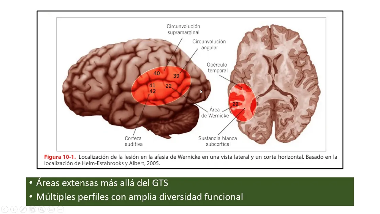
Neuropsicología de las Afasias 2
3.3K
Effective aphasia rehabilitation follows key principles: (1) Use it or lose it - use deficit functions to prevent further loss; (2) Train the deficit function directly; (3) Be specific about which functions to train; (4) Repetition is essential - single exposures are insufficient; (5) Intensity should be graduated from low to high; (6) Transfer training to other contexts; (7) Consider interference from other factors. Prominence (familiarity and relevance) of training tasks affects recovery outcomes. Brain imaging shows that aphasia recovery involves reorganization of language networks, with patients showing increased connectivity in alternative networks (often right hemisphere) to compensate for damaged left-hemisphere networks. Rehabilitation training induces neuroplastic changes, strengthening connectivity between remaining language circuits. These changes can be quantified and tracked over time, demonstrating how the brain adapts to language deficits through neuroplasticity.
Click to watch →
Lesion Site
Core Symptoms
Lesion Site
0:11- 1
The lesion is located in the arcuate fasciculus.
- 2
These fibers connect Broca's area to Wernicke's area.
- 3
Localization also includes the parietal lobe or insula.
The Cortical Dysfunction and Dual-Stream Model
While the classic Wernicke-Geschwind model taught for the USMLE attributes conduction aphasia strictly to a disconnection of the arcuate fasciculus, modern cognitive neuroscience challenges this view. Contemporary research and neuroimaging indicate that conduction aphasia is often caused by cortical lesions rather than pure white matter tract damage, specifically localizing to the left temporoparietal junction (area Spt). According to the Dual-Stream Model of speech processing, conduction aphasia is better understood as a deficit in auditory-motor integration rather than a simple transmission failure along a single fiber pathway. Studies show that selective damage to the arcuate fasciculus does not reliably produce conduction aphasia, and many patients with the condition have intact white matter tracts but cortical damage. This shifts the understanding of conduction aphasia from a simple 'disconnection' syndrome to a complex breakdown in a cortical network responsible for translating acoustic speech signals into motor articulatory plans.
Anatomy and functions of Broca's area (inferior frontal gyrus) and Wernicke's area (superior temporal gyrus).
The basic vascular supply of the dominant cerebral hemisphere, specifically the divisions of the middle cerebral artery (MCA).
Fundamental neurological definitions of fluent versus non-fluent speech production.
The physiological pathway of normal language processing from auditory perception to vocal articulation.
Differential diagnosis of transcortical aphasias (transcortical motor, sensory, and mixed) and how they differ from conduction aphasia regarding repetition.
Clinical bedside assessment techniques for language, specifically testing repetition using standardized phrases (e.g., 'No ifs, ands, or buts').
The study of other white matter tract disconnection syndromes in neurology, such as alexia without agraphia.
Speech-language pathology rehabilitation strategies and neuroplastic recovery patterns for patients with arcuate fasciculus lesions.
2.7K views26likes3:30@allornonelawOriginal Release: 2014-04-20
Hello friends welcome back to all and unlaw today I'm going to talk about conduction ephesia okay conduction ephesia I'm going to talk briefly and this is for usml Pearl okay so I'm going to talk up briefly what where is the legion remember the legion over here is if this is a frontal lobe and this is a brain okay okay and this if this is a broas area and this is what you call V area broadman area and broas area 44 and this is 22 right ver area Bros are frontal low V on what do you call Superior temporal Gus right there is a connecting fiber over here you can see I'm connecting it and if there's any Legion over here Legion over here then it will produce what you call conduction phasia remember conduction aasia okay right let's see what happens if this is involved okay so remember this is the known as AR fasiculus that fibers okay it's called as orchit or sometimes it's Legion in the par lob okay or Ficus okay so let's see what happens if there's any Legion in this the words over here if there's any Legion in the uh what do you call in the in these fibers words comprehended correctly okay but remember but cannot be passed on for speech or writing remember this is really very important talking point I'm talking about words are comprehensive comprehended com comprehended okay correctly they comprehended correctly but cannot be passed on for speech or writing cannot be passed on for speech or writing okay got it and the other feature of this conduction apesia is they will have a trouble repeating statements you can see trouble repeating statements or words or a sentence is very common in vnic area in broa involvement or conduction of Asia or vesiculas or the fibers involved in the connecting broas in the verik area so trouble repeating statement is uh what do you call in usml examination is no way important because this is very common in all of them so it it won't help you to differentiate it from whether it's a brok up or Nik or fibers connecting both centers right so the important point over here is in conduction Aphasia is the words are comprehended correctly but remember cannot be passed on for speech or writing okay guys so thank you so much for watching this video take care
Up Next

14:55
EMG Basics, Muscle Hypertrophy, Denervation & Rigor Mortis
@MedicosisPerfectionalis
195.2K views•2021-09-20

48:47
Graphic Medicine: Comics for Collaborative Healthcare Communication
@nationalpatientadvocate
189 views•2023-12-04

10:28
Neuroanatomy: Central and Peripheral Nervous System Divisions Explained
@AKLECTURES
136.2K views•2014-09-20

3:05
Stages of Labor and Vaginal Birth | Childbirth Animation
@nucleusmedicalmedia
52.1M views•2017-08-18
Related Study Plans & Knowledge Roadmaps
Structured learning paths in Medicine
medicine
Abnormal LFTs: Diagnostic Workup & Pathology
01Liver Anatomy and Basic Physiology
02Introduction to Liver Function Tests (LFTs)
03Hepatocellular vs. Cholestatic Patterns
+ 3 more learning modules
12h6 modules
Explore Roadmap
medicine
Analyzing Blood Smears: RBC, WBC & Platelets
01Introduction to Blood Components & Smear Preparation
02Normal Morphology of Blood Cells
03Erythrocyte Pathologies & Morphological Variants
+ 2 more learning modules
12h5 modules
Explore Roadmap
medicine
Mechanical Ventilation: Waveforms & Alarms
01Anatomy and Physiology of Breathing
02Introduction to Mechanical Ventilation & Core Modes
03Ventilator Settings and Waveform Interpretation
+ 2 more learning modules
12h5 modules
Explore Roadmap
medicine
Neuro Exam: Localization & Stroke Syndromes
01Foundations of Neuroanatomy and Vascular Supply
02The Neurological Examination: Step-by-Step
03Principles of Neurological Localization
+ 3 more learning modules
18h6 modules
Explore Roadmap