






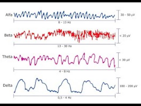



![S2E3 Prof. Stefano Romagnoli - EEG for beginners [ITA]](https://i.ytimg.com/vi/z1rSC6u3RGI/maxresdefault.jpg)
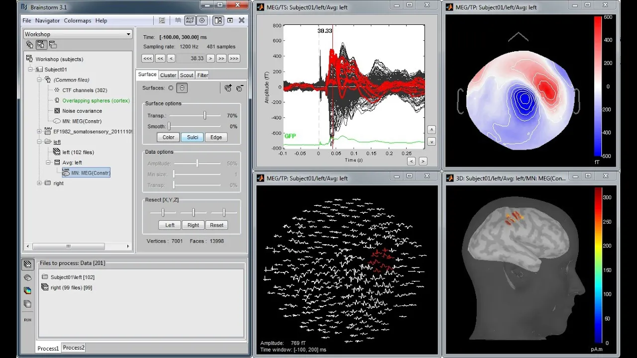


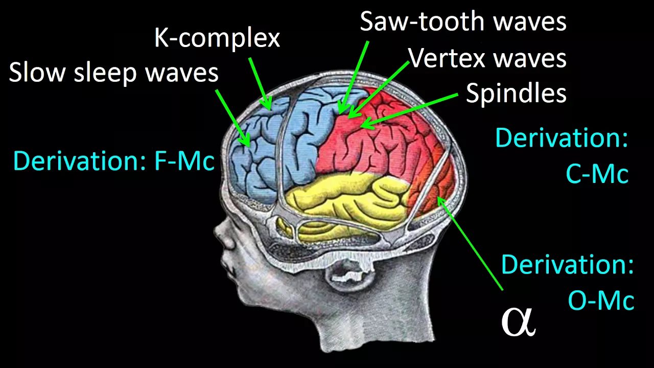










This video explains how EEG patterns change through the five stages of normal sleep: (1) Drowsiness shows decreased muscle artifact, slow rolling eye movements, loss of alpha rhythm, and excess theta activity; (2) Stage 1 sleep features posts (positively charged, occipital maximal) and vertex sharp waves (negatively charged, central maximal); (3) Stage 2 sleep includes K-complexes (high-voltage, arousal-related) and sleep spindles (11-15 Hz, 1-1.5 sec duration, central maximal); (4) Slow wave sleep (stages 3-4) displays diffuse high-voltage delta activity (>20% predominance); (5) REM sleep shows low-voltage background, alpha/beta activity, minimal muscle artifact, and rapid complex oblique eye movements.
Normal Sleep EEG: Stages, Transients & Waveforms
Added:Fundamentals of neurophysiology, including how postsynaptic potentials generate electroencephalographic (EEG) signals.
Familiarity with the International 10-20 system of electrode placement and basic EEG montages.
Understanding of basic awake EEG rhythms, specifically alpha, beta, theta, and delta frequencies.
Ability to distinguish physiological artifacts (such as eye blinks, muscle activity, and cardiac pulse) from cerebral activity.
Polysomnography (PSG) setup and scoring guidelines according to the American Academy of Sleep Medicine (AASM).
Identification of abnormal sleep EEG patterns, including sleep-onset REM periods (SOREMPs) and sleep-induced epileptiform discharges.
Developmental changes in sleep architecture and EEG waveforms from neonatal stages through advanced age.
Clinical evaluation of sleep disorders such as parasomnias, narcolepsy, and sleep apnea using EEG data.
109.2K views1.2Klikes16:52Original Release: 2015-06-08
in this video we will review EEG in normal sleep specifically we will look at different stages of normal sleep and look at the components of each of these stages including drowsiness stage one sleep stage two sleep slow wave sleep and REM sleep in order to understand the progression into different stages of sleep it is useful to first start with normal wakefulness this is a patient with a normal background EEG during wakefulness there are several clues that this patient is awake which we have reviewed in Prior videos you can see prominent myogenic artifact in the frontal and temporal regions on both sides this is likely related to summated compound muscle action potentials from the frontalis and temporalis muscles which have some ongoing tone during wakefulness in addition best seen in the posterior aspects of the midline electrodes there is a normal posterior dominant Rhythm or Alpha rhythm in this case the alpha rhythm is 11 Herz which is well within the normal range thirdly we can see some eye movements including an eye blink here and a quick horizontal eye movement here at the end of the page all of this suggests normal wakefulness likely with eyes closed at least initially as we scroll forward we can see some eye opening with prominent eye movements more muscle artifact and a blocking of the posterior dominant Rhythm as we move forward we see some eye closure and the posterior dominant Rhythm becomes more augmented and easier to see then over the next 10 or 15 seconds we start to see a few changes first we see decreased myogenic or muscle artifact which might suggest that this patient is relaxing and possibly going into a decreased state of arousal this is one of the earliest indications that drowsiness might be starting the second very prominent thing that we can see on this recording are prominent very slow undulations within the temporal change bilaterally if you look carefully at any particular second you can see that there is a large electropositivity at F7 and at the same time there is a large electro negativity at f8 we have reviewed eye movements in a previous video but this would suggest that the positively charged cornea is causing positive deflection at F7 on the left side and there is a negatively charged retina causing negatively charged deflection at f8 on the right side and so this is an eye movement to the left judging by the rate of change and the time course of this undulation which takes between two and 3 seconds these eye movements are slow these are Slow Rolling eye movements which are a normal manifestation of drowsiness as we move forward we can see the alpharithm begins to drop out compare the first half of this page to the second half and you can see that while there is a reasonably welldeveloped alpharithm in the first half of the page this is not seen very well in the second half another thing that you are starting to see in the second half of the page is some excessive Theta activity this is probably best appreciated in some of the midline electrodes such as right here so to review some of the earliest signs of drowsiness or early sleep include decreased muscle or myogenic artifact Slow Rolling horizontal eye movements loss of the normal Alpha Rhythm and excess of theta activity as you begin to read eegs you will see that there will be some fluctuations in the rate of drowsiness and it is uncommon for people to drop off to full sleep right away often as on this page you will see some deeper drowsiness which is interspersed with some bursts of diffuse higher frequency activity seen on the second half of the page here I have moved forward several epochs 2 or 3 minutes later into the stage of sleep you can notice that there is much less muscle artifact suggesting that the patient is in a deeper stage of restfulness you can also notice that there is no posterior dominant Alpha Rhythm and there is much less higher frequency Alpha and beta activity than we saw on previous parts of this recording in addition I would like to focus your attention to the posterior aspect of the head where we see several slightly sharply contoured waveforms which I will point out here you can see that these waveforms have a sharp contour and they are upwardly deflected in the posterior channels of each chain this would suggest that they are either electr negative more anteriorly or they are electropositive posteriorly we can sort this out further by changing montages here I have switched to an average reference Montage if we look at the same waveform on the right side we can see that there is a downward deflection at O2 with reference to the average suggested that this is a waveform with positive polarity maximal in the occipital region if we think about the elements of these waveforms we can come up with the name of this particular phenomenon these have positive polarity they are maximal in the occipital region they are sharply contoured transients and they are occurring during sleep if we put this together we can see that these are posts posts are normally seen in stage one or early sleep although they can persist into stage two sleep as well posts are often the first indication that a patient is transitioning from drowsiness to stage one sleep you might have noticed when thinking about the normal wakefulness this video that posts have a very similar morphology to Lambda waves except that they occur during sleep although the exact generator of posts is unknown patients with prominent Lambda waves often have prominent posts it is useful to think about posts as the Sleep State correlate of Lambda waves as they have very similar morphology location and polarity I have switched to the EEG of another patient for a better clearer example of the next common stage 1 sleep transient again we know that this patient is asleep for several reasons including the absence of prominant myogenic artifact the absence of any normal clear well modulated posterior dominant Rhythm and the presence of sleep transients I would focus your attention to the center of this screen you can see that there are slightly high voltage sharply contoured waveforms occurring in a run which are best seen in the central chains both on the left on the right and in the midline you can see that the quote unquote phase reversal or maximum negativity for these sharply contoured waveforms occurs at C3 CZ and C4 again this confirms that these are sharply contoured waveforms which have negative polarity and are maximal in the central part of the head also known as the vertex these waveforms are called vertex sharp waves and they are often seen in late stage 1 sleep and can persist throughout stage two sleep as well I have switched to a transverse Montage to demonstrate that these waveforms are best seen in the center of the head on this transverse Montage the central chain of electrodes goes from left to right across the center of the head and you can see that the vertex sharp waves are highest voltage in this region it is often useful to look at sleep using a transverse Montage as many other sleep transients as you will see are maximal in the central head region I have switched the Montage again this time to an average reference Montage simply to confirm that these waveforms are highest voltage and negatively charged at CZ C3 and C4 aka the vertex region of the head to summarize the two main components of stage one sleep are posts and vertex waves I have returned to our first patient to demonstrate the next sleep transient that is important to recognize again Focus your attention to the center of this screen here you can see a relatively high voltage relatively broadly contoured long duration waveform that is frontal and Central maximal with a relatively diffuse use field this high voltage waveform has two large phases but these waveforms can often have more phases here we see an initial high voltage negativity followed by a subsequent positivity in addition superimposed on this waveform you can see some high frequency fast activity all of the characteristics I described are characteristics of a k complex a complex is one of the two characteristic findings seen during stage two sleep here is another example of a more complicated C complex which you can see has several phases the origin of the name of K complex is the is the subject of some dispute however there is some evidence to suggest that the K might stand for knock K complexes are generated during arousals and often generated during external stimulation such as when a technologist knocks on the wall or the door it is common to see K complexes preceding an arousal if you look at this part of the Sleep recording you can see that there is a string of two k complexes that then precede an arousal which is indicated by The increased muscle artifact and the loss of sleep transients here I have switched to a transverse Montage which as I said before can be a useful way of looking at sleep Transit which are often maximal in the midline you can see that the K complex has a slightly different location than the vertex waves which were maximal at CZ these K complexes are slightly more frontal maximal although they do have a broad field here we can see the other main component of stage two sleep focusing again on the center of the page you see a rhythm lasting approximately 1 to 1 and 1/2 seconds consisting of undulating 14 Hertz rhythm maximal in the center of the head this is a sleep spindle sleep spindles are the other Hallmark of stage two sleep to summarize the components of spindles they are usually medium voltage midline maximal and have a duration of at least half a second and usually 1 to 1 and 1/2 seconds and a frequency of 11 to 15 Hertz usually averaging 12 to 14 Hertz this is a routine EEG during sleep in a normal 9-month-old baby you can see in the center of the page another sleep spindle in this case the spindle is maximal at C3 CZ and C4 if we look forward in this child we can see that at times the spindles are either maximal on one side as seen here or on the other side as seen here these are what are called asynchronous sleep spindles which is a normal phenomenon between the ages of 3 months and 18 months some degree of asynchronous spindles can also be seen up to 2 years of age the other thing to notice about these spindles is that they are much longer than sleep spindles in adults in this case this spindle lasts almost 3 seconds some EEG Learners have used the word asymmetric rather than asynchronous but the major distinguish factor is that asymmetric would suggest that the spindles are either exclusively or almost always seen on one side of the head and not on the other asynchronous spindle should be seen in approximately equal quantities on both sides of the head but at any one time they might be maximal on one side of the head or the other so to summarize the two major components of stage two sleep are spindles and K complexes as we scroll forward we can start to identify some of these sleep transients coexisting in this stage 2 sleep recording at the start of the page we see a k complex as we move forward we see a slightly bluntly contoured vertex wave near the end of the page we see a run of posts and then we see some spindles superimposed on some Delta activity the posts can occur in rhythmic runs which sometimes appear sharply contoured at the end of the page the vertex wave can appear more sharply contoured than on previous examples and sometimes the spindles can be less welld developed and have a broader field extending into the frontal head regions I have switched back to the recording of the other patient to show a later stage of sleep on this recording you do not see vertex waves K complexes or spindles but there is no muscle artifact and this patient is asleep here what you do see are diffuse that is they affect the entire head high voltage slow frequency Delta activity with some superimposed higher frequency Alpha and beta activity you can see that this higher voltage Delta activity predominates during this entire Epoch this is what's called slow wave sleep previously called stage three and stage four sleep this is a deeper stage of sleep than the stage one or two that we saw previously by definition slow wave sleep is defined when greater than 20% of the Sleep background is predominated by high voltage Delta activity finally we have reached the stage of sleep which we will discuss last here in comparison to the slow wave sleep you can see that the background is much lower voltage and that the main waveforms seen are in the alpha and beta range perhaps seen best when focusing on the midline in addition there is very little or almost no muscle artifact finally we see some very complicated eye movements which have both a horizontal and vertical component we can see that they have a horizontal component because there is an opposite polarity in the right and left temporal region for example looking at this particular eye movement we can see that they have a vertical component because there is a synchronous deflection in the frontal polar region on the left and the right therefore we would call these oblique eye movements if you look at this set of three eye movements you can see that they are all in slightly different directions because they all have slightly different polarity in the temporal and frontal regions you can also see that the initial eye deflection is very quick lasting only about 100 milliseconds therefore we can conclude that this patient is having rapid slightly chaotic oblique eye movements taken together with the fact that we know this patient is asleep and that there is very little muscle or movement artifact we would conclude that this patient is in rapid eye movement sleep or REM sleep many eegs have difficulty distinguishing REM sleep from wakefulness but I would point out that there is no normal anterior to posterior gradient of the alpha Rhythm there is no muscle and the eye movements are somewhat complex and therefore are less likely to represent voluntary eye movements to summarize we have reviewed the five different stages of normal sleep including drowsiness stage one stage two slow wave sleep and REM sleep being aware of the normal components of sleep allows us to recognize abnormalities which will be reviewed in subsequent videos
Up Next

16:16
Sleep Neuroscience: Stages, EEG & Brain Mechanisms
@labfrontofficial
144 views•2025-04-27

21:55
Deciphering Neural Circuits Controlling Anorexia in Mice | Richard Palmiter
@AllenInstitute
2.3K views•2011-10-10

4:10
Vagus Nerve (CN X): Anatomy, Nuclei & Functions Explained
@Alilamedicalmedia
305.2K views•2022-10-31

13:03
How Exercise Benefits Your Brain: Science Explained
@TED
11.4M views•2018-03-21
Related Study Plans & Knowledge Roadmaps
Structured learning paths in Neuroscience
neuroscience
Appetite Regulation: Brain Circuits & Hormones
01Foundations of Endocrinology and Brain Anatomy
02Introduction to Energy Homeostasis
03Peripheral Signals: Leptin, Ghrelin, and Insulin
+ 2 more learning modules
14h5 modules
Explore Roadmap
neuroscience
Neuroeconomics: Value, Risk & Brain Mechanisms
01Foundations of Neuroscience and Brain Anatomy
02Introduction to Neuroeconomics and Subjective Value
03Dopaminergic Prediction Errors & Reinforcement Learning
+ 3 more learning modules
18h6 modules
Explore Roadmap
neuroscience
Spatial Navigation: Cells, Grids & Brain Maps
01Foundations of Brain Anatomy and Memory
02Place Cells and Head-Direction Cells
03Grid Cells: The Brain's Coordinate System
+ 2 more learning modules
12h5 modules
Explore Roadmap
neuroscience
BCIs: Neural Decoding & Prosthetic Control
01Neuroscience Foundations: How the Brain Signals Intent
02Signal Acquisition: Recording the Brain's Activity
03Neural Decoding: Translating Brain Waves into Data
+ 2 more learning modules
25h5 modules
Explore Roadmap