









![eGurukul 4.0 - CNS [EEG Waves & Disorders] | Dr. Ashish Kumar [Physiology]](https://i.ytimg.com/vi_webp/tq7IyM2YCx8/maxresdefault.webp)
















Sleep is regulated by a complex neural network involving the reticular activating system, which promotes wakefulness through neurotransmitters like norepinephrine, serotonin, histamine, and acetylcholine, while the ventrolateral preoptic nucleus (VLPO) acts as a master switch to initiate and maintain sleep through GABAergic inhibition; this flip-flop switch operates within a broader framework of circadian rhythms and homeostatic sleep pressure, with additional regulatory mechanisms governing the transition between non-REM and REM sleep, and these neural pathways are modulated by various medications and connected to the autonomic nervous system through neurovisceral integration.
Sleep Neuroscience: Stages, EEG & Brain Mechanisms
Added:Basic neuroanatomy, particularly the functions of the brainstem, hypothalamus, thalamus, and cerebral cortex.
Fundamentals of neuronal excitability, synaptic transmission, and key neurotransmitters (such as GABA, glutamate, acetylcholine, and monoamines).
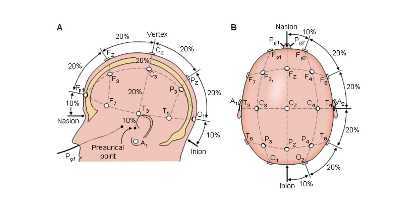
Principles of Electroencephalography (EEG), including how electrical activity of the brain is recorded and the concept of neural oscillations.
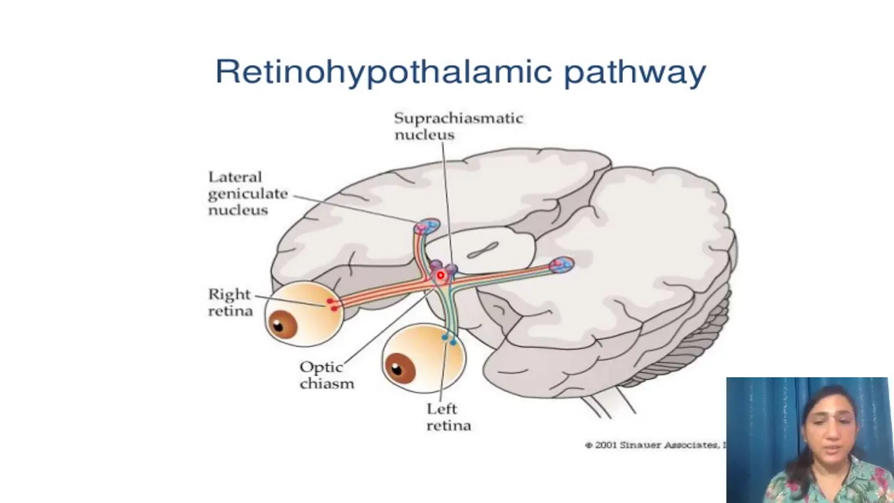
An introduction to circadian rhythms and the role of the suprachiasmatic nucleus (SCN) in regulating daily biological cycles.
Pathophysiology of sleep disorders, such as narcolepsy (loss of orexinergic neurons), insomnia, and REM sleep behavior disorder.
The cognitive and physiological functions of sleep, specifically synaptic plasticity, memory consolidation, and the glymphatic clearance system.
Pharmacology of sleep and wakefulness, focusing on how anesthetics, stimulants, and sedative-hypnotics modulate sleep architecture.
Advanced neuroscientific techniques used in sleep research, such as optogenetics, chemogenetics, and polysomnography data analysis.
144 views0likes16:16Original Release: 2025-04-27
So let's first delve into the core of our topic, the neuroscience of sleep, and this section will explore the intricate neural mechanisms that govern sleep.
So I think many, you've probably seen this.
This is a hypnogram showing an idealized breakdown of the stages of sleep through the night, and you can see that human cycles through non-REM sleep and REM sleep.
So non-REM sleep and non-REM is divided into stage one, two and three, and one and two, and then three.
And these stages cycle approximately every 90 minutes in an old Tridion rhythm, amounting about four to five non-REM REM cycles per night.
And there's typically more N3 sleep in the early night and more REM sleep later in the latter half of the sleep period.
Now how do we distinguish these sleep stages?
And this is primarily done with the EEG or electroencephalogram and it's part of a broader PSD assessment.
And this slide shows the characteristic EEG patterns.
So we notice the progression of non-REM sleep.
As we move from wakefulness with its alpha waves to deeper sleep, the EEG becomes increasingly synchronized.
And so Stage N1 shows lower amplitude mixed frequencies, mainly theta waves at four to seven hertz.
Stage N2 is defined by sleep spindles, those brief bursts of faster activity, and K complexes, the large distinctive waves, which is by biphasic in nature.
And then stage N3, which is deep sleep, is characterized by high amplitude, slow delta waves.
And REM sleep, however, is completely different.
You know, the EEG de synchronizes resembling wakefulness with its higher frequencies and lower amplitude waves.
We also see theta waves during the tonic phase and unique saw tooth waves, during the phasic phase of REM.
And of course you have rapid eye movement and muscle atonia, basically a paralysis of your muscles, which is measured by electrooculogram and the EMG respectively.
And these are also crucial for identifying REM.
So these distinct EEG patterns are the key to objectively defining and analyzing each stage of sleep.
So we go from the patterns of wakefulness and sleep to understanding the underlying mechanism.
And this slide introduces the reticular activating system, a critical network for wakefulness and arousal.
And the foundation of our modern understanding comes from the pioneering work of Moruzzi and Magoun, which was done in 1949 and pictured here, and their key discovery was that the ascending reticular activating system located in the upper pons and midbrain locations were critical for arousal.
And basically what they did was they took anesthetized cats, stimulated this region, and that would shift the EEG of the cat from slow synchronized waves of sleep, just like what we saw, to the desynchronized active pattern of wakefulness, even if the direct sensory input was blocked.
And so this demonstrated that the, ascending reticular activating system has a powerful intrinsic ability to promote wakefulness.
This diagram shows projections from key brainstem nuclei releasing neurotransmitters such as norepinephrine from the Locus Coeruleus (LC), serotonin from the Raphe nucleus, dopamine from the substantia nigra and the ventral tegmental, and the histamine from the tuberomammillary nucleus, and acetylcholine also from the tegmental areas of the midbrain.
So this widespread activation is what underlies the alert and awake state.
Now this slide illustrates a simplified model, the sleep-wake switch, highlighting the key brain regions and pathways involved.
And let's first focus on the wake promoting side, shown on the left, and we see the familiar players from our previous, slide, the brainstem nuclei, including the Locus Coeruleus (LC), the Dorsal Raphe, the basal forebrain, all standing per projections upwards.
And this, activates our awareness and arousal.
And we also note, important players like the orexin neurons and these areas collectively activate the cortex of the thalamus, maintaining wakefulness.
Now looking at the right, we see the sleep promoting side, and the key player here is the ventrolateral preoptic nucleus, or VLPO, and also the median preoptic nucleus in the hypothalamus.
And the VLPO is kind of like a master switch.
It's the sleep promoting centers that generally inhibit all the wake promoting centers to promote onset and maintenance of sleep.
So this slide illustrates sort of the flip-flop nature of the sleep wake switch, showing the reciprocal relationship between the wake promoting and sleep promoting neuronal activity.
And this graph plots the firing rates of key neuronal populations across transitions from wake to sleep.
On the left, we see the transition from wakefulness to the light non-REM sleep, and you notice the firing rates of wake promoting neurons shown in blue for acetylcholine, gray for the histamine, green for norepinephrine from the Locus Coeruleus (LC).
sort of decrease as, this animal transitions into sleep.
Simultaneously, the firing rate of the neurons in the sleep promoting preoptic area, which releases GABA, is shown in red increases.
The right graph shows the reverse, the transition from non-REM sleep back to wakefulness.
And as the animal wakes up, the GABAergic sleep-promoting neurons decrease their firing, while the wake-promoting, cholinergic, histaminergic, and noradrenergic neurons increase their activity.
So this reciprocal inhibition where wake promoting neurons suppress sleep-promoting neurons and vice versa, the hallmark of a flip flop switch.
And this is shown here basically in a much more, easy to visualize diagram.
And again, you could see that the monoamines and acetylcholine are on the wake side, GABA in from the VLPO and MnPO, is on the right associated with sleep.
Now this flip flop model, while useful conceptually maybe is an oversimplification in some ways.
In reality, the dynamics of this neurotransmitters are much more nuanced, and this slide shows actual measurements of the monoamine levels, not just firing rates across wakefulness, REM and non-REM.
And you could look at serotonin, histamine and norepinephrine.
And let's just take a look at the serotonin, for instance.
and blue is here marked, by wake.
The white areas is non-REM, and red is REM.
And you could see that the levels of the monoamines are highest during wakefulness and generally lower during non-REM sleep.
And, but they don't completely disappear.
Instead, we see a gradual decrease in serotonin, histamine as the, as the animal progresses through non-REM sleep.
and this is important to demonstrate that the sleep weight transition isn't a binary on off switch for these monoamines, it's more of a dimmer switch, a gradual modulation of their levels.
And so far we presented the sleep weight Flip-Flop Switch as a relatively isolated system, but in reality, it's beautifully integrated with other crucial regulatory processes.
And we still have a basic balance between these two sides.
However, now we add Orexin, a neuropeptide produced in the lateral hypothalamus and orexin acts to stabilize the wake state promoting wakefulness and preventing inappropriate transitions to sleep.
And we know that individuals who lack the, ability to secrete Orexin has narcolepsy, because then they, you know, have this unstable switching back and forth between the wake and sleep.
In addition, the entire system is modulated by two major forces.
First, the circadian rhythm, our internal biological clock, exerts a, a time of day dependent influence, promoting wakefulness during the day and sleep at night.
And second, the Homeostatic sleep drive, which builds up with prolonged wakefulness, primarily through the accumulation of substances like adenosine.
And adenosine acts on A1 receptors to increase the sleep pressure.
So these modulators act on both the wake and sleep centers, and it's a dynamic balance constantly adjusted by internal factors like orexin and external cues conveyed by the circadian homeostatic drives.
And so this is just a quick summary of the representations here during wakefulness, the monoamines acetylcholine, orexin, is elevated.
During non-REM, GABA, is elevated.
All right.
So now we've established the basic flip flop switch between wakefulness and sleep, which is primarily driven by the balance between the monoamines and acetylcholine and the GABA neurons.
Now let's add another level: the switch within sleep between non-REM and REM sleep.
And notice the lower portion of the diagram.
We have a second seesaw representing this non-REM REM balance during non-REM sleep, as we have seen, the VLPO, MnPO are active, inhibiting the wake- promoting centers, but within the state there's also reduced activity from the REM- promoting cholinergic neurons of the PPT and LDT in the brainstem.
Norepinephrine and serotonin contribute, to sort of go against, this REM sleep.
And so the transition to REM sleep often involves a shift in this balance.
Activity decreases in the noradrenergic and serotonergic neurons further, and there's a surge in activity from in these cholinergic PPT/LDT neurons.
And this cholinergic drive combined with the continued inhibition of the wake promoting centers by the GABA results in the unique state of REM sleep, which is, you know, represented by desynchronized EEG, muscle atonia and dreaming.
And so we have a nested system, a primary Flip-Flop switch between wake and sleep and a secondary Flip-Flop switch within sleep governing the alternation between non-REM and REM.
And so this slide sort of details the brain circuitry behind the non-REM REM sleep switch.
We have REM-on regions, like the cholinergic, LDT and PPT and REM-off areas like the ventrolateral periaqueductal gray (vlPAG) and the LPT, which are GABAergic.
Orexin, from the lateral hypothalamus, I don't see that here, is involved.
It suppresses REM, while MCH, or melanin-concentrating hormone, may promote it.
So crucially, the ascending projections from the LDT/PPT are responsible for the key REM EEG features, such as theta waves, via connections to the septo-hippocampal region.
The descending portion from the SLD, another part of the REM- on areas, cause muscle atonia of the REM sleep by inhibiting motor neurons, primarily using GABA and glycine.
And so this sort of provides a summary of that.
You have the monoamines and acetylcholine elevated during wakefulness.
You have GABA elevated in both stages of sleep, but then you have acetylcholine and MCH elevated during REM sleep with some minor activation of monoamines during the non-REM sleep.
And so this slide builds on our previous look at monoamine levels, now adding acetylcholine dynamics across the sleep stages.
And we've already seen that serotonin histamine are highest during wake, decrease in non-REM, but have, levels in, in REM.
And now if you look at acetylcholine, this shows a biphasic pattern.
It's high during wakefulness, as expected.
It decreases significantly during non-REM, but crucially, it increases again during REM sleep reaching levels compared to or even exceeding wakefulness.
So you may be wondering like, why am I spending so much time on these neurotransmitter systems and the flip flops switches?
It's because I think it's relevant to how, many of the common medications affect sleep and wake wakefulness.
And let me give you some examples of this.
On the wake side, we see that MAO inhibitors increased monoamine levels, which can enhance alertness, but worsen insomnia.
Anti-histamines, like mirtazapine or doxepin, block histamine, a key wake-promoting monoamine, and this is why they're used often as sleep aids.
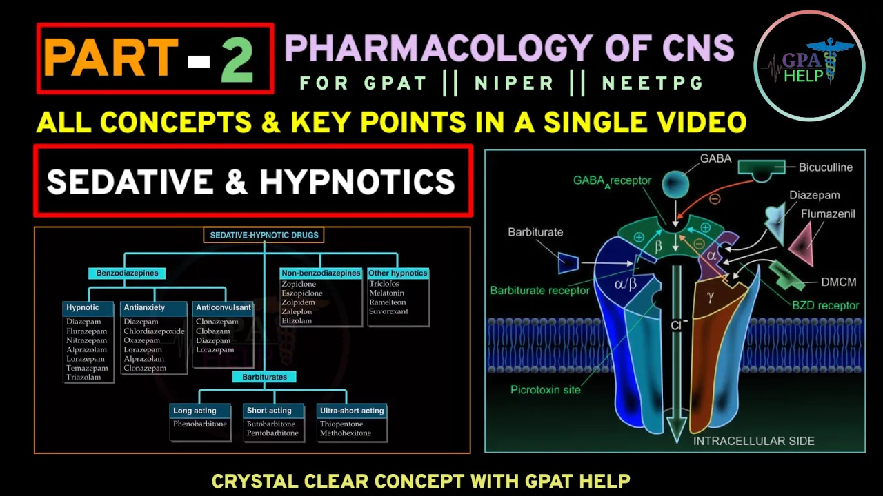
On the sleep side, we have benzodiazepines and alcohol with both enhance the effects of GABA.
And with thus promoting and strength strengthening the sleep side of the switch.
And if you look at the non-REM REM balance, like SSRIs and many anti-psychotics, this increases the norepinephrine and serotonin that favors non-REM and then also suppresses REM.
And finally, Alzheimer's disease characterized by a loss of cholinergic neurons, often leads to significant deficits in REM sleep as well.
And so this slide illustrates the connection between our sleep centers and our broader central autonomic network, as explained by the NEUROVISCERAL integration theory.
As you can see, sleep centers are primarily located in the hypothalamus, midbrain, and pons region that are also linked to the stress, motor control and behavior, and the endocrine system.
So this anatomical overlap underscores that sleep is not just the process of shutting down.
Instead, it's a deeply integrated with the autonomic nervous system and, and which is the system responsible for regulating heart rate, digestion, and stress response.
And so the Neurovisceral integration theory posits that these brain regions collaborate to coordinate our internal body state with our behaviors and environmental responses, and this integration is bidirectional.
We have bottom up influences where the signals from the body, such as stress, hormones, and pain can affect sleep.
But there's also top down influences where practice like meditations, which can impact the prefrontal and cingulate cortex, can modulate the activity of these lower brain regions and in turn influence sleep.
So in essence, sleep is a dynamic pattern process influenced by both influences on both bottom end, top down, factors.
Up Next

28:35
Neurophysiology II: Brain Areas & Sleep-Wake Cycle | Intro Guide
@Mbbsnaija
110 views•2023-08-15

21:55
Deciphering Neural Circuits Controlling Anorexia in Mice | Richard Palmiter
@AllenInstitute
2.3K views•2011-10-10

4:10
Vagus Nerve (CN X): Anatomy, Nuclei & Functions Explained
@Alilamedicalmedia
305.2K views•2022-10-31

13:03
How Exercise Benefits Your Brain: Science Explained
@TED
11.4M views•2018-03-21
Related Study Plans & Knowledge Roadmaps
Structured learning paths in Neuroscience
neuroscience
Appetite Regulation: Brain Circuits & Hormones
01Foundations of Endocrinology and Brain Anatomy
02Introduction to Energy Homeostasis
03Peripheral Signals: Leptin, Ghrelin, and Insulin
+ 2 more learning modules
14h5 modules
Explore Roadmap
neuroscience
Neuroeconomics: Value, Risk & Brain Mechanisms
01Foundations of Neuroscience and Brain Anatomy
02Introduction to Neuroeconomics and Subjective Value
03Dopaminergic Prediction Errors & Reinforcement Learning
+ 3 more learning modules
18h6 modules
Explore Roadmap
neuroscience
Spatial Navigation: Cells, Grids & Brain Maps
01Foundations of Brain Anatomy and Memory
02Place Cells and Head-Direction Cells
03Grid Cells: The Brain's Coordinate System
+ 2 more learning modules
12h5 modules
Explore Roadmap
neuroscience
BCIs: Neural Decoding & Prosthetic Control
01Neuroscience Foundations: How the Brain Signals Intent
02Signal Acquisition: Recording the Brain's Activity
03Neural Decoding: Translating Brain Waves into Data
+ 2 more learning modules
25h5 modules
Explore Roadmap