

















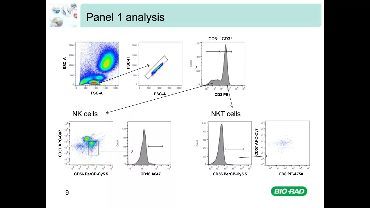



Flow cytometry is a cell analysis technique that uses hydrodynamic focusing to pass cells one-by-one through a laser beam, where forward scatter light (detected at acute angles) measures cell size proportionally and side scatter light (detected in perpendicular directions) measures cell complexity/granularity; these two parameters combined in a 2D histogram allow rapid analysis of thousands of cells per second to determine cell phenotype and health status.
Flow Cytometry Explained | Principles of FACS Analysis
Added:Basic cell biology, specifically understanding cell morphology (size and internal complexity) and cell surface receptors.
The physics of light and optics, including wavelength, light scattering, and the electromagnetic spectrum.
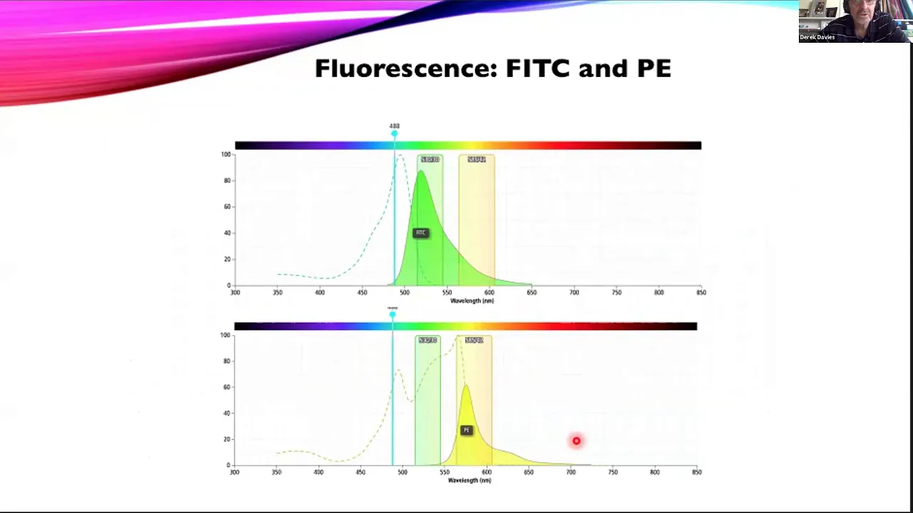
The principles of fluorescence, including how fluorophores absorb light at an excitation wavelength and emit it at a longer wavelength.
The concept of antigen-antibody specificity and how fluorescently-conjugated antibodies are used to label specific cell types.
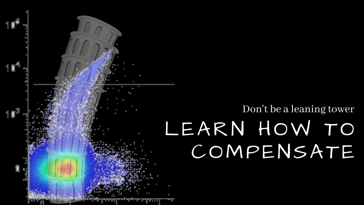
The principles of spectral overlap and how to perform fluorescence compensation in multi-color flow cytometry panels.
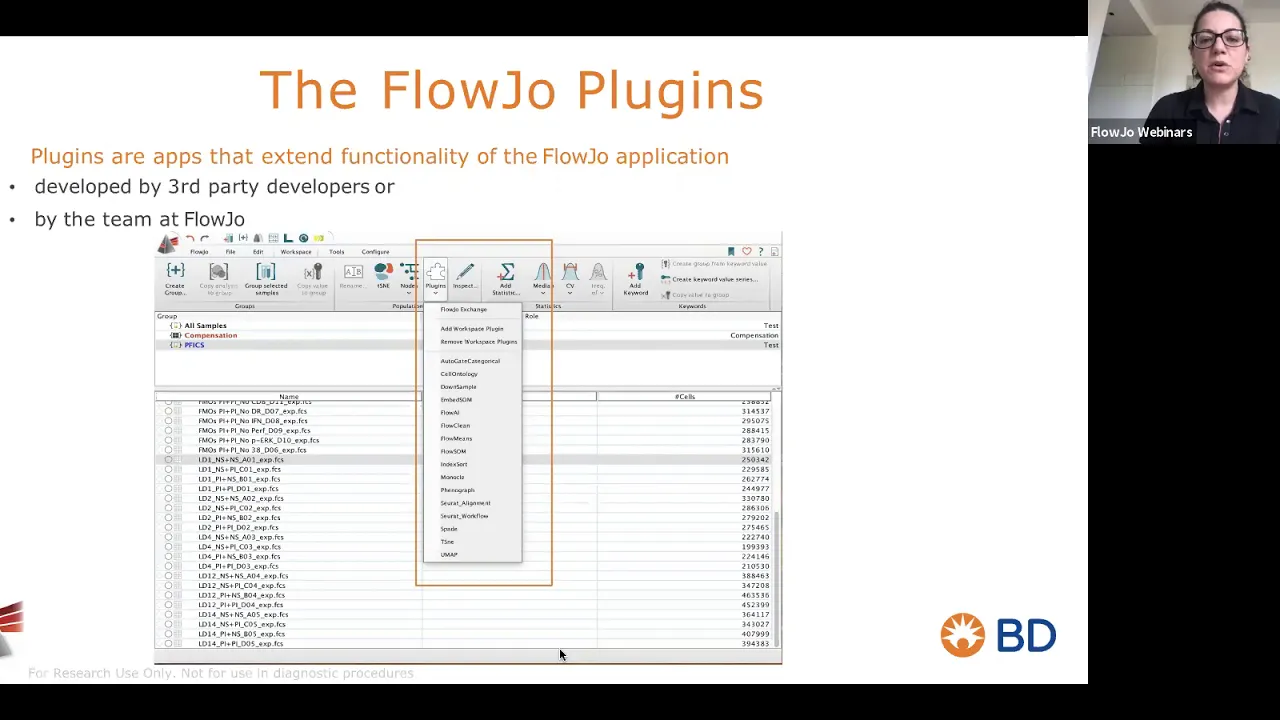
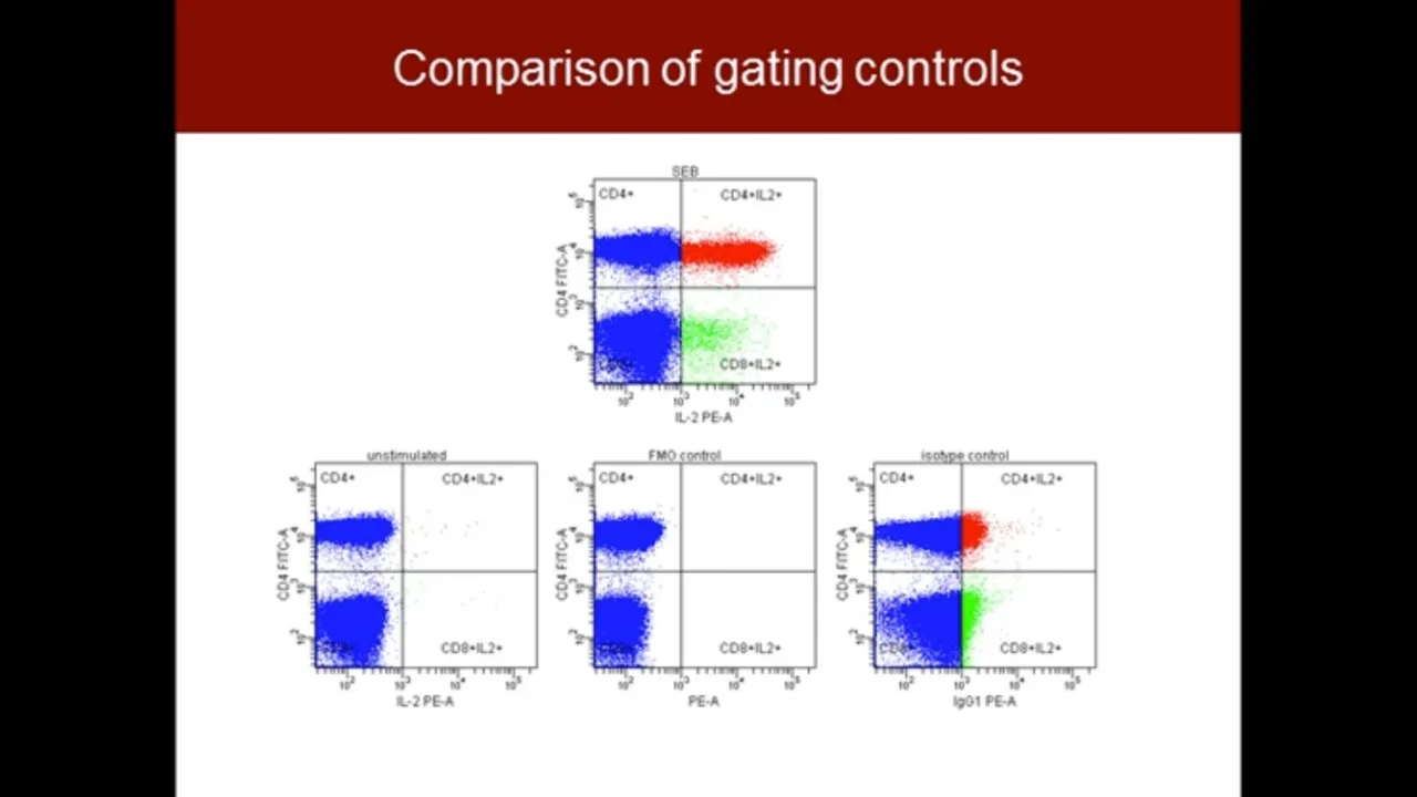
Advanced data analysis and gating strategies using specialized software (e.g., FlowJo) to isolate specific cell populations.
Design of multi-parameter flow cytometry panels, including choosing the right fluorophore-antibody combinations for low and high-abundance markers.
Clinical and research applications of flow cytometry, such as immunophenotyping, cell cycle analysis, apoptosis assays, and sorting specific stem cell populations (FACS).
169.6K views3.5Klikes10:29Original Release: 2018-04-20
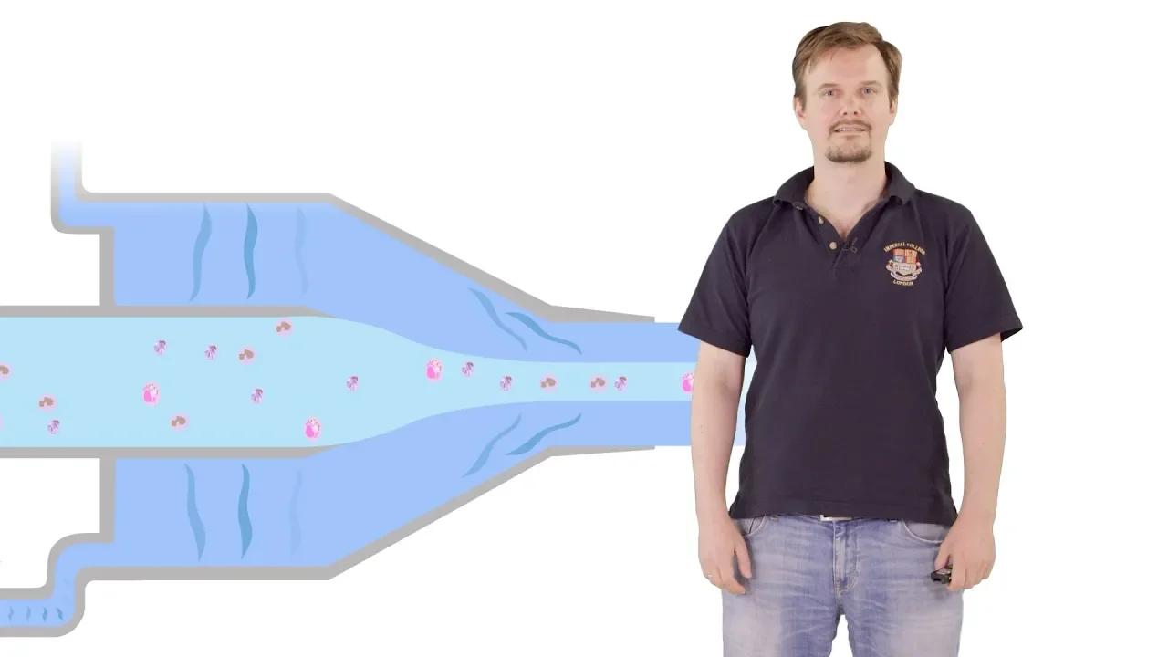
hello guys in this video I'm going to speak about a very important or very very interesting mechanism called the flow cytometer and facts actually I did two videos and this video I'm going to mention specifically flow cytometer and in the next video I'm going to mention the facts mechanism fluorescence activated cell sorting lists are speaking about flow cytometer flow cytometer is cell counting mechanism so it's used to count the cells and together to analyze the phenotype and the health situation of the cell how does it work in general first in general in flow cytometer i have something called the flow cell in the flow cell we add our cell mixture then I have the sheath fluids the sheath fluids will apply something called the hydrodynamic focusing the hydrodynamic focusing or the sheath fluids will send the cells one by one through the flow cytometer tube it's very important to to pass the cell what the cells one by one through the through the tube because here I have something called the laser beam and this the cells should pass through the Laser laser beam one by one this point where when where the cells pass through the laser beam is called the interrogation point now when the cell pass through the laser beam there are two events happening first if the forward scattering which is the light scattered in an acute angle to the forward direction and the side scattering glass which is the light scattered in unison in every other direction in side directions these two events or this light scatter scattering is detected by specific detectors called the forward detector and sight detectors and these detectors are connected to a PC or a data analysis and aisle analyzer which is going to analyze the data obtained from the detectors now this is in general now I'm going to speak in more in details saying that this is the sat flow cytometer of flow cytometry tube and then the cells pass one by one through the laser beam when the cell paths through the laser beam the cells scattered the light in a forward direction which is in a the light scattered in an acute angle this forward scattered light is directly proportional to the size of the cell why because the cells scatter as like a particular amount of light in the forward direction but when the cell is bigger the cell the forward scattered light will be larger or like yeah more and more Scott more forward scattered light will be detected and when the cell is smaller the cell will scatter less light in the forward direction so the forward scattered light is directly proportional to the size of the cell now the detector detects the pulse of photons which are the light scatters the forward scattered light is a pulse of photons which are detected by the detector and the detector then is as I told you before is connected to a PC or computer the PC will convert the intensity of the light detected by the detector into voltage and then I will get a specific amount of voltage for every cell passing through the laser beam the PC will give me a data similar to this so every time a cell pass through the laser beam I will get a specific peak like high voltage peak for large cells and low voltage Peaks for small cells the data or the forward scattered light data at the end be similar to this time versus voltage and then the peak will be proportional to the size of the cell this is forward scattered light the second events happening in the full earth flow cytometer is the site scattered light so awareness when a cell paths through the laser beam the cell will scatter the light in side direction and this side scattered light is directly proportional to the complexity on granulizer the granularity of the cell why because the simple cells scattered light in the side direction yes but when we have granules inside the cell so when the cell is complex from inside these little granules will also scatter light in different directions and in this case I will get more site scattered light like this so this is a complex cell from inside with it contains small granules inside and then these granules will also scatter the light in different directions and in this case I would get a higher intensity of the site scattered light similarly to what we saw in forward scattered light the the detector our detectors are connected to the PC which is going to convert the intensity of the light into voltage and then I'll get something similar to this so the side scattered like that I will be similar to the forward scattered light data so for the the the complex cells will give me a high peak and the simple cells will give me a small peak now in order to analyze the cells what I have to do is that I have I can do I can combine both forward scattered light and side scatter light data together in in this case I'll get something called the 2-dimensional histogram the 2-dimensional histogram it's name it's two-dimensional so I have side scatter and forward scatter data all together in this case where I get is something similar to this so I have here four quadrant if the cell appear in the lower quadrants it means that it has low signal on the side scatter detectors which means that the cell is simple if the cell appears in the upper in the upper half it means that it gives a high signal or the slight scatter detectors so it means that the cell is complex if the cell appear on the right half it means that the cell give high signal on the forward scatter which means that the cell is big and if the cell appears here it means that the cell is small so if the cell appears for example in this quadrant it means that the cell is small and it's simple if the cell appears here it means that the cell is small but it's complex here it means that the cell is big and complex and here it means that the cell is big but simple I will give you a very good example of this which I have an example about the analysis of a sample of white blood cells so what we see here is an analysis of white blood cells this is like scatter and this forward scatter light what we see here is a group of cells which are complex very cupric complex cells because they give a high signal on the side scatter detector and they are medium inside they are not so big and they are not small so there are Windom inside obviously these cells are granulocytes because granulocytes or like as in a few buzzer few of neutrophils they are medium inside that they are very complex cells here I have bigger cells because they give almost like they give more or less high signal on the forward scatter but they give low signal on the side scattered so they are big but they are not too complex they're simple cells and these are monocytes because monocytes are big cells but they are not complex here we have very simple cells because they give very low signal on the side scatter and they are like smaller than monocytes and granular granulocytes so obvious obviously these are lymphocytes because the lymphocytes are smaller in size and they are simple cells of course this is a very useful technique used to to count the cells and to analyze the phenotype and the health situation of the cells and what's very special about this technique is that we can analyze many thousands of particles and cells per second it's like it's an easy technique it's not very hard to do to perform and it's precise it's very used nowadays in in all the laboratories this is everything I wanted to tell about flow cytometer which is very important to know if you want to know more about facts mechanism in the next video I would speak about fire effects mechanism so if you know you want to know more about facts mechanism then you want to you need to watch the next video if you liked this video please don't forget to Like share subscribe the channel and if you have any questions leave it in the comments I will answer you and see you in the next video bye
Up Next

5:57
Frog Gastrulation: Xenopus Morphogenetic Movements Explained
@animatedbiologywitharpan
80.5K views•2023-04-07

50:12
Bacterial Communication: Quorum Sensing and Biofilm Formation Explained
@JHUAAP
12.6K views•2011-06-28

5:35
Enteric Nervous System Explained: The Gut's Brain | Neurobiology Lecture
@alumniu6029
438 views•2018-09-12

7:08
Bacteriophages: Earth's Deadliest Killers and Future Antibiotics
@kurzgesagt
34.6M views•2018-05-13
Related Study Plans & Knowledge Roadmaps
Structured learning paths in Biology
biology
RNAi: Biogenesis, RISC & Gene Silencing
01Molecular Biology Foundations: DNA to Protein
02Introduction to RNA Interference (RNAi)
03Molecular Biogenesis of miRNA and siRNA
+ 2 more learning modules
10h5 modules
Explore Roadmap
biology
Mycorrhizae: Signaling, Biophysics & Ecology
01Foundations of Mycorrhizal Symbiosis
02Molecular Signaling and Root Colonization
03Biophysics and Bioenergetics of Nutrient Exchange
+ 1 more learning modules
11h4 modules
Explore Roadmap
biology
Circadian Clocks: TTFL, SCN & Periphery
01Foundations of Molecular Biology & Gene Expression
02Introduction to Chronobiology and Actograms
03The Core TTFL: Molecular Architecture of the Clock
+ 3 more learning modules
14h6 modules
Explore Roadmap
biology
Population Genetics: Math, Drift & Selection
01Genetic & Probability Foundations
02The Hardy-Weinberg Principle and Baseline Models
03Mathematical Modeling of Natural Selection
+ 3 more learning modules
18h6 modules
Explore Roadmap