














![Anti-Arrythmics Pharmacology [Action Potential, Arrythmias, Waughan Williams Classification]](https://i.ytimg.com/vi_webp/1YXMzaCBYQg/maxresdefault.webp)
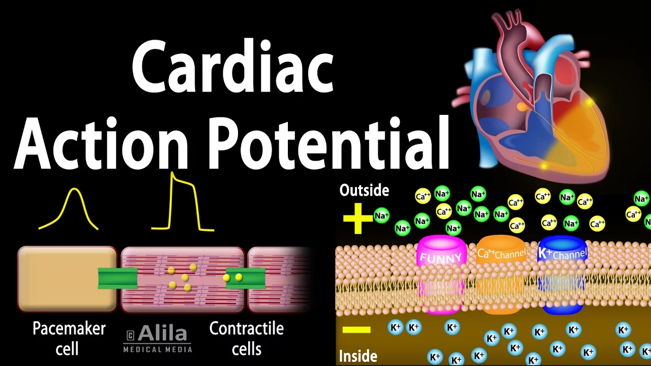
The SA node generates action potentials through a unique pacemaker mechanism where the membrane potential oscillates from -60 mV to -40 mV (phase 4/pre-potential) due to funny currents (If channels) and transient calcium channels, then rapidly depolarizes (-40 mV to +30 mV, phase 0) via L-type calcium channels, followed by repolarization (phase 3) through potassium efflux; the autonomic nervous system modulates this process by having sympathetic stimulation (via beta-1 receptors and cAMP) increase heart rate by enhancing funny current and calcium influx, while parasympathetic stimulation (via muscarinic M2 receptors) decreases heart rate by increasing potassium conductance and reducing calcium influx.
SA Nodal Action Potential & ANS Effects: Cardiac Physiology
Added:Basic cellular electrophysiology, including resting membrane potential, electrochemical gradients, and the function of voltage-gated ion channels.
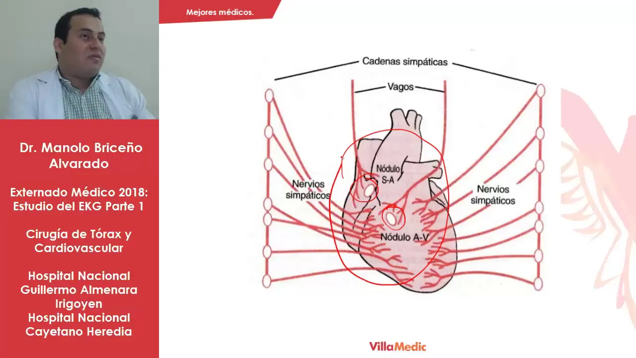
The general anatomy of the cardiac conduction system, specifically the location and role of the Sinoatrial (SA) node as the primary pacemaker.
Fundamentals of the Autonomic Nervous System (ANS), specifically distinguishing between sympathetic (fight-or-flight) and parasympathetic (rest-and-digest) pathways.
The concept of neurotransmitters and cardiac receptors, particularly acetylcholine (muscarinic receptors) and norepinephrine (beta-adrenergic receptors).
The mechanisms of cardiac contractile cell action potentials (ventricular myocytes) and how they differ from pacemaker action potentials.
How cellular pacemaker activity correlates to the electrical vectors and waveforms observed on a standard Electrocardiogram (ECG/EKG).
Clinical pathology of cardiac rhythm disorders, such as sinus bradycardia, sinus tachycardia, and sick sinus syndrome.
Cardiovascular pharmacology, exploring how drugs like beta-blockers, calcium channel blockers, and atropine alter SA nodal action potentials to regulate heart rate.
2.5K views38likes20:24Original Release: 2025-06-21
[Music] [Music] Okay, welcome back guys. Now in the second video, let's discuss about the action potential of an SA node. Okay, we have discussed that SA is a pacemaker of the heart. It is going to undergo self depolarization.
See, try to understand something like this. There are two sets of students.
Okay, so one is self motivating. Okay, he's going to motivate by himself and he will come and motivate the other person.
The other type of students they cannot selfotivate. They need to have an external stimulation. Now in the same way our pacemaker cells, they will undergo self depolarization.
automatically they will undergo activation.
Okay, that's why they are called as a pace makers. They control the heart rate out of which the SA node is the pacemaker of the heart because it's producing the maximum number of action potentials. So now let's see SA load action potential how the action potential of the Senode happens. Okay.
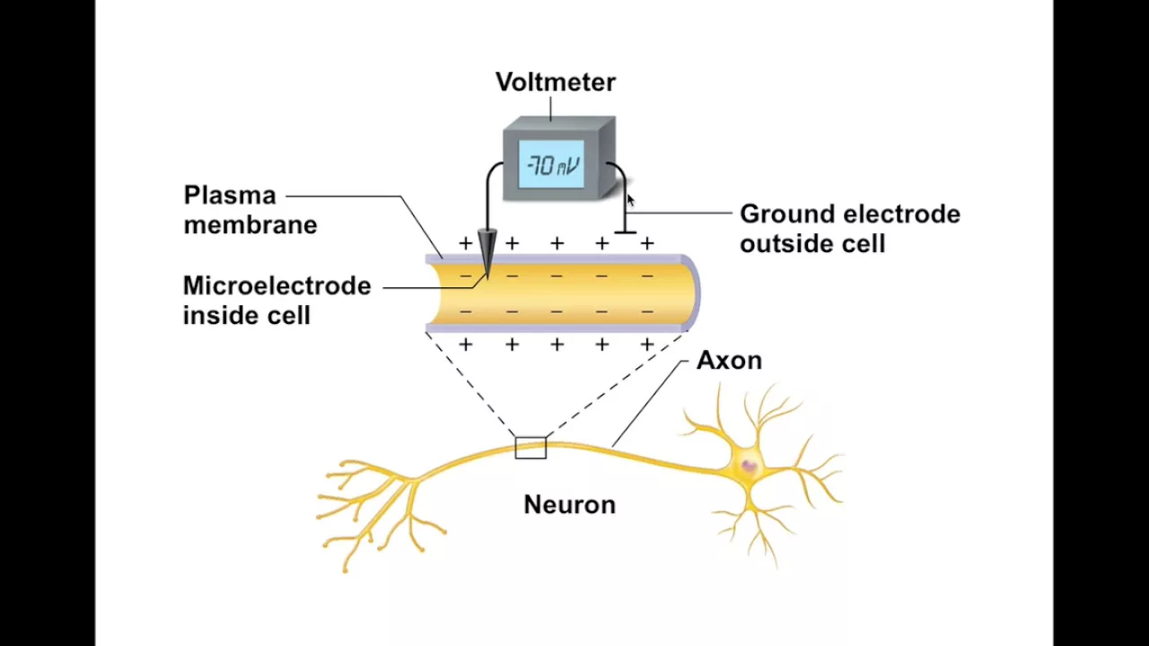
See the Sodal has a pacemaker potential or restless membrane potential. Okay.
Which is at -60 m volts. Okay guys please look at here in this graph what I'm showing you is the SA nodal action potentials. Now see let's start from the minus 60. The SA node. Okay that one particular cell is having a membrane potential of minus60 but it is not constant. And it is not constant. See what is happening. This potential is moving towards positive side. Am I right or not? Now -60 it is gradually gradually becoming -40. -60 it is becoming -40 means getting little bit positivity into the cell. Right? From -60 it is getting -40.
So now cell is moving from -60 to -40 or I can say the membrane potential is moving towards positive side or towards threshold. Now you can ask me sir why this is happening? Why the membrane potential is not resting constantly at minus 60? Why it is moving towards threshold? Now let me show you here.
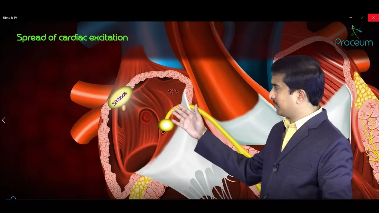
Imagine this is SA node.
Okay this is S node. Now this acode because of the sodium potassium ATBs okay there are channels called as sodium potassium ATBs. What the sodium potassium ATPs are doing they're throwing the sodium ions out and bringing the potassium ions in. Okay see more sodium are going out more positivity more positivity is going out.
Cell is all the time losing the positive ions and getting only less number of positive ions in.
So more loss of positivity. So cell will become negative inside. Negative inside.
See this only contribute for a very less change only -5 m volts not - 60 - 5 m volt it will create a negative membrane potential of minus 5 only. But what I want to put into your mind is sir on this essay node there are these channels.
Please look at these channels. What are these channels? These channels are called as the leaky potassium channels.
They are leaky potassium channels.
Remember all our body cells have highest concentration of potassium. Potassium concentration is more inside the cell.
So what potassium will love to do?
Potassium will move out from high concentration to low concentration. All the time 24x7 potassium is going out which means positive ions are going out.
When the positive ions are going out what happened to the potential inside the cell? It will become minus. So now it is at minus 60.
Okay - 60 negativity is there inside the cell. Why? Because positive ends are moving up.
But what happens? Okay. What happens to this SC nal cell is s on this sea nodal cell there are some special channels present. Okay. There are some special channels present which are not seen with the ventricular meioy. These are not present in the ventricular meioite or atliotes. Now what are these channels is these channels are called as the leaky sodium channels. Leaky sodium channels.
Okay. They are if I if channels are H CN channels are also called as L E A KY leaky sodium channels or funny channels okay funny sodium channels what they will do is for every 0.8 8 seconds. What happens is sodium is coming in. Sodium is coming in. Okay. Sodium is coming in. Now when the sodium is coming into the cell, what exactly is happening? Positive ions are coming in. Then little bit more little by little, little by little, little by little positivity is coming in. So what happens now? From -60 the membrane potential will move to minus 40. Okay.
Now this is restless membrane potential.
Right? Resting means if it is there constantly at minus60 that is called as a resting membrane potential but this is a pacemaker potential which is not resting it is restless all the time from - 60 it will go to -40 because of the inward movement of the sodium mainly but also remember one more important point there are also some channels called as the calcium channels which are TT type calcium channels T type T means transient transient calcium channels Even through via this transient calcium channels, some amount of calcium will also come. Some amount of calcium will also come. Now calcium is also a positive charge. Sodium is also a positive charge. So as the positive charges are coming into the cell, as the positive charges are coming into the cell into the SA node, now cell will undergo activation. Activation. What kind of activation sir? The membrane potential is moving from -60 to -40. Important point is see whenever the SA nodal potential inside SA node whenever minus40 is attained or achieved immediately on the cell okay on the cell surface some new channels are going to open okay already these channels are there but they are closed now whenever there is minus40 now these channels will open these channels are called as calcium channels okay voltage gated calcium channels okay these calcium Calcium channels are sensitive. Okay, these calcium channels are sensitive to what? They are sensitive to voltage. Which voltage? The the voltage inside the cell. The voltage inside the SA node. Now, whenever minus40 is attained, immediately these calcium channels are going to open which will cause entry of calcium into the cell. Okay. Which will cause entry of let me show the calcium with yellow color.
they will cause entry of calcium into the cell. So lots and lots of calcium will enters into the cell. Okay.
Important point is so these channels these are not the three type calcium channels. These are L type calcium channels. Longstanding okay longstanding calcium channels calcium comes into the cell. Cell will undergo depolarization depolarization.
Okay. Now let's see all these events in the graph guys please concentrate from -60 okay from -60 the memory potential have to go to the -40 -40 so what is this stage called as what is this area which I'm showing you this is the phase 4 of the action potential this phase 4 is actually restless membrane potential or pacemaker potential because of the influx of the sodium through the IF channels or HCN channels are funny sodium channels are leaky sodium channels. So this current is also called as a funny current. Okay, this current is also also called as a funny current.
See please concentrate slow influx of the sodium because of the slow influx of the sodium this moment is happening from -60 to -40. Now once -40 is attained see this red color graph which I'm showing you this red color graph is nothing but the depolarization phase the phase of phase zero. Okay, phase zero is the depolarization. Now depolarization is because of what? Depolarization is because of the rapid influx of the calcium through the L type calcium channels through the L type calcium channels. Now later what happens? Later what happens? Now after depolarization after some time okay when the positive potentials are attained now what happens is see this calcium influx decreases.
Calcium is not coming into the cell.
Calcium is not coming into the cell.
Now what is continuing all the time potassium is going out potassium is going out going out going out going out.
So what happens as the positive charges are not coming in and positive charges are constantly going out. So what happens again the membrane potential will again come back to minus60. It will come back to -60. Okay. So this is phase three. Phase three this is depolarization. Sorry not sorry not repolarization this is repolarization phase of repolarization it's because of the outward movement of potassium or it's because of the flux of potassium okay due to a flux of potassium repolarization will occur so a calcium entry into the cell decreases okay so how many phases are there this is phase four there's a pre- potential or memory potential there is a depolarization and there is a repolarization okay now after this let's see what are the questions which you need to know for your exam. Sir, how many phases are there? Pre- potential, depolarization which is a phase zero and repolarization phase 3. What are important points? See this pre- potential is actually oscillating.
Okay, it's oscillation of the resting membrane potential in the SA node. It's like oscillating means moving. See what is happening in the pre- potential phase. In the pre-pot potential phase, the resting membrane potential I shouldn't say resting membrane potential but okay just for con convenience the membrane potential is moving from -40 m volts to -60 m volts and what are the ions responsible why this is happening so this is happening because of the funny current first important point this is because of the funny currents okay so what is this funny current this funny current is because of the entry of sodium ions into the cell because of the influx of sodium M also because of influx of calcium ions via transient calcium channels okay via transium calcium channels. Important point is during this phase potassium flux also decreases. Okay. So there is a decline in potassium current. The potassium and whatever are going out of the cell.
Okay. Actually decreases. So positive charges are not going out rather now positive charges are coming in. Sodium is coming in trickling into the cell.
Calcium is trickling into the cell.
Okay. So these are the events which happens in the pre- potential. Now during depolarization, deolarization is all the time phase zero. Now deolarization is because of what? It is primarily because of the inward calcium currents through the L type calcium channels and repolarization.
Depolarization is primarily by the flux of potassium ions through the delay rectifier potassium channels. Okay. So these are the points which I want you to know. Now after see after seeing this now let's discuss about the effect of autonomic nervous system on pacemaker potential. How autonomic nervous system is going to affect the pacemaker potentials. Now how many types of atomic nervous system do you do you know guys there is autonomic nervous system in the sense see autonomous activity means involuntary activity. Your heart rate is not under your control. Okay there are two types of autonomic nervous system.
Sympathetic nervous system parasympathetic nervous system.
Sympathetic nervous system increases the heart rate. Parasympathetic nervous system decreases the heart rate. How how this will happen? Now we'll see seen sympathetic nervous system. Whenever there is sympathetic activity in your body, what it will do? It increases the heart rate. So increasing heart rate is called as a positive chronotropic action. Chronotropy is the word used for heart rate. If I say positive chronotropy, heart rate increases. So sympathetic nervous system is a is acting how it is acting it is a positive chronotropic agent sympathetic nervous system by producing the epinephrine it increases the heart rate what it will do sympathetic nervous system releases neurotransmitter epinephrine or adrenaline now this neurotransmitter what it will do in on in the node in the SA node it increases the c levels the cyclic AM levels are activated what the cyclic AMP will do so cyclic AM opens the funny currents opens the leaky sodium channels. Whenever the leaky sodium channels are activated, more and more funny currents are going to enter into the node. So what will happen now?
See, please concentrate guys in this graph. This green color graph is a normal graph normal. Okay, there is no sympathetic activity and no parasympathetic activity. This is a normal action potential in a person who is absolutely calm and relaxed. There is no sympathetic activity, no parasympathetic activity. Now whenever sympathetic activity happens what I have explained to you CAP level increases in the AC node. Whenever CP level increases now more and more funny currents are coming into the cell when more funny currents are coming into cell it will take very less time it will take very less time for the moment from -60 to -40. Okay see for every 0.8 seconds threshold is getting attained for every 0.8 seconds one deolarization will happen and repolarization will happen.
But now just for every 0.3 seconds now what will happen deolarization will happen. So what I'm trying to put into your mind is see sir this pre- potential phase see it's now taking 0.8 seconds from starting from -60 to enter into 40 actually here this is - 60 okay now let's write it down from - 60 from -60 to go to -40 normally take how many how many how many seconds 0.8 8 seconds.
Okay, let's take it something like that.
But now this moment it's taking so whenever there is sympathetic activity now this event is happening in a very less time very less time the slope is increased or not slope is now increased from when compared to this now the slope is increased because of the fast movement of leaky fast movement of sodium through the leaky sodium channels okay or funny sodium channels. So what happens now in a very fast time action potential is completed. Okay. Now exact opposite thing is the parasympathetic nervous system. Parasympathic nervous system by producing the neurotransmitter restine.
What it will do? It will decrease the heart rate. So negative chronotropy.
What it will do? What it will do? How it decrease the heart rate? It opens the potassium channels. When the potassium channels are open, potassium is going out of the cell because intracellular potassium concentration is more when compared to outside. So potassium all the time wants to leave the cell.
Potassium wants to leave the cell. So potassium is a positive charge. So continuously positive charges are going out going out going out. Positivity is going out. So now cell is inactive.
Okay. Cell is inactive. Now it will take a lot of time for the moment of meant potential from -60 to -40. See that's what I have shown you in the image. See in this orange color graph now it is taking a lot of time. Why? Because s is having negativity. Okay. Now it will take a lot of time for the moment for the threshold to attain. So what happened to the heart rate? Heart rate decreases. So remember parasympathetic nervous system opens potassium channels leading to increased effects of potassium ions which will decrease the slope of pre- potential. Now it's taking a lot of time for the pre- potential to reach the threshold.
Okay. So we have discussed the effect of autonomic nervous system on pacemaker potential. Okay. Sympathetic nervous system increases the slope completes the action potential in very fast phase.
Parasympathetic nervous system opens the potassium channels decreases the slope of decreases the slope of pre- potential. So if prepotential is taking a lot of time so heart rate decreases.
Okay. Now after this let's see some important terminologies which you need to know. Okay. See what is chronotropy?
Chronotropy all the time should remind you heart rate. Chronotropy means heart rate rate. Okay. Inotropy. Toby means contractility.
See this is heart rate. How many times the heart is contracting and how much powerfully the heart is contracting.
That's a power of contraction. The power of contraction is myioardial contractility. If I say a positive chronotropic positive chronotropic drug for example. Okay. See dioxin a positive chronotropic drug. What it will do? It doesn't increase the heart rate but it increases the power of contraction. The heart is now contracting more powerfully. Okay. So, otropy means we are talking about the myioardial contractility. Droootropy. Droootropy means cardiac conduction velocity with how much speed the conduction is happening in the heart. And bathamotropy is cardiac exitability. How easy the heart is getting excitable. How easily we can exite it. Okay. So that's the bathamotropy. Okay. So I want you to know at least chronotropy inotropy.
These two are important terminologies which are like know kept on coming.
Chronotropy and iotropia are important.
Okay. Now after this let's try to clinically integrate with the pharmacology guys there is a drug called as iabradin iva i bradin. Now name itself you can clearly say the bradin.
Braden means what? Bradin bradic cardia decreases the heart rate. Actually this ivabraden is a drug which decreases the heart rate decreases the heart rate. So when we are going to use it in all those conditions whenever there is so much heart rate we have to decrease the heart rate. So you can use a drug called as evabin. Now how this evabin is going to work sir? See i evabin it is a funny current channel blocker. Funny sodium channels or leaky sodium channels are funny currents. Okay, hen channels. Now these hen channels will be blocked by iabradin.
Now whenever the funny sodium channels are blocked now sodium is coming into the cell. Now the entry of sodium into the cell the funny current entry into the cell is taking a lot of time. It's taking a lot of time. Now what happens the slope is decreased. So from -60 from -60 to -40 okay from -60 to -40 it's taking a lot of time it's taking a lot of time for the pre- potential to move towards the threshold potential. So what happens automatically the action potential duration increases. Action potential duration increases. So heart rate decreases. Heart rate decreases. So that's how I braiden is going to work.
It's useful. It's useful to reduce the heart rates in sinus tachicardias.
Whenever there is tachic cardia you have to decrease the heart rate by giving drugs like braiding. So it also blocks the funny channels in the visual have visual apparatus actually these funny channels are not only present in the heart not only present in the cord but these funny channels okay hen channels are also present in the eyes. So by using this drug iradin VA remember like VA means visual acuity is going to be decreased. Now just try to remember see the name the name iabraden VA is there right? Eva Braden VA means visual acuity VA s U visual okay visual acuity is decreased means the sharpness of the vision or the vision is going to be affected that's a side effect of this drug okay so with this we have discussed some important topics regarding theal action potentials and some clinical correlations okay hope the video is helpful now see you in the next video with a new topic thank you
Up Next

20:51
Baroreceptor Reflex: Regulation of Blood Pressure
@MedicosisPerfectionalis
29.6K views•2024-06-18

48:47
Graphic Medicine: Comics for Collaborative Healthcare Communication
@nationalpatientadvocate
189 views•2023-12-04

10:28
Neuroanatomy: Central and Peripheral Nervous System Divisions Explained
@AKLECTURES
136.2K views•2014-09-20

3:05
Stages of Labor and Vaginal Birth | Childbirth Animation
@nucleusmedicalmedia
52.1M views•2017-08-18
Related Study Plans & Knowledge Roadmaps
Structured learning paths in Medicine
medicine
Abnormal LFTs: Diagnostic Workup & Pathology
01Liver Anatomy and Basic Physiology
02Introduction to Liver Function Tests (LFTs)
03Hepatocellular vs. Cholestatic Patterns
+ 3 more learning modules
12h6 modules
Explore Roadmap
medicine
Analyzing Blood Smears: RBC, WBC & Platelets
01Introduction to Blood Components & Smear Preparation
02Normal Morphology of Blood Cells
03Erythrocyte Pathologies & Morphological Variants
+ 2 more learning modules
12h5 modules
Explore Roadmap
medicine
Mechanical Ventilation: Waveforms & Alarms
01Anatomy and Physiology of Breathing
02Introduction to Mechanical Ventilation & Core Modes
03Ventilator Settings and Waveform Interpretation
+ 2 more learning modules
12h5 modules
Explore Roadmap
medicine
Neuro Exam: Localization & Stroke Syndromes
01Foundations of Neuroanatomy and Vascular Supply
02The Neurological Examination: Step-by-Step
03Principles of Neurological Localization
+ 3 more learning modules
18h6 modules
Explore Roadmap