![Anatomical Position and Directional Terms [Anatomy MADE EASY]](https://i.ytimg.com/vi/t6-ueqFK1IE/maxresdefault.jpg)






















![[#알쓸범잡2] 모든 범죄는 흔적을 남긴다...! 김상욱 교수가 들려주는 과학수사로 해결한 사건모음📁 | #나중에또볼동영상](https://i.ytimg.com/vi/kzGOPlh_SMU/maxresdefault.jpg)

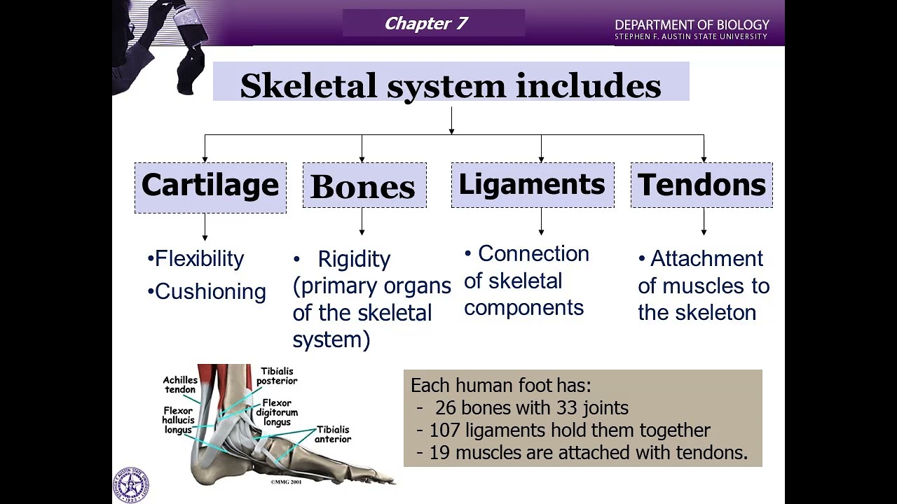
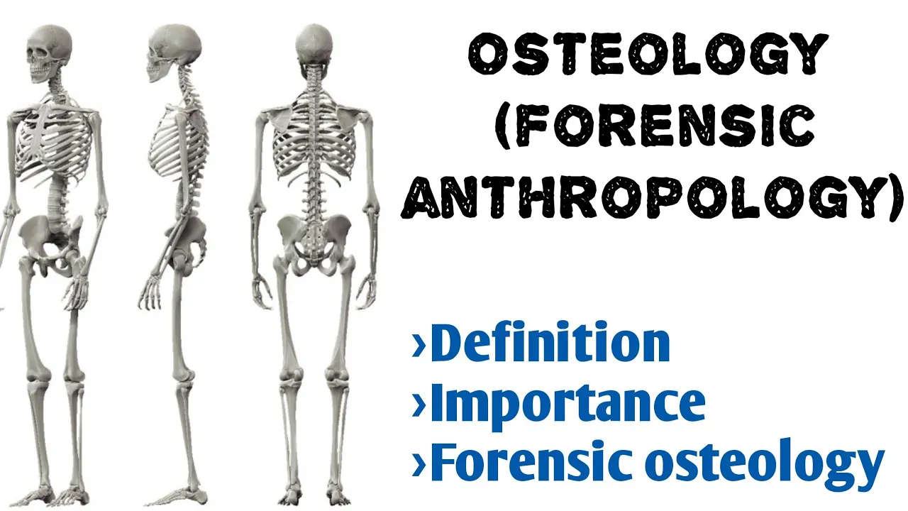
Human osteology is the study of bones essential to biological anthropology, enabling comparison of skeletal remains across species and understanding evolutionary adaptations. The human skeleton is divided into two primary parts: the axial skeleton (skull, ribcage, and vertebral column) and the appendicular skeleton (limbs and pelvis). Key anatomical terminology includes directional terms like proximal/distal (relative to trunk), medial/lateral (relative to sagittal midline), anterior/posterior (relative to coronal plane), and superior/inferior (above/below). The skull contains eight cranial bones held by fibrous sutures (coronal, sagittal, and lambdoidal) that don't fully fuse until age two, with the foramen magnum's position indicating locomotion type. The vertebral column consists of cervical (7), thoracic (12), lumbar (5), sacral (5 fused), and coccygeal (3-5 fused) vertebrae, protecting the spinal cord. The appendicular skeleton includes the pectoral girdle (clavicles and scapulae), upper limb bones (humerus, radius, ulna, carpals, metacarpals, phalanges), and lower limb bones (femur, tibia, fibula, tarsals, metatarsals, phalanges), all arranged symmetrically on both sides of the body.
Human Osteology: Axial & Appendicular Skeleton Explained
Added:With some relevant information on evolution understood, let’s now review some information regarding human anatomy, so that we can dive into the meat of this anthroplogy series. As we will be discussing a wide variety of skeletal remains, we must recall some things about the human skeleton.
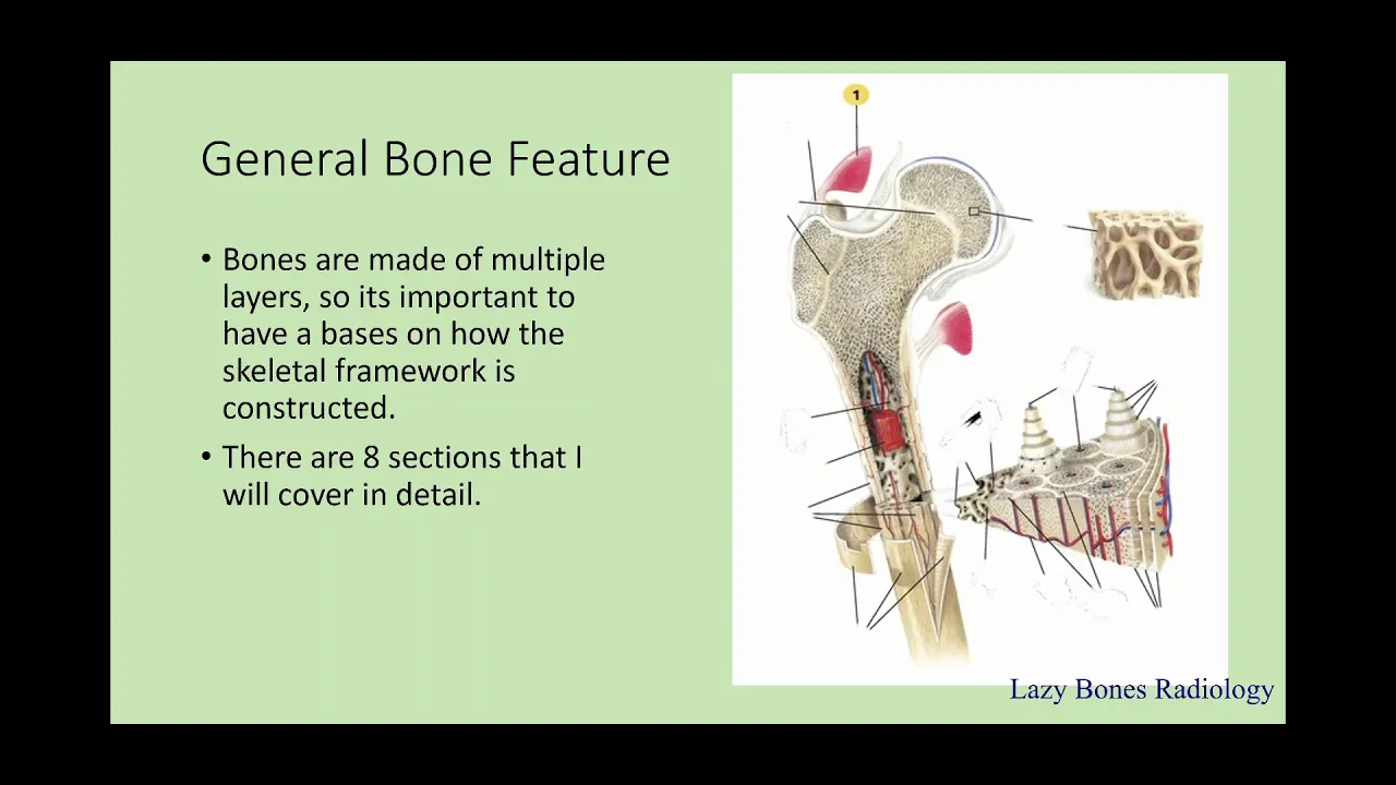
This was covered in a basic way over in the Anatomy & Physiology series, but now let’s revisit the subject with a little more context. Osteology, the study of bones, is vitally important to Biological Anthropology. Whether we are considering forensic work, paleoanthropology, or the functional morphology of other living primates, knowing the nature of the human skeleton is critical. Within the context of human evolution, we need to see how our bones have changed over time, and how this relates to things like diet and locomotion.
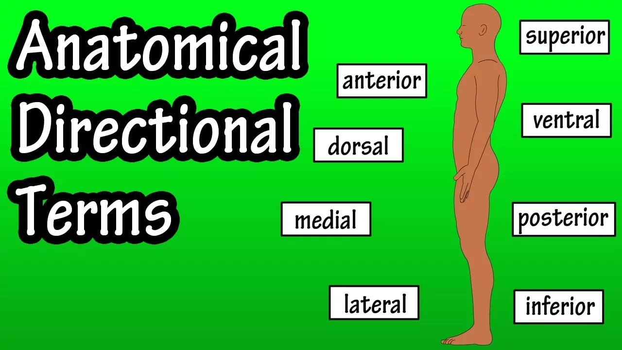
Osteology generally concerns not just the structure of the skeleton, but its function as a whole, and with consideration to each bone. The human skeleton is navigated assuming the following anatomical position: upright with the thumbs pointing outward and the palms forward. Anatomical planes and directions help orient bones in relation to one another. The transverse plane cuts the skeleton into top and bottom, the sagittal plane into left and right, and the coronal plane into front and back. Distal and proximal refer to a bone’s position or surface in the limbs with relation to the trunk: The phalanges, or fingers, are distal to the humerus, or upper arm. Likewise, the humerus is proximal to the phalanges. Medial and lateral refer to a bone’s position or surface in relation to the sagittal midline of the body: The ulna, a forearm bone, is medial to the radius.
Likewise, the radius is lateral to the ulna. Anterior and posterior refer to a bone’s position or surface in relation to the coronal plane: The sternum, or breast bone, is anterior to the vertebral column. Likewise, the vertebral column is posterior to the sternum. Lastly, inferior and superior refer to a bone’s position or surface as “above” and “below”: The skull is superior to the vertebral column. Likewise, the vertebral column is inferior to the skull.
The skeleton can also be divided into two primary parts: the axial skeleton and the appendicular skeleton. The axial skeleton contains the skull, ribcage and the vertebral column, while the appendicular skeleton contains the limbs and pelvis. The skull contains numerous bones, some of which are paired in two mirrored parts, and some of which are not. The bones of the skull are held together by fibrous joints, known as the sutures of the skull. These sutures do not fully close until age two in humans, which is why babies have a “soft spot” on their head, that is effectively exposed brain. The largest sutures of the skull include the coronal suture, which joins the frontal bone with the parietal bone, the sagittal suture, which joins the two parietal bones, and the lambdoidal suture, which joins the parietal bone with the occipital bone. The bones of the brain case thus include the non-paired frontal, occipital, sphenoid, and ethmoid bones, and the paired parietal and temporal bones, for a total of eight cranial bones. At the base of the skull, where the spinal cord exits, is a hole called the foramen magnum. This hole’s angle and position can be used to determine the locomotion of ancient hominins, whether they are bipedal or quadrupedal.
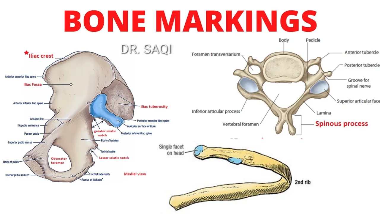
The bones of the face can also be paired or unpaired. Unpaired bones include the vomer and the mandible, or jaw, and the paired bones include the nasals, lacrimals, palatines, inferior nasal conchae, the maxilla, and the zygomatics, or cheekbones. The mandible and maxilla house the teeth, which are not considered to be bones but are important to know about. Humans, like all primates, have four types of teeth that are defined by their cusping: incisors, canines, premolars, and molars. The human dental formula is the same as all catarrhine primates: 2:1:2:3, which indicates that in each quadrant of the mouth we have two incisors, one canine, two premolars and three molars. The most posterior molars, the wisdom teeth, cause crowding in some humans and must be removed. The vertebral column protects the spinal cord, which exits the skull via the foramen magnum, and is composed of several types of vertebrae: Cervical, Thoracic, Lumbar, Sacral and Coccygeal. Within the cervical vertebrae, the Atlas (C1) is the first and allows the head to “nod” up and down, while the Axis (C2) is second and allows the head to “shake” left and right. There are seven cervical vertebrae, twelve thoracic vertebrae, five lumbar vertebrae, five fused sacral vertebrae known collectively as the sacrum, and three to five fused coccygeal vertebrae, colloquially known as the tailbone. Attaching to the thoracic vertebrae, humans have 12 paired ribs. “True” ribs connect directly to the sternum, “False” ribs via cartilage, and “Floating” ribs do not connect at all. The sternum is composed of three smaller bones: The manubrium is most superior, then the body, and the xyphoid process is most inferior.
Moving on, the appendicular skeleton includes the limbs and pelvis. The appendicular skeleton is symmetrical, so the bones on the right side of the body have a mirror counterpart on the left side.
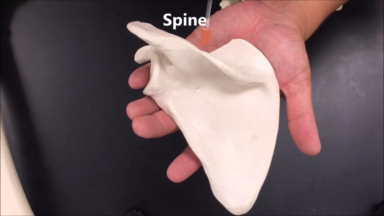
The pectoral girdle connects the upper limbs to the axial skeleton, and is composed of two scapulae, or shoulder blades, and two clavicles, or collarbones. The scapula in humans is highly mobile, and articulates with the clavicle through the acromion process. Both bones also articulate with the humerus; the bone of the upper arm. The forearm is composed of two bones: the radius and the ulna. The proximal end of the ulna creates the olecranon, or elbow, and in anatomical position is medial to the radius. The radius, naturally, is lateral to the ulna and articulates distally with the carpals, or wrist bones, specifically, the scaphoid and lunate bones. From lateral to medial, the other carpal bones include the trapezium, trapezoid, capitate, hamate, triquetrum, and pisiform bones. The carpals articulate with the metacarpals, or palmar bones, which articulate with the phalanges, or finger bones. A single finger bone is called a phalanx, and there are proximal, middle, and distal phalanxes that are separated by the finger joints.
The pelvis can be separated into two halves, which are individually known as os coxae. A single os coxa is composed of three fused bones: the ilium, or blade of the pelvis, the ischium, and the pubis. The femur, the largest bone in the body, articulates with the pelvis by the acetabulum, or socket of the hip. The lower leg is composed of the tibia and the fibula. The tibia, or shin bone, articulates proximally with the femur, while the fibula articulates with the tibia instead. The tarsals, or ankle bones, meet the tibia by way of the talus bone, which articulates with the calcaneus, or heel bone. From medial to lateral, the remaining tarsals include the cuneiforms and navicular, as well as the cuboid bone. The metatarsals make up the plantar portion of the toes and they articulate with the phalanges, or toe bones.
As we move forward in this series, we will see how these bones change over time; going from a more basal “ape-like” condition in the Miocene, to the familiar large-brained, bipedal hominins that would eventually yield humans. So let’s continue and dive back into the history of hominins.
Up Next
Bones Tell Us: Sexing & Aging Skeletons in Bioarchaeology
@RosieCrawford
1K views•2021-04-28

Bacterial Communication: Quorum Sensing and Biofilm Formation Explained
@JHUAAP
12.6K views•2011-06-28

Protein Structure Explained: Primary to Quaternary | Biochemistry
@ProfessorDaveExplains
1.4M views•2016-08-27

Bacteriophages: Earth's Deadliest Killers and Future Antibiotics
@kurzgesagt
34.6M views•2018-05-13
Related Study Plans & Knowledge Roadmaps
Structured learning paths in Biology