












![[손해사정사 2차 무료강의] 의학이론 3강 기본이론 골격계 김빛나 교수](https://i.ytimg.com/vi/zbCThhUPyJc/maxresdefault.jpg)



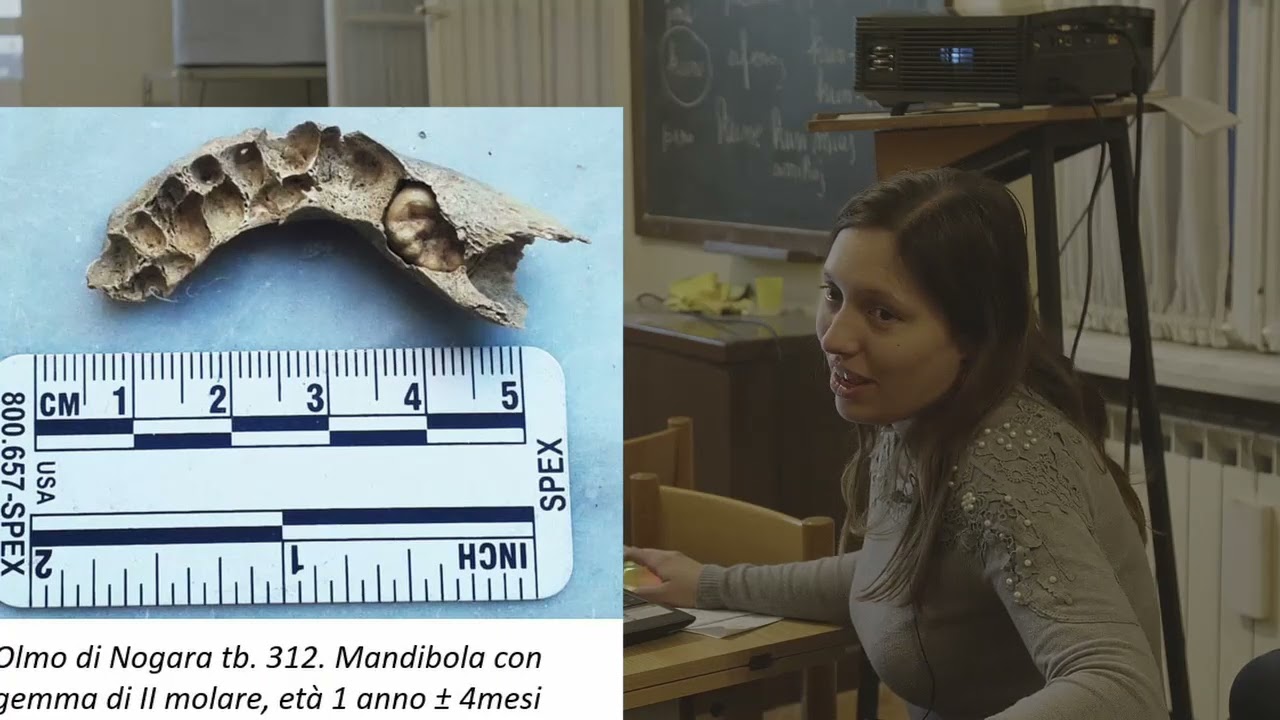


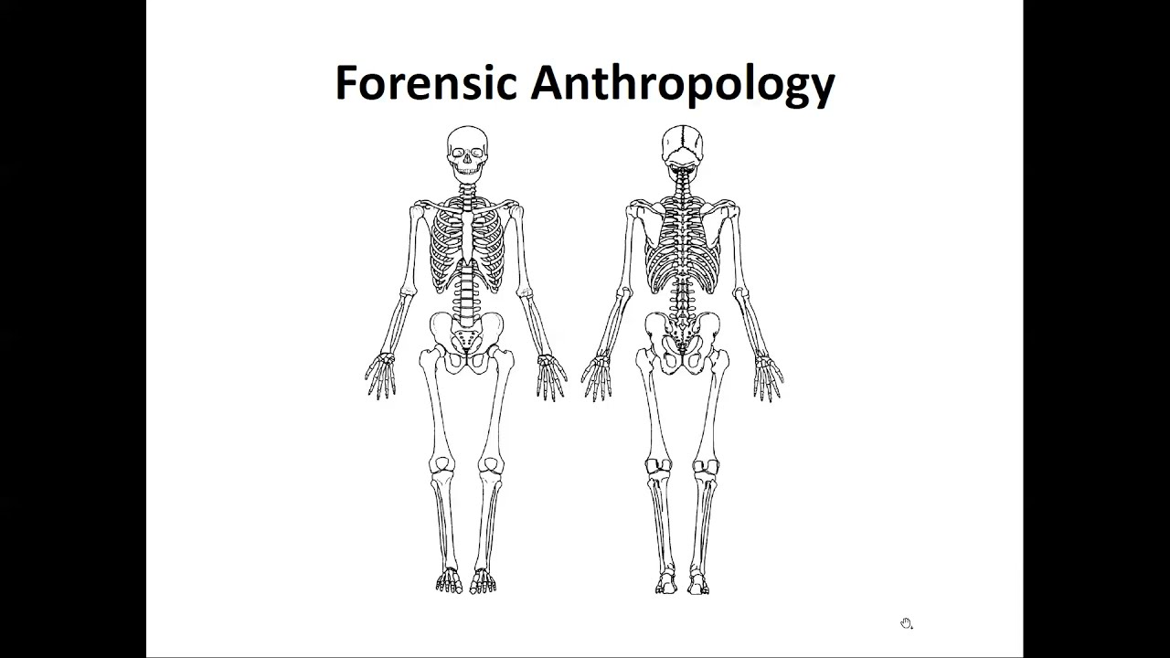

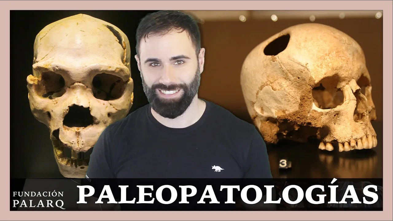















The pelvis and skull are the most informative skeletal elements for determining sex and age in bioarchaeology. For sex determination, the pelvis shows the most pronounced differences: females have a wider, shallower subpubic angle (82.7° average) and larger sciatic notch (82.7°) compared to males (65.3°), while skull features like the glabella (vertical in females, sloping in males), supraorbital ridge (more robust in males), and mastoid process (longer in males) provide additional indicators. For aging, cranial sutures fuse progressively with age, teeth show wear patterns and linear enamel hyperplasia indicating childhood stress, and pelvic methods include the auricular surface (wear and densification), pubic symphysis (fusion phases), and acetabular method (increased porosity with age). These methods have limitations, particularly for individuals over 40, where degeneration rates vary significantly based on diet and lifestyle.
Bones Tell Us: Sexing & Aging Skeletons in Bioarchaeology
Added:today we are learning all about bones now bones have been used throughout history in the archaeological record as tools as building materials and for jewelry but what can they actually tell us about the people themselves [Music] the leg bones connected to the knee bone the knee bones connected to the thigh bone the side bones connected to the hip bone now shake them skeleton bones woo so for this video i have just picked out my two favorite elements of the skeleton and that is the skull and the pelvis to talk to you about what they can teach you about that human and the general population there are a lot of things to learn from the rest of the skeleton too and a lot of that is in my dissertation so if you want to learn more about that go and watch my dissertation video i'll link that in the pinned comments but for time reasons we're just going to focus on the pelvis and the school and in particular sexing and aging skeletons so up first we have the school now i have spent eight years drawing this out for you so we have the school from the front and the school from the sides we can learn loads from the school we can learn about sex age nutrition and trauma now when we're talking about the skull we are referring to the whole head so we're looking at the entire picture if we are talking about the crania then we are simply talking about the top half and we are not including the mandible at the bottom the mandible is the lower jaw bone the one that you can move up and down like that you can't actually move the maxilla that is attached to the top now one thing archaeologists do when they find a skeleton is try and work out whether it is female or male and to do this you can look at the skull i'll talk about the other features that we need to look at as well and i will say that if you look at obviously the bigger picture that is much better because one method is not reliable on its own if you are doing it with the skull which we're going to talk about now the main things you need to look at are the mandible the nuchal crest which is actually slightly to the right the supraorbital ridge the glabella and the mastoid process which is that little sticky-outy thing that i'm circling now now this really will be a whistle-stop tour so i'm gonna link all of my references in the description so the glabella is this part of the forehead here the bit between your eyes and the top of your head in women the glabella is generally more vertical in males it kind of slopes a bit more so from my picture i would probably say this was a female bella cause can you see it's almost a right angle before it turns and obviously not all skulls follow a pattern and sometimes it's not obvious enough to define which is why you need all the other methods too the supraorbital ridge is if you feel on your face where kind of your eye sockets are and your brow bones are that sticks out slightly in men it sticks out more and it's a bit more robust tends to be anyway whereas in women again it's kind of flatter less pronounced the mastoid protest is another way of doing it it is this kind of sticky outfit here underneath your ear you can kind of feel it if you're like this but it is much less pronounced in women the male mastoid process tends to be longer than the female one passi atal 2015 looked at 70 different indian crania and on average the female mastoid process was 5 millimeters shorter than the males moving on to the mandible which is this lower jaw in males this spit here which is called the mental protuberance is more flared than in women because of the muscle attachments there's also usually a stronger jaw as you can see mine is quite sloppy in males that tends to be a bit more angular it also often has a much thicker bottom margin so you'll see that the skull will kind of flare out a bit at the bottom and the bottom will be really thick and that is because of these jaw muscles that they would have attached and generally been bigger in males however there are a lot of limitations to using the school for sexing including that with age female skeletons get much more robust and much more similar to male skeletons because of the reduction in estrogen in say the menopause it also is quite rare to have a full skull come out or a full mandible there is a lot of fragmentation in the archaeological record which makes it difficult to find all of the features and you maybe can only find one as well as the women starting to have more kind of masculine bone features when they get older men under the age of 30 quite often have more feminine facial features and especially in children the sex hormones don't happen until puberty so you can't sex children from the school it just you just can't unless you use the new dna techniques but we're not going to go over that in this video now it is more difficult and less reliable to use the skull for aging but we are going to go through it anyway quickly so the first one is to look at the closures of the cranial sutures which are these bits of the wiggly lines we have quite a few of those on our head on our crania and with age they fuse up so in children they're really really pronounced their bones might not even quite stay they might not stay together essentially and that's because they're not fused whereas with age our bones fuse i'll put one of the methods of doing this on the screen now so that's what that is and basically the stages go from completely unfused to complete obliteration and obliteration means when it's completely locked together that's kind of say that's childhood that's complete obliteration now complete obliteration does tend to happen between 36 and 75 years which is a big big age gap obviously so once you get kind of over 30 over 35 with this method it's not particularly accurate and you really need to look at the other elements of the skeleton which to be honest they're probably better to look at first anyway now the teeth are another super super interesting element of the school that you can use for aging in children the teeth are the most accurate method of aging a skeleton because that is when they grow and we know roughly the the growth patterns of teeth and which ones come first when they should be partially erupted etc in adults we can look at the eruption of the third molar if it's a particularly young adult if the third molar or wisdom teeth has erupted then they are probably over the age of 21 22 and then after that we're looking at degeneration of the teeth so the more worn down they are perhaps the older but if you're gonna use that method for aging older people then you really do need to know all about the lifestyle all about the diet because they could have been eating lots of food you know that would wore down their teeth so it's difficult unless you know everything else about the society which usually in archaeology we don't so here we have some teeth these are terrible drawings i'm so sorry we have a molar and we have our front teeth which is our canine or our incisors now the condition we're going to talk about is linear enamel hyperplasia and this one is quite a solid indicator of childhood stress which is probably down to poor nutrition linear enamel hyperplasia shows up as a sort of ring on the tooth so where the enamel is thinner than the rest of it so on a molar it could be here and there will be lots of rings on teeth depending on you know how many periods it is if it's thicker than it is a longer period of stress what it is essentially showing is a period of weaker or slower enamel growth now what is really interesting is that because we are pretty aware of the sort of order and speed that teeth grow and start to erupt we can quite easily estimate what age this stress happened in childhood now it has to be in childhood because it happens when the enamel is forming and if if an adult skeleton has these rings on the teeth then it happened in childhood it didn't happen in adulthood saying that if it is in the third molar then we can assume that this period of stress occurred kind of late teens if we have a child skeleton that still has its deciduous teeth which is milk teeth then we can probably assume that this stress happened pre-birth so when it was still inside the mother which then in turn leads it to suggest that the woman that was carrying the child was also struggling with things like nutrition or maybe a bout of illness which was some reason affected the growth of the child's teeth now what archaeologists can do is look at the patterns of leh across a population so if one population has a really high incidence of enamel hyperplasia at three four years old then it can possibly be put down to the introduction of a more adult diet so post breastfeeding when a child is waning in some societies in the hunter-gatherer period that was at three or four years old and it's a lot younger now but we're talking way back and because breast milk is good for passing on immunity and all of the nutrients needed for a young child's growth once they are weaned off that then there can be a period of stress where the body kind of adapts to this new diet evidence for this is also seen in long bone stunting if you want a really in-depth discussion of stunting long bone growth and um the teeth then you can have a look at my dissertation video because that's what i did my dissertation on i'll link that also in the pinned comment another indicator of health in the school this time in the cranial is criminal or vitalia or also known as hyper that you um you had you try again porotic hyperostosis now parotic hyperostosis is a condition where red blood cell production is larger which causes the marrow expansion responsible for lesions which essentially in skull you have what looks a bit like dots it kind of looks spongy and porous and i'll put a proper picture of it on the screen now parotic hyperostosis is thought to be caused by iron deficiency and anemia it's possible this isn't the case basically people aren't completely sure there are a lot of convicting conflicting theories at the moment i think the most widely spread reason is iodine to anemia if it is found in the eye sockets then it is called cribra or battalia but it is a form of parotic hyperostosis it looks the same it like a sponge but in the back of the eye sockets and that is more common in children than just on the head we are now going to move on to the pelvis and i'm going to do it in the same structure as before so we're going to do sexing and then aging the pelvis is i think by far the most useful bone in the body for building a biological profile of the person who it belonged to and that is because of some very pronounced differences in sex and also pronounce differences that happen to the bone as you get older so here we have uh oh now here we have our pelvises i didn't draw these i printed them out so the pelvis on the top left of the screen is male and the pelvis on the bottom right is female and the main features of the pelvis that we are looking at are the subpubic angle or the pubic arch which is this area here so according to angel 1969 the male subpubic angle is deeper and more narrow whereas the female is wide and shallow and that is all to do with childbirth and how the pelvis opens up to let the baby pass through another thing to look out for is the angle of sciatic notch the sciatic notch is this bit of the pelvis which is on either side in men it is much smaller more of an acute angle in a women it is more of an obtuse angle it is more of a v-shape in men and a u-shape in women now a study done by cinc and pateri in 1978 found that out of 200 random pelvises of known sex the average angle for female pelvises was 82.7 degrees whereas men was 65.3 degrees there was however a 10 degree overlap in the range so it does need a scale and you do need to look at the other features in order to do it so you could combine it with features from school or just the other aspects of the pelvis like with the skull you can also age a skeleton from the pelvis and there are three ways of doing this the first one is with the auriculus surface the second one is with the pubic symphysis and the third one is the acetabula method so for method number one we are looking at the auricular surface which is where the pelvis joins the sacrum here i'll put a picture which is kind of highlighting it better on the screen now so in older individuals the auricular surface will show more signs of wear degradation and densification there will also be a breakdown of the marginal lipping which is this here which joins the two together now the pubic symphysis is here and here there are two so can you see where they join together they actually look kind of like that so it's the end of the bone and they join together i'll try and find a better picture of that and put it on the screen the most accurate method of this is tsuji brooks and they have phases so as you get older your pubic symphysis goes from looking a bit like this to more like this so essentially in young adulthood the pubic symphysis is more billowed which is this sort of texture that you can see this is because it is unfused and with age as it fuses the rim becomes complete and defined around the outside and the surface is no longer billowed i'll put all of the phases on the screen so you can see them the third method which is the acetabula method is looking at this whole kind of area which is the iliac fossa and in particular this kind of surface so i've written the number three on here but essentially it's like all of the surface and with age it seems it's a very new method but it does seem that iliac fossa gets kind of more porous with age there also becomes more of a sharpness of the rim between the iliac fossa and the iliac crest now the iliac crest is this kind of area on the outside which i've highlighted there this method is sometimes said to be more accurate however it's just not used widely enough and often this surface is broken that being said is much less susceptible to damage than the pubic symphysis and quite often well more often than not the pubic symphysis doesn't survive in the archaeological record so you have to turn to the other two methods the main problem with the pubic symphysis method is that it underestimates the age of over 40s and therefore creates a huge underrepresentation of over 40s and in particular over 50s essentially aging anyone over the age of about 40 becomes really difficult because growth has completely stopped everything's completely stopped fusing and you just have to start looking at degeneration which happens at completely different rates to lots and lots of different people and all depends on diet and nutrition and activities during life so i hope you enjoyed my video i hope you learnt a lot and if i have got anything wrong or if you've found some new papers please do let me know in the comments i love to learn as much as you do i'm obviously not an expert i just did my degree in it give me a massive thumbs up if you have a skull or a pelvis don't forget to subscribe bye
Up Next

Immune System Made Easy- Innate & Adaptive Immunity Explained
@osmosis
460.6K views•2024-03-10

Bacterial Communication: Quorum Sensing and Biofilm Formation Explained
@JHUAAP
12.6K views•2011-06-28

Enteric Nervous System Explained: The Gut's Brain | Neurobiology Lecture
@alumniu6029
438 views•2018-09-12

Bacteriophages: Earth's Deadliest Killers and Future Antibiotics
@kurzgesagt
34.6M views•2018-05-13
Related Study Plans & Knowledge Roadmaps
Structured learning paths in Biology