The p53 protein acts as a critical tumor suppressor that determines whether a cell with damaged DNA dies, undergoes growth arrest, or repairs itself; it works by binding to specific DNA sequences called p53 response elements in gene promoters, recruiting RNA polymerase to activate genes involved in DNA repair, cell cycle arrest, or apoptosis, thereby preventing the development of cancer when approximately half of all human cancers involve p53 dysfunction.
p53 Tumor Suppressor Protein: DNA Damage Response Explained
Added:Deep Dive
Basic eukaryotic cell cycle phases (G1, S, G2, M) and the general concept of restriction checkpoints.

CELL CYCLE & CHECK POINTS II APOPTOSIS II RPSC 1st GRADE BIOLOGY II UG LEVEL I NEW TOPIC UG LEVEL
1.4K
Eukaryotic cells undergo cell division through Interphase (G1, S, G2) and M Phase. G1 (14 hours) synthesizes enzymes for DNA replication. S phase (6 hours) replicates DNA with histone synthesis. G2 (3 hours) prepares for mitosis. M Phase (1 hour) includes prophase, metaphase, anaphase, and telophase. Checkpoints ensure proper progression: G1, G1-S, S-G2, and G2-M transitions. The G0 phase is a resting state where cells exit the cycle but can return to G1 if DNA repair is successful.
Click to watch →

Cell cycle checkpoints | DNA damage checkpoint | spindle assembly checkpoint | Cell biology
90.4K
Cell cycle checkpoints are regulatory mechanisms that ensure proper cell division by monitoring cellular conditions at critical transition points; the G1/S checkpoint (restriction point) assesses environmental conditions and DNA damage before S phase entry, using ATM/ATR sensors to activate p53 which then inhibits cyclin-CDK complexes via p21; the G2/M checkpoint monitors for DNA damage and unreplicated DNA before mitosis, also using ATM/ATR-Chk1 signaling to block cyclin B-CDK1 activation; and the spindle assembly checkpoint during metaphase ensures proper chromosome alignment by using MAD proteins to inhibit the anaphase-promoting complex until equal tension is achieved, preventing faulty chromosome segregation.
Click to watch →
AP Biology Review: CRAM Session
109.3K
Cell cycle phases: G1 (growth, preparation), S (DNA synthesis), G2 (preparation for division). Checkpoints: G1 checkpoint (decides whether to divide), M checkpoint (metaphase ensures proper chromosome alignment for equal segregation).
Click to watch →
Stroll Through the Playlist (a Biology Review)
2.6M
The cell cycle includes G1 (growth), S phase (DNA synthesis/replication), G2 (preparation for division), and M phase (mitosis and cytokinesis). G1, S, and G2 together form interphase when the cell is not dividing. Checkpoints control progression through the cycle. If requirements aren't met, cells either repair damage or undergo apoptosis (programmed cell death). Key regulatory proteins include Cdk, cyclin, and p53. Cancer cells fail to respond correctly to checkpoints, dividing uncontrollably.
Click to watch →

Cell Cycle basic concepts explained in Amharic : Mitosis Vs Meiosis
5.7K
The cell cycle is a series of phases that cells go through before dividing. G1 (Gap 1) is the first phase where cells grow and prepare for division. S (Synthesis) is the second phase where DNA replication occurs. G2 (Gap 2) is the third phase where cells continue to grow and prepare for division. M (Mitosis) is the final phase where the cell actually divides into two daughter cells. Each phase has specific checkpoints to ensure the cell is ready to proceed.
Click to watch →
Deep Dive
The fundamental structure of DNA and common sources/types of DNA damage, such as UV radiation and double-strand breaks.
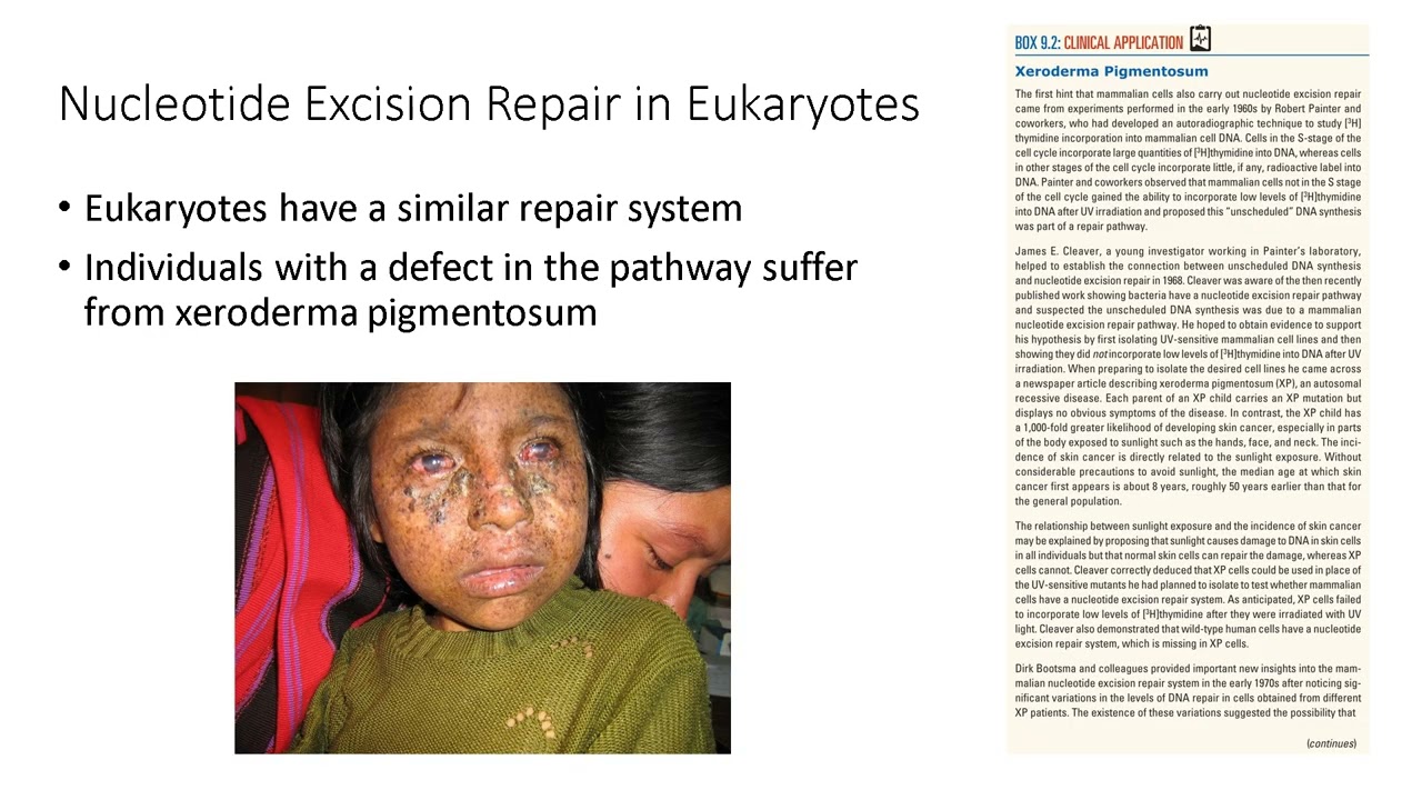
Lecture 9 DNA Damage and Repair Mechanisms Explained
317
This comprehensive section covers the fundamental principles of DNA damage and repair. It begins with defining mutations as any DNA sequence change, ranging from point mutations to frameshift mutations. DNA damage originates from endogenous sources (reactive oxygen species from metabolism) and exogenous sources (UV light, ionizing radiation, chemical agents). UV light causes cyclobutane pyrimidine dimers and 6-4 photoproducts that distort DNA structure. Ionizing radiation generates reactive oxygen species causing single-strand breaks, base modifications, and double-strand breaks. Chemical agents include alkylating agents (forming DNA adducts and cross-links) and aflatoxin B1 (causing liver cancer). The Ames test detects mutagens using histidine-deficient Salmonella. Direct reversal mechanisms include photoreactivation (using photolyase and blue light) and O6-methylguanine DNA methyltransferase (a suicide enzyme). Base excision repair addresses small lesions through glycosylase-mediated base removal, AP site processing, and gap filling. Nucleotide excision repair handles bulky distortions by removing 24-32 nucleotide segments. Defects in nucleotide excision repair cause xeroderma pigmentosum, demonstrating the clinical importance of these pathways.
Click to watch →
Pokroky v biologii 2024 (3.1) Martin Kuthan: Dolování genomů (PřF UK 10.2.2024)
1.4K
UV radiation causes characteristic DNA damage including cyclobutane pyrimidine dimers, which form covalent bonds between adjacent pyrimidines. Ionizing radiation (X-rays, gamma rays) causes more severe damage including single and double-strand breaks, as well as generating secondary radicals that chemically damage DNA. These types of damage are more difficult to repair than spontaneous damage and can lead to cell death or cancer if not properly repaired.
Click to watch →

NEET 2022 | Molecular Basis of Inheritance | DNA की संरचना | DNA की पैकेजिंग | Biology NEET 2022
105.2K
This section covers the fundamental structure of DNA discovered by Watson and Crick in 1953. The DNA double helix consists of two anti-parallel strands running in opposite directions (5' to 3' and 3' to 5'). Each strand has a backbone made of alternating phosphate groups and deoxyribose sugars, with nitrogenous bases projecting inward. The most important feature is the specific base pairing through hydrogen bonds: adenine (A) always pairs with thymine (T) through two hydrogen bonds, while guanine (G) always pairs with cytosine (C) through three hydrogen bonds. The instructor emphasizes this complementary base pairing as the fundamental principle of DNA structure and replication. The physical dimensions are explained: one complete turn of the helix is 3.4 nanometers long and contains exactly 10 base pairs, with the distance between adjacent base pairs being 0.34 nanometers.
Click to watch →

DNA damages : an overview
719
Environmental factors cause distinct patterns of DNA damage with specific biological consequences. UV light at 260 nm wavelength is absorbed by aromatic DNA bases, causing adjacent pyrimidines (especially thymines) to form cyclobutane pyrimidine dimers. This structural distortion prevents DNA polymerase from reading the template properly, causing replication fork arrest or random nucleotide incorporation. Double strand breaks represent catastrophic damage where both DNA strands are severed, typically by ionizing radiation or nucleases, preventing replication and inevitably causing cell death. Base analogs like 5-bromo-2'-deoxyuridine resemble natural bases but pair incorrectly, inducing mutations through incorrect base pairing. These environmental and chemical sources demonstrate how external factors systematically challenge genetic stability.
Click to watch →

【講義サンプル動画】実戦シリーズ 生命科学 第1講 前半 | 医学部学士編入対策講座 河合塾KALS
868
DNA damage occurs through various mechanisms with different repair requirements. Radiation is classified into particle radiation (alpha particles as helium nuclei, beta particles as high-speed electrons) and electromagnetic radiation (X-rays from electron transitions, gamma rays from nuclear transitions). Double-strand breaks are the most severe DNA damage because both strands are severed, making repair difficult. Single-strand breaks are easier to repair using the intact complementary strand as a template. UV radiation causes thymine dimers, where adjacent thymine bases become covalently bonded, creating structural distortions. Homologous recombination evolved as a repair mechanism for double-strand breaks, converting them into two single-strand breaks that are easier to repair. This mechanism is evolutionarily conserved across all domains of life.
Click to watch →
Deep Dive
The functional distinction between proto-oncogenes and tumor suppressor genes in cancer biology.
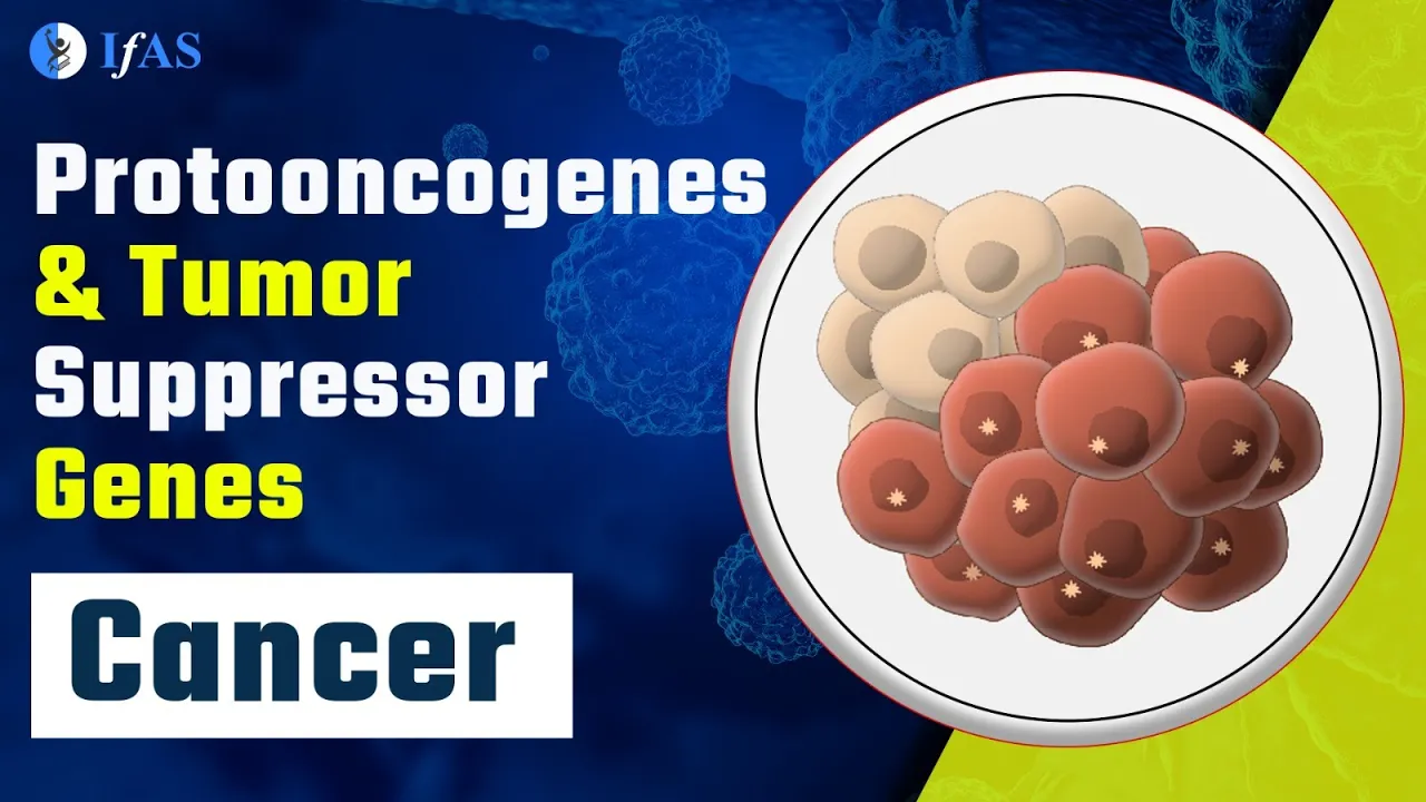
PROTOONCOGENES & TUMOR SUPPRESSOR GENES I CANCER I FAST REVISION SERIES I CSIR2023 I CSIR
13.1K
Proto-oncogenes are normal genes that promote cell proliferation and require gain-of-function mutations in at least one allele to become oncogenes, while tumor suppressor genes inhibit cell proliferation and require loss-of-function mutations in both alleles to contribute to cancer development; this distinction is fundamental to understanding cancer biology, with examples including p53 (tumor suppressor) and RAS (proto-oncogene), and the two-hit hypothesis explaining that sporadic cancers require two mutations in tumor suppressor genes while inherited forms require only one.
Click to watch →

Proto oncogens | How proto oncogenes lead to cancer? | The biology of protooncogenes | USMLE
11.6K
Proto-oncogenes are normal genes that regulate cell growth, proliferation, and survival by producing proteins that promote cell division. Tumor suppressor genes counteract this by suppressing growth and division. The balance between these two gene classes determines cancer risk. Gain-of-function mutations in proto-oncogenes or loss-of-function mutations in tumor suppressors lead to uncontrolled cell growth and tumor formation. Proto-oncogenes code for diverse signaling pathway components including cell surface receptors (EGFR, ERBB2), intracellular kinases (BCR-Abl, B-Raf), and transcription factors (MYC, N-MYC). Understanding how these genes normally regulate the cell cycle provides the foundation for comprehending cancer biology.
Click to watch →
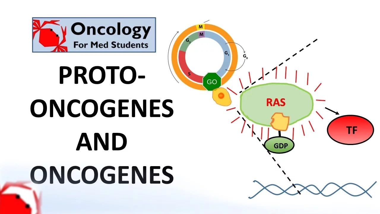
7. Proto-oncogenes and Oncogenes
205.8K
Tumor suppressor genes normally prevent cell growth and division; their loss-of-function leads to cancer. Proto-oncogenes normally promote cell growth; their gain-of-function as oncogenes promotes cancer. Using a car analogy, tumor suppressors are like brakes on the cell cycle, while oncogenes function like an accelerator pedal. Tumor suppressors require two mutations (two hits) to cause cancer, whereas oncogenes need only one mutation.
Click to watch →
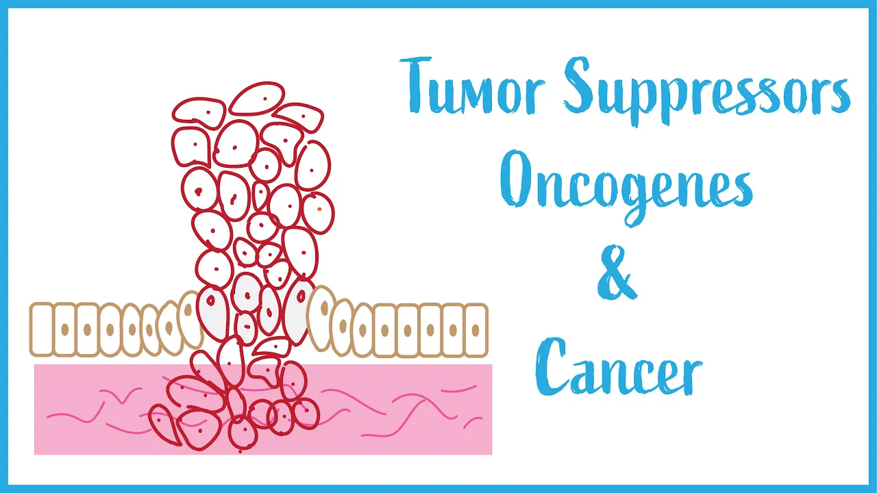
Tumor suppressors, Oncogenes and Cancer Biology (USMLE Essentials)
20K
Tumor suppressor genes encode proteins that control cell division by acting as gatekeepers (preventing cell cycle progression) and caretakers (repairing DNA). Loss-of-function mutations increase cancer risk by allowing uncontrolled cell division. The prototypical example is p53, the guardian of the genome. In contrast, oncogenes function as powerful inducers of cell division and survival, coding for receptors and signal transduction proteins. Unlike tumor suppressors, oncogenes require gain-of-function mutations to promote carcinogenesis. When proto-oncogenes become mutated, they become permanently activated oncogenes that drive cancer development.
Click to watch →
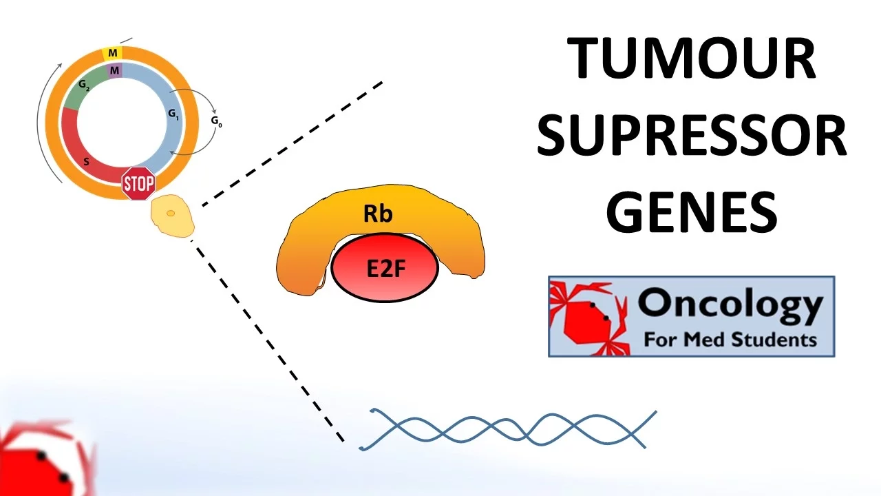
6. Tumour Suppressor Genes (Retinoblastoma and the two hit hypothesis, p53)
197.8K
Genes involved in cancer development are divided into two categories: tumor suppressor genes and proto-oncogenes. Tumor suppressor genes inhibit cell growth and division, while proto-oncogenes promote cell growth and division. When proto-oncogenes mutate, they become oncogenes that can drive cancer development.
Click to watch →
Deep Dive
The role of transcription factors in regulating gene expression and cellular responses.

Week 12- lecture 4- transcription factors
137
Transcription factors are DNA-binding proteins that regulate gene expression at the transcriptional level by binding to specific DNA sequences called cis-acting elements (promoters, enhancers, silencers) located near or within genes; these factors can be activated by cellular signals (like hormone binding) and then bind to DNA to either activate or repress transcription, allowing cells to respond to different conditions by turning genes on or off.
Click to watch →

RÉGULATION DES EXPRESSION DES GENS
6.3K
Transcription factors are proteins that bind regulatory sequences to control gene expression. Activators bind enhancers and recruit RNA polymerase to initiate transcription. Repressors bind silencers and block transcription. Some transcription factors are ligand-activated receptors that enter the nucleus upon binding hormones or other signaling molecules. This allows extracellular signals to regulate gene expression, enabling cells to respond to environmental changes.
Click to watch →

Chp1 (part4) Robbins Basic Patho | Cellular Activation | Signal Transduction Pathways
5K
Transcription factors control DNA to RNA conversion. Conformational changes allow phosphorylation and nuclear translocation, exposing specific DNA sequences for protein expression. DNA binding domains permit specific binding to short sequences in promoter regions. Growth-promoting transcription factors include Myc and Jun, while p-53 triggers growth arrest. The signal transduction pathway converts extracellular signals (input) into cellular responses (output) through a complex network of biochemical events. This represents the fundamental mechanism by which cells translate environmental signals into functional changes.
Click to watch →

GPCR-cAMP-PKA pathway|G protein coupled receptor|Protein kinase A- Cyclic AMP|CSIRNET|gate|IITJAM|
69.6K
Cellular responses to signals are ultimately controlled at the level of gene transcription. Regulatory proteins called transcription factors must be activated before they can bind to DNA and initiate RNA synthesis. Inactive transcription factors are often held in check by inhibitory proteins or sequestered in inactive complexes. Upon receiving appropriate signals, these factors become activated and translocate to the nucleus, where they recruit RNA polymerase to specific gene promoters. This transcriptional control mechanism allows cells to coordinate complex responses involving multiple genes and proteins, enabling sophisticated physiological adaptations.
Click to watch →

Regulation Of Gene Expression In Eukaryotes | B.Sc. Zoology 3rd Semester | Avantika Ma'am |
47.5K
Transcription factors are proteins that bind to DNA and regulate gene expression by activating or repressing transcription. Different cell types express different genes despite having identical genomes, achieved through differential gene expression controlled by transcription factors. Growth factors activate signaling cascades that lead to transcription factor activation, enabling cells to respond to environmental signals. This mechanism allows cells to develop specialized functions by expressing specific gene subsets.
Click to watch →
Prerequisite Knowledge
- Concept 01Basic eukaryotic cell cycle phases (G1, S, G2, M) and the general concept of restriction checkpoints.
- Concept 02The fundamental structure of DNA and common sources/types of DNA damage, such as UV radiation and double-strand breaks.
- Concept 03The functional distinction between proto-oncogenes and tumor suppressor genes in cancer biology.
- Concept 04The role of transcription factors in regulating gene expression and cellular responses.
Subsequent Learning
- Step 01The biochemical regulation of p53, focusing on the MDM2-p53 negative feedback loop and ubiquitin-proteasome degradation.
- Step 02The detailed molecular pathways of apoptosis, specifically the intrinsic pathway involving Bcl-2 family proteins, cytochrome c release, and caspases.
- Step 03Clinical genetics of TP53 mutations, including Li-Fraumeni Syndrome and its hereditary cancer risks.
- Step 04Modern therapeutic strategies targeting the p53 pathway, such as MDM2 inhibitors (Nutlins) and p53 reactivation therapies.
Stress & p53
Gene Targeting
Gene Activation
Stress & p53
0:01Playing Section
- 1
Environmental stress like UV light damages DNA.
- 2
p53 protein decides cell death, arrest, or repair.
- 3
Half of all cancers have p53 switched off.
Non-Canonical Mechanisms of p53 Tumor Suppression
While the classical paradigm dictates that p53 prevents cancer primarily by orchestrating cell cycle arrest, DNA repair, and apoptosis in response to acute DNA damage, modern research challenges this dogma. Studies using mutant mouse models have demonstrated that the capacity of p53 to trigger these traditional responses is actually dispensable for its tumor-suppressive capabilities in many contexts. Instead, an emerging counter-perspective suggests that p53's critical cancer-preventative roles lie in 'non-canonical' functions. These include the regulation of cellular metabolism, suppression of reactive oxygen species (ROS), restriction of stem-cell-like self-renewal, and the induction of ferroptosis (an iron-dependent form of cell death). Consequently, this perspective argues that p53 acts less as an acute responder to catastrophic DNA damage and more as a continuous coordinator of metabolic homeostasis and cellular fitness.
Deep Dive
The biochemical regulation of p53, focusing on the MDM2-p53 negative feedback loop and ubiquitin-proteasome degradation.

ACB intrinsic apoptosis and p53
468
p53 regulates its own levels through a negative feedback loop with Mdm2. Under normal conditions, p53 induces mdm2 expression; Mdm2 then binds p53 and targets it for degradation via ubiquitination, keeping p53 levels low. In response to DNA damage, ATM/ATR phosphorylate both p53 and Mdm2 at specific sites, disrupting their interaction. This breaks the negative feedback loop, allowing p53 to accumulate and activate its target genes. Once damage is repaired, p53 returns to baseline levels through normal degradation pathways.
Click to watch →
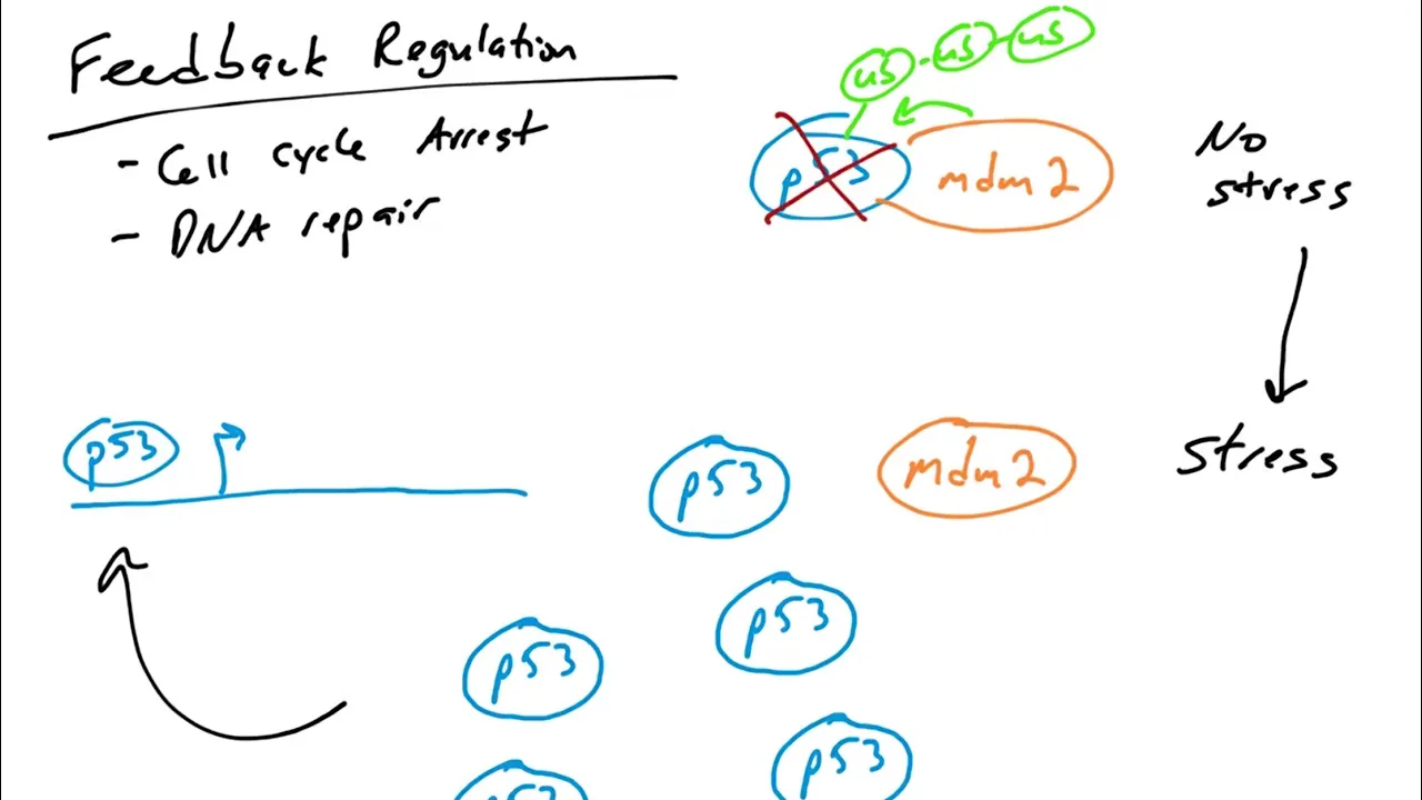
p53 Target Genes: feedback loop regulation of p53 by mdm2
3.7K
p53, a tumor suppressor protein, is regulated by MDM2 through an auto-regulatory feedback loop: under normal conditions, MDM2 (an E3 ubiquitin ligase) binds to p53 and ubiquitinates it, targeting p53 for degradation; however, when cells experience stress such as DNA damage, p53 escapes MDM2-mediated destruction, accumulates in the nucleus, and activates genes involved in cell cycle arrest and DNA repair; once repairs are complete, p53 itself induces MDM2 expression, which then re-establishes p53 degradation, returning the cell to normal conditions.
Click to watch →
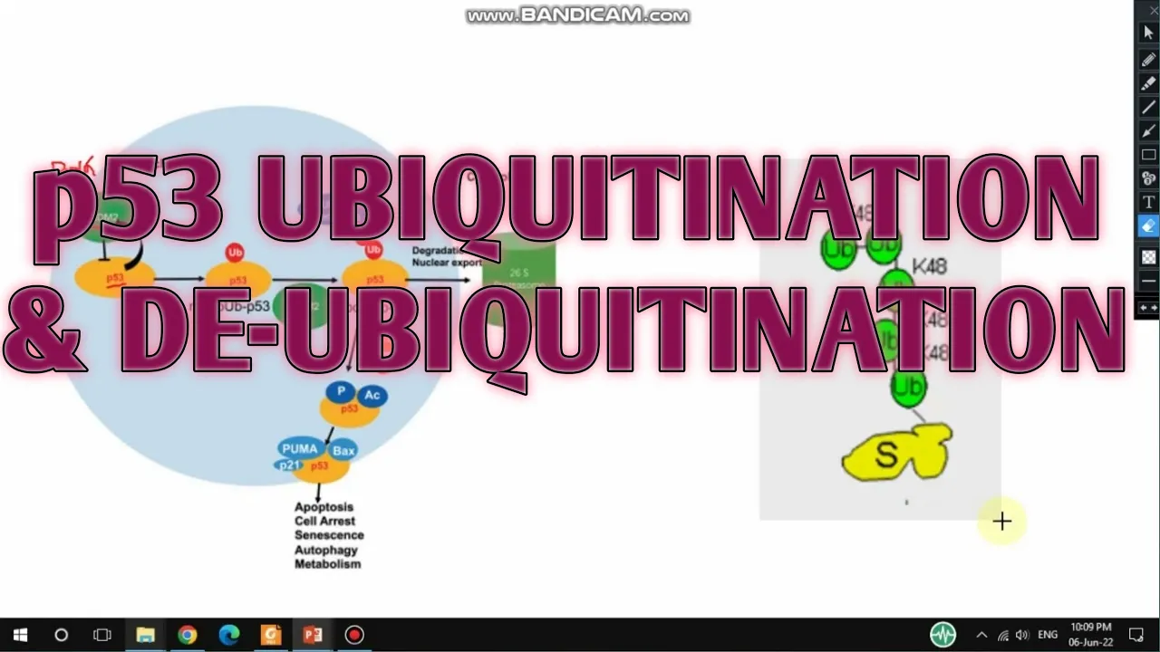
p53 ubiquitination and deubiquitination II MDM2-mediated p53 ubiquitination II xgene and proteinx
705
p53, a tumor suppressor protein, is normally kept at low levels by MDM2-mediated ubiquitination and proteasomal degradation; however, upon DNA damage, deubiquitinating enzymes reverse this process, stabilizing p53 and enabling its activation to induce cell cycle arrest via p21 and apoptosis through proteins like Bax and PUMA, thereby creating a negative feedback regulatory loop where p53 activation initially increases MDM2 expression, which subsequently degrades p53 to maintain cellular homeostasis.
Click to watch →
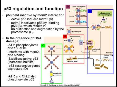
p53 basics
1K
p53 is a tetrameric transcription factor that regulates cell cycle arrest, apoptosis, and DNA repair through target genes. Under normal conditions, p53 exists in an unphosphorylated form that is rapidly degraded by the ubiquitin-proteasome system via MDM2-mediated ubiquitination. This creates a negative feedback loop where p53 activates its own degradation pathway. The MDM2 protein acts as a ubiquitin ligase that binds to p53 and marks it for proteasomal degradation. This self-regulation maintains low baseline p53 levels in healthy cells, keeping the protein in a surveillance mode waiting for DNA damage signals.
Click to watch →
Cell Biology | Cell Cycle Regulation
731.8K
MDM2 is an E3 ubiquitin ligase that negatively regulates p53 activity. Under normal conditions, MDM2 binds to p53 and promotes its degradation, keeping p53 levels low. When DNA damage occurs, ATM phosphorylates MDM2, causing it to dissociate from p53. This releases p53 from inhibition, allowing it to activate its downstream targets. The MDM2-p53 feedback loop provides tight control over p53 activity, ensuring that p53 is only activated when necessary for cellular responses to stress.
Click to watch →
Deep Dive
The detailed molecular pathways of apoptosis, specifically the intrinsic pathway involving Bcl-2 family proteins, cytochrome c release, and caspases.
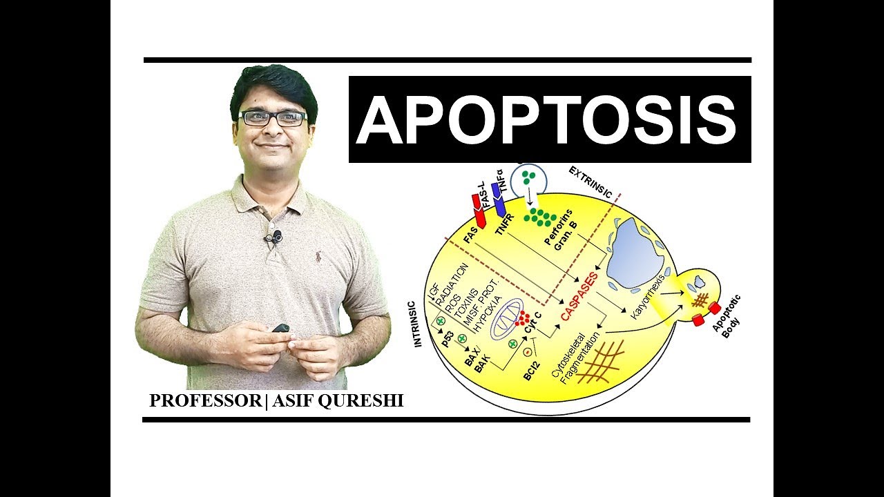
Apoptosis | Pathology | CellDeath | MBBS | PharmD | NLE | NEET | FCPS | USMLE | MRCP| DrAsifLectures
5.2K
The intrinsic pathway (also called mitochondrial pathway) involves cytochrome c release from mitochondria. Under normal conditions, BCL-2 family proteins inhibit cytochrome c release, preventing apoptosis. When apoptosis is triggered by decreased growth factors, radiation, reactive oxygen species, toxins, misfolded proteins, or hypoxia, p53 activates pro-apoptotic BAX/BAK proteins. These proteins make the mitochondrial membrane permeable, releasing cytochrome c which then activates caspases, leading to cell death.
Click to watch →
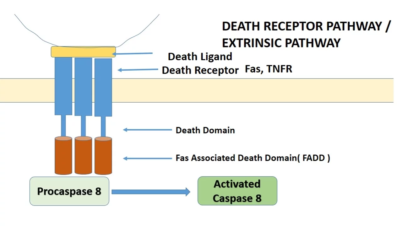
APOPTOSIS PART 1: Definition, Causes & Mechanism/Pathways
189.3K
The intrinsic pathway involves mitochondrial outer membrane permeabilization. Cytochrome c release from mitochondria combines with Apaf-1 to form apoptosomes that activate caspase 9, which then activates executional caspases. SMAC/DIABLO proteins neutralize IAP inhibitors, allowing caspase activation. The Bcl-2 family regulates this balance: anti-apoptotic members (Bcl-2, Bcl-XL, Mcl-1) prevent cytochrome c release, while pro-apoptotic members (Bax, Bak, Bcl-XS) promote it. Sensor proteins (Bad, Bim, Puma) act as arbiters, responding to cellular stress by modulating the balance between survival and death signals.
Click to watch →

Pathology & Immunology Webinar (USMLE Step 1) | MedSchoolBro
23.2K
Apoptosis is an energy-dependent, programmed cell death that differs from necrosis (always pathological). Key morphological features include cell shrinkage, chromatin condensation, membrane blebbing, and formation of apoptotic bodies with intact cell membranes. DNA laddering at 180 bp fragments indicates apoptosis. The intrinsic mitochondrial pathway is triggered by internal stress (cell injury, DNA damage, decreased hormone stimulation), leading to Bcl-2 inactivation and release of cytochrome C from mitochondria. Pro-apoptotic proteins Bax and Bak promote cytochrome C release, while anti-apoptotic Bcl-2 and Bcl-xL inhibit it. Bcl-2 overexpression causes follicular lymphoma via translocation 14;18.
Click to watch →
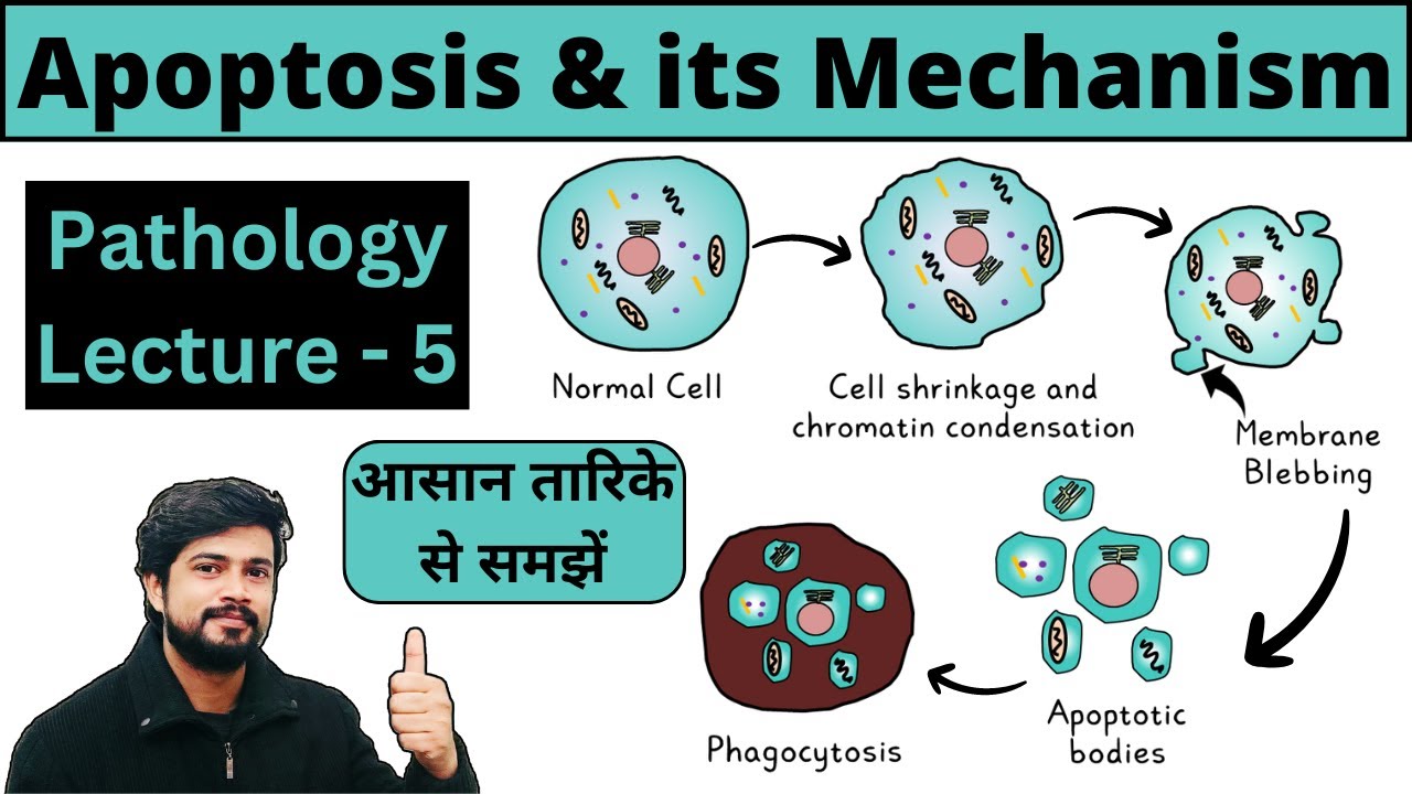
Apoptosis - Programmed Cell Death | Mechanism - Intrinsic & Extrinsic Pathway | Pathology Lecture
79.5K
In the intrinsic pathway, cellular stress causes pro-apoptotic BCL-2 family proteins to overcome anti-apoptotic proteins, leading to mitochondrial outer membrane permeabilization. Cytochrome c is released from mitochondria into the cytoplasm, where it binds to APAF-1 and caspase-9 to form the apoptosome complex. Caspase-9 activation initiates the execution phase of apoptosis, triggering the cascade that destroys the cell.
Click to watch →

Apoptosis, pathology, morphology, pathway, initiation, intrinsic, extrinsic, execution, degradation
12.1K
Apoptosis is a regulated form of cell death essential for eliminating faulty cells without causing inflammation, occurring through three phases: initiation (via intrinsic mitochondrial pathway or extrinsic death receptor pathway), execution (caspase activation), and degradation (formation of apoptotic bodies phagocytosed by macrophages); the intrinsic pathway involves BCL-2 family proteins controlling mitochondrial permeability and cytochrome C release, while the extrinsic pathway uses death receptors like TNFR-1 and Fas to trigger caspase activation.
Click to watch →
Deep Dive
Clinical genetics of TP53 mutations, including Li-Fraumeni Syndrome and its hereditary cancer risks.
Breast Cancer | Clinical Medicine
67.4K
Li-Fraumeni syndrome results from TP53 mutations, causing multiple cancers including breast, brain, lymphoma, leukemia, adrenal carcinoma, and sarcoma. Patients present with young-onset aggressive cancers, often triple-negative breast cancer. This is a high-risk genetic condition requiring comprehensive screening.
Click to watch →
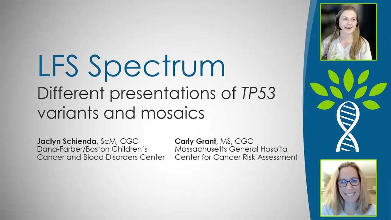
Li-Fraumeni Syndrome LFS Spectrum: Different Presentations of TP53 Variants & Mosaics
1.2K
This extensive section presents the modern clinical framework for understanding Li-Fraumeni syndrome as a spectrum rather than a single condition. Four main categories are defined based on TP53 status and cancer history: Phenotypic LFS (classic clinical criteria without identifiable TP53 mutations), Classic LFS (carriers with TP53 mutations and typical cancer patterns), Attenuated LFS (carriers with TP53 mutations but no personal cancer history and less typical family histories), and Incidental LFS (carriers with TP53 mutations but neither personal nor family history consistent with classic LFS). The section then explores mosaicism—mutations present in some but not all cells—explaining key types including CHIP (blood-only mutations), somatic mosaicism (tumor-confined), constitutional mosaicism (multiple tissues), and gonadal mosaicism (reproductive cells). The section concludes by addressing why identical TP53 mutations produce vastly different clinical outcomes, attributing variation to modifier genes, environmental factors, and gender differences, while emphasizing the multifactorial nature of cancer risk in LFS.
Click to watch →

Grand Round 08 06 2026
115
This segment provides comprehensive coverage of Li-Fraumeni syndrome, a hereditary cancer predisposition syndrome caused by germline TP53 mutations. The syndrome was described in 1969 by Frederick Lee and José Fraumeni through analysis of 648 pediatric rhabdomyosarcoma cases. The clinical criteria include patients under 45 with sarcoma or first-degree relatives with cancer before age 45. The TP53 gene, located on chromosome 17, functions as the 'guardian of the genome' by interrupting cell division for DNA repair and inducing apoptosis if damage is irreparable. Mutations are found in 30-50% of human tumors. In Brazil, the R337H mutation is 15 times more common than in other countries, affecting an estimated 300,000 individuals.
Click to watch →

Encore Open Mics with Dr. Stites - Li-Fraumeni Syndrome and Precision Prevention
120
Li-Fraumeni syndrome (LFS) is a rare hereditary cancer syndrome caused by mutations in the TP53 gene, often called the 'guardian of the genome.' First identified in 1969 and named in 1982, it was linked to TP53 in 1990. The syndrome follows autosomal dominant inheritance, meaning each child has a 50% chance of inheriting the mutation. TP53 normally prevents cancer by triggering cell death in damaged cells; when mutated, it fails to stop cancer development. LFS significantly increases risks for specific cancers: childhood adrenocortical carcinoma, sarcomas, and brain tumors before age 18; and breast cancer, lung cancer, and gastric cancer in adulthood. The syndrome is one of the few inherited forms of lung cancer.
Click to watch →

What Causes Cancer?
106.4K
Li-Fraumeni syndrome is a cancer predisposition syndrome caused by germline mutations in TP53. Patients inherit one pathogenic variant copy of TP53, giving them very strong predisposition to many different cancers including bone cancers, breast cancers, brain tumors, leukemias, soft tissue cancers, and several others. These individuals typically develop cancer before age 45.
Click to watch →
Deep Dive
Modern therapeutic strategies targeting the p53 pathway, such as MDM2 inhibitors (Nutlins) and p53 reactivation therapies.

Ronald Hoffman, MD - Targeting The MPN Stem Cell: What is Missing?
208
MDM2 antagonists (nutlins) reactivate p53 by blocking its degradation, inducing apoptosis in malignant stem cells. Clinical trials show 60-80% survival rates at 12-24 months in refractory patients. PROTACs offer improved tolerability by degrading MDM2 every three weeks versus daily dosing. Selinexor inhibits XPO1, retaining p53 and anti-apoptotic proteins in the nucleus. Unlike JAK2 inhibitors, selinexor reduces stem cell burden in transplant models, representing a paradigm shift toward direct stem cell targeting.
Click to watch →
81st Symposium - Targeting Cancer - Guillermina (Gigi) Lozano
675
The p53 family includes p63 and p73, but these show minimal mutations in human cancers, with p63 typically downregulated in epithelial cancers. Recent findings reveal oncogene addiction to mutant p53 in certain lymphomas—removing mutant p53 causes tumor regression. MDM2 inhibitors aim to reactivate p53 by blocking its degradation, but clinical results remain mixed due to fluctuating p53 levels. Future strategies may focus on disrupting mutant p53 interactions with other proteins to inhibit metastasis, representing a shift from attempting complete p53 restoration toward targeting specific oncogenic pathways driven by mutant p53.
Click to watch →

Optimizing the Management of Dedifferentiated Liposarcoma
209
The p53-MDM2 pathway represents a key therapeutic target in cancer. While p53 mutations occur in 50% of cancers, the remaining 50% have intact wild-type p53 but often show MDM2 overactivation that silences functional p53. Research on 8,000 sarcoma patients confirmed p53 and MDM2 mutations are mutually exclusive. Approximately 20% of all solid tumors exhibit MDM2 amplification with wild-type p53. MDM2 inhibitors have been investigated for 25 years, beginning with nutlin, but initial trials failed due to severe thrombocytopenia preventing further dosing. A critical breakthrough came from recognizing that intermittent dosing (3 days on, 11 days off) dramatically reduced toxicity while maintaining efficacy, transforming an abandoned class of drugs into viable therapeutic candidates.
Click to watch →
Targeting tumor protein 53 (p53): Is there anything we can do therapeutically?
867
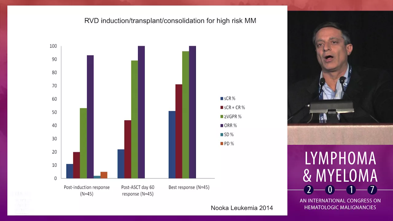
Monoclonal antibodies provide incremental benefit in p53-deleted myeloma, with elotuzumab adding significant efficacy over lenalidomide alone and daratumumab combinations showing additive effects. However, none completely overcome the adverse outcomes associated with 17p deletion. Direct p53 pathway targeting represents the future direction, utilizing MDM2 inhibitors (nutlin-like compounds) that block the MDM2-p53 interaction to release p53's tumor-suppressive functions. These drugs require retention of functional p53 to work, distinguishing them from other targeted therapies. Selective MDM2 inhibitors are currently in clinical development, particularly in combination with bortezomib, which may increase cellular dependence on p53 function. The overarching principle is that p53 abnormalities fundamentally alter treatment selection, requiring upfront risk-adapted approaches rather than reactive adjustments. New drug combinations continue to improve outcomes, as illustrated by successful treatment of a 46-year-old patient with smoldering myeloma using pomalidomide-daratumumab-dexamethasone achieving sustained remission.
Click to watch →

G. Selivanova - Small molecules reactivating p53 as anti-cancer drugs andresearch tools
372
p53 is a tumor suppressor transcription factor that induces cell cycle arrest and apoptosis in response to cellular stress, but is mutated in approximately 50% of human cancers; researchers have developed small molecules like Prima-1 (APR-246) that reactivate mutant p53 by stabilizing its DNA-binding domain, and compounds like Nutlin and Rita that reactivate wild-type p53 by blocking its interaction with MDM2, offering promising new approaches for cancer therapy by restoring the p53-mediated apoptotic pathway in cancer cells.
Click to watch →
Stress & p53
Gene Targeting
Gene Activation
Stress & p53
0:01- 1
Environmental stress like UV light damages DNA.
- 2
p53 protein decides cell death, arrest, or repair.
- 3
Half of all cancers have p53 switched off.
Non-Canonical Mechanisms of p53 Tumor Suppression
While the classical paradigm dictates that p53 prevents cancer primarily by orchestrating cell cycle arrest, DNA repair, and apoptosis in response to acute DNA damage, modern research challenges this dogma. Studies using mutant mouse models have demonstrated that the capacity of p53 to trigger these traditional responses is actually dispensable for its tumor-suppressive capabilities in many contexts. Instead, an emerging counter-perspective suggests that p53's critical cancer-preventative roles lie in 'non-canonical' functions. These include the regulation of cellular metabolism, suppression of reactive oxygen species (ROS), restriction of stem-cell-like self-renewal, and the induction of ferroptosis (an iron-dependent form of cell death). Consequently, this perspective argues that p53 acts less as an acute responder to catastrophic DNA damage and more as a continuous coordinator of metabolic homeostasis and cellular fitness.
Basic eukaryotic cell cycle phases (G1, S, G2, M) and the general concept of restriction checkpoints.
The fundamental structure of DNA and common sources/types of DNA damage, such as UV radiation and double-strand breaks.
The functional distinction between proto-oncogenes and tumor suppressor genes in cancer biology.
The role of transcription factors in regulating gene expression and cellular responses.
The biochemical regulation of p53, focusing on the MDM2-p53 negative feedback loop and ubiquitin-proteasome degradation.
The detailed molecular pathways of apoptosis, specifically the intrinsic pathway involving Bcl-2 family proteins, cytochrome c release, and caspases.
Clinical genetics of TP53 mutations, including Li-Fraumeni Syndrome and its hereditary cancer risks.
Modern therapeutic strategies targeting the p53 pathway, such as MDM2 inhibitors (Nutlins) and p53 reactivation therapies.
100.3K views2.4Klikes5:41@WEHImoviesOriginal Release: 2020-06-26
p53 tumor suppressor your body's cells and tissues are under constant stress from the environment such as DNA damage from UV light emitted from the Sun damage to your DNA causes cells to follow one of three outcomes either the cell dies stopped growing permanently or slows down to repair before resuming normal growth the protein p53 plays an essential role in determining this fate p53 plays a critical role in keeping cells healthy on our bodies cancer free half of all human cancers of p53 switched off p53 is found in the nucleus of each cell responding to DNA damage p53 activates distinct sets of genes that drive processes such as cell death DNA repair or growth arrest and this is what determines the outcome of the cell the activation of genes involved in DNA repair has been linked to p53 x' ability to suppress cancer in the nucleus DNA is wound around histone proteins to create chromatin genes are encoded in your DNA p53 targets specific genes controlling whether they are turned on or off via their DNA promoter the DNA promoter contains a part specific for p53 known as the p53 response element the stars of the gene is found within the core promoter p53 binds its response elements and helps bring RNA polymerase to the start of the gene RNA polymerase is the enzyme responsible for reading and transcribing for gene code this p53 target gene is currently off RNA polymerase is not reading the gene code the amount of p53 increases in response to UV induced damage from the Sun [Applause] the highly dynamic p53 protein is made of four identical parts each part contains a DNA binding domain p53 binds DNA all four DNA binding domains combine the response elements in the promoter of target genes to turn the gene on p53 does two things first p53 makes the gene more accessible by recruiting proteins that remodel the DNA second p53 brings RNA polymerase to the now accessible target gene now switched on the gene will be read and transcribed by RNA polymerase activating pathways such as soda growth arrest or DNA repair
Up Next

50:24
Cancer Genetics: Oncogenes, Tumor Suppressors, and Mutations | MIT 7.013
@mitocw
41.8K views•2014-01-15

50:12
Bacterial Communication: Quorum Sensing and Biofilm Formation Explained
@JHUAAP
12.6K views•2011-06-28
3:32
Proteasome Function & Protein Recycling Explained | Molecular Animation
@WEHImovies
42.5K views•2024-05-24

7:08
Bacteriophages: Earth's Deadliest Killers and Future Antibiotics
@kurzgesagt
34.6M views•2018-05-13
Related Study Plans & Knowledge Roadmaps
Structured learning paths in Biology
biology
RNAi: Biogenesis, RISC & Gene Silencing
01Molecular Biology Foundations: DNA to Protein
02Introduction to RNA Interference (RNAi)
03Molecular Biogenesis of miRNA and siRNA
+ 2 more learning modules
10h5 modules
Explore Roadmap
biology
Mycorrhizae: Signaling, Biophysics & Ecology
01Foundations of Mycorrhizal Symbiosis
02Molecular Signaling and Root Colonization
03Biophysics and Bioenergetics of Nutrient Exchange
+ 1 more learning modules
11h4 modules
Explore Roadmap
biology
Circadian Clocks: TTFL, SCN & Periphery
01Foundations of Molecular Biology & Gene Expression
02Introduction to Chronobiology and Actograms
03The Core TTFL: Molecular Architecture of the Clock
+ 3 more learning modules
14h6 modules
Explore Roadmap
biology
Population Genetics: Math, Drift & Selection
01Genetic & Probability Foundations
02The Hardy-Weinberg Principle and Baseline Models
03Mathematical Modeling of Natural Selection
+ 3 more learning modules
18h6 modules
Explore Roadmap